





Abstract
Dear Sir,
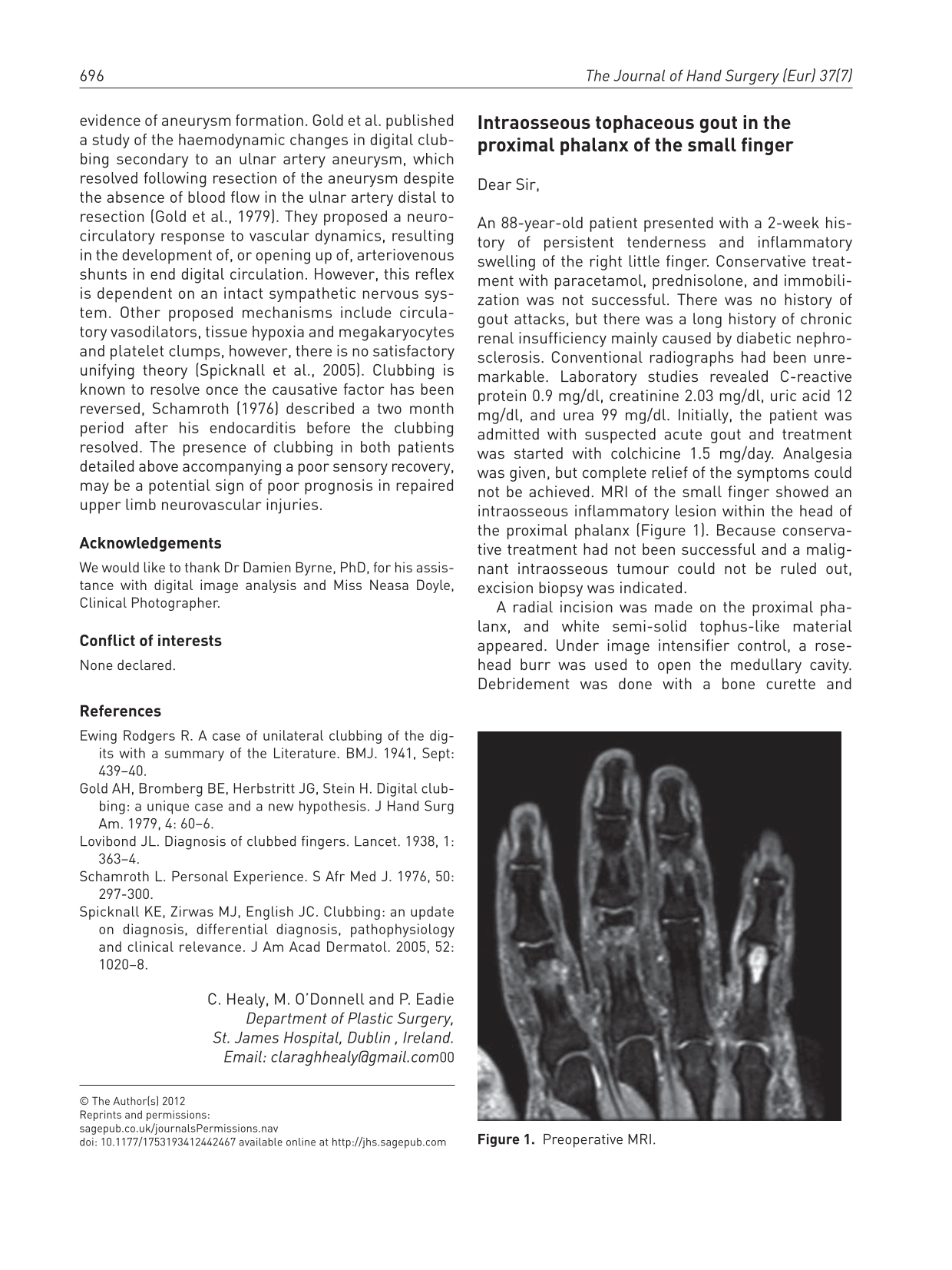
An 88-year-old patient presented with a 2-week history of persistent tenderness and inflammatory swelling of the right little finger. Conservative treatment with paracetamol, prednisolone, and immobilization was not successful. There was no history of gout attacks, but there was a long history of chronic renal insufficiency mainly caused by diabetic nephrosclerosis. Conventional radiographs had been unremarkable. Laboratory studies revealed C-reactive protein 0.9 mg/dl, creatinine 2.03 mg/dl, uric acid 12 mg/dl, and urea 99 mg/dl. Initially, the patient was admitted with suspected acute gout and treatment was started with colchicine 1.5 mg/day. Analgesia was given, but complete relief of the symptoms could not be achieved. MRI of the small finger showed an intraosseous inflammatory lesion within the head of the proximal phalanx (Figure 1). Because conservative treatment had not been successful and a malignant intraosseous tumour could not be ruled out, excision biopsy was indicated.

Preoperative MRI.
A radial incision was made on the proximal phalanx, and white semi-solid tophus-like material appeared. Under image intensifier control, a rose-head burr was used to open the medullary cavity. Debridement was done with a bone curette and bacteriological cultures were obtained. Radiographs confirmed complete excision of the lesion. No autologous bone graft was inserted in view of the small defect. There was complete relief of symptoms after the operation. Bacteriological cultures were negative, but histopathology confirmed the suspected diagnosis of intraosseous tophaceous gout.
At follow-up examinations 11 days and 5 weeks postoperatively, the wound was healed and had no signs of infection. At the last review 17 weeks after operation, the incision remained satisfactory with some tenderness reported on firm palpation. MRI showed consolidating bone and no signs of recurrence (Figure 2). Local recurrence is not expected, provided the patient continues to take antihyperuricaemic medication.
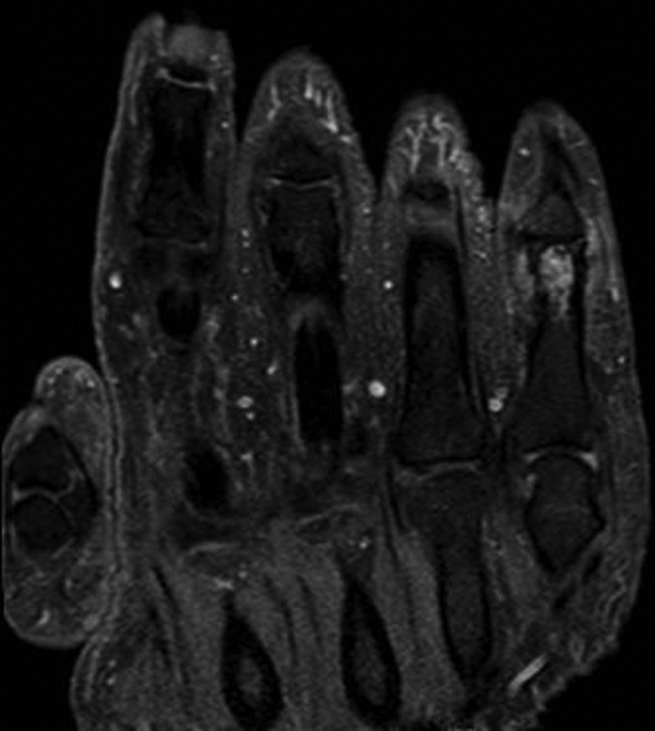
MRI 17 weeks after operation.
In gout there is a deficiency of xanthine oxidase. Inefficient renal tubular elimination of the accruing products of purine metabolism results in accumulation of urate crystals in blood and tissues, accompanied by an inflammatory reaction. The prevalence ratio of men to women is 3:1 (Lawrence et al., 2008), peaking between aged 40 and 60 years (Smith et al., 2010). Chronic urate accumulations in interstitial tissue can form subcutaneous tophi; far less commonly, they occur in bone tissue as intraosseous tophi. MRI can be used to detect interstitial inflammatory reaction and tophaceous gout. Intraosseous tophi, however, are hard to distinguish from intraosseous tumours (Liu et al., 2003); in the latter case, surgical biopsy and histopathological diagnosis are mandatory.
In spite of its low incidence, tophaceous gout should be considered in the differential diagnosis of intraosseous lesions, especially if there is a history of hyperuricaemia and renal insufficiency.
Footnotes
Acknowledgements
K.-U. Eckardt, Department of Nephrology, and A. Okuducu, Department of Pathology, Klinikum Nürnberg Süd, Nürnberg, Germany.
Conflict of interests
None declared.