





Abstract
Dear Sir,
A 75-year-old woman presented with a painless soft tissue swelling on the flexor aspect of the proximal phalanx of the left index finger. She had first noticed the slowly growing lesion 6 months previously. Flexion and extension of the finger were normal. Radiography of the finger did not show any bone lesions. Preoperative pulmonary function evaluation, chest radiography, and laboratory tests were normal.
Her past medical history was significant. She had had an open carpal tunnel release on the left side 10 years before, 5 years after a malunited fracture of the left distal radius. When the carpal tunnel was opened, the flexor tendons were found to be surrounded by hypertrophied tenosynovium, which necessitated flexor tenosynovectomy. At the time of surgery, the diagnosis was not clear; however, histopathology of the flexor tenosynovium showed multiple noncaseating, epithelioid granulomatous lesions consistent with sarcoidosis. The symptoms resolved after surgery and she had no other signs of systemic sarcoidosis. After surgery she did not take any medication to reduce the risk of recurrent sarcoid tenosynovitis. Ten years later, she had not had any signs of local recurrence at the carpal tunnel region.
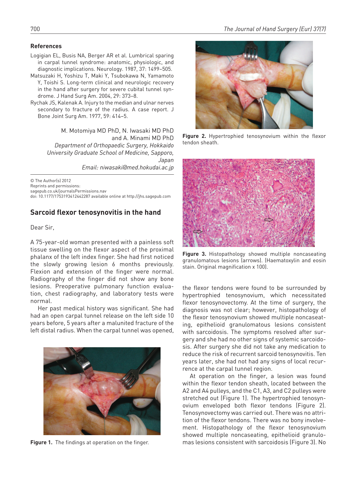
At operation on the finger, a lesion was found within the flexor tendon sheath, located between the A2 and A4 pulleys, and the C1, A3, and C2 pulleys were stretched out (Figure 1). The hypertrophied tenosynovium enveloped both flexor tendons (Figure 2). Tenosynovectomy was carried out. There was no attrition of the flexor tendons. There was no bony involvement. Histopathology of the flexor tenosynovium showed multiple noncaseating, epithelioid granulomas lesions consistent with sarcoidosis (Figure 3). No acid-fast organisms were demonstrated on Ziehl-Neelsen staining. She refused to take steroid treatment. One year after operation, she had not developed any signs of local recurrence or systemic involvement of the disease.

The findings at operation on the finger.
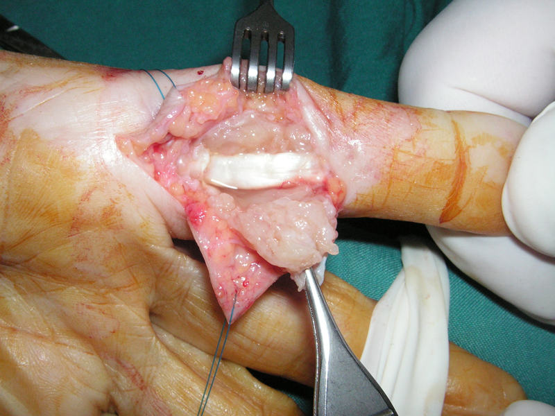
Hypertrophied tenosynovium within the flexor tendon sheath.
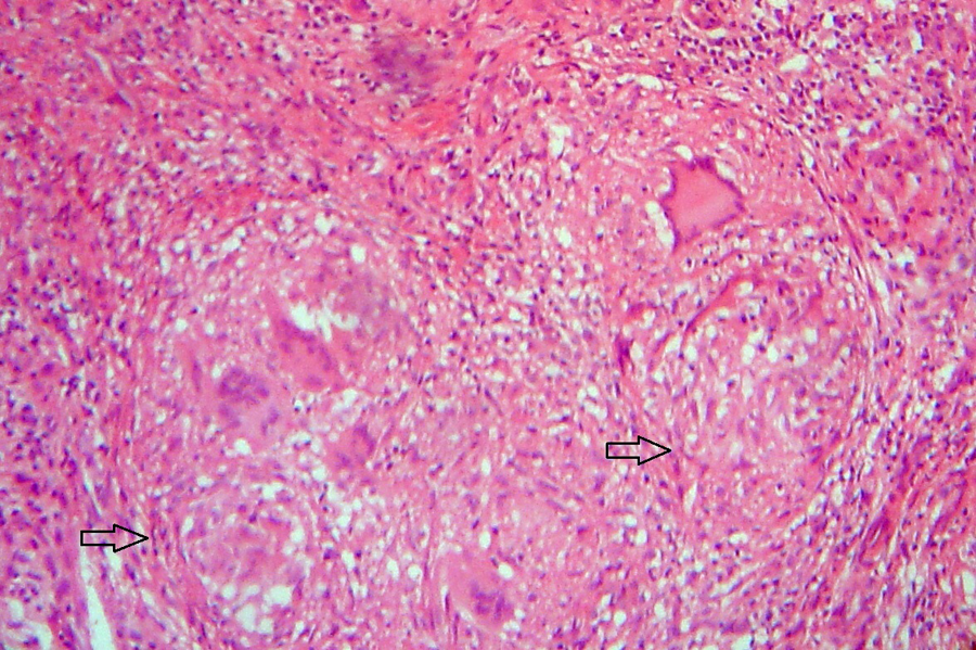
Histopathology showed multiple noncaseating granulomatous lesions (arrows). (Haematoxylin and eosin stain. Original magnification × 100).
Sarcoidosis is a systemic granulomatous disease with an unknown aetiology. Skeletal involvement has been reported up to 30% of patients with sarcoidosis (Altman et al., 2011; Katzman et al., 1997; Merle et al., 1986). Hand involvement is not common and isolated flexor tenosynovitis is a rare manifestation of the disease (Katzman et al., 1997; Merle et al., 1986; Wang et al., 2005). However, tenosynovitis in the hand may be the first manifestation of systemic sarcoid disease (Altman et al., 2011; Katzman et al., 1997).
Although systemic steroid therapy has been recommended to prevent recurrence and progression of sarcoid disease (Merle et al., 1986; Wang et al., 2005), the prognosis and functional outcome of sarcoidosis involving the hand are not clear (Altman et al., 2011; Merle et al., 1986). It might be a self-limiting disease (Altman et al., 2011). Bony involvement may cause pathological fractures that are difficult to treat, and sarcoid tenosynovitis may be complicated by tendon rupture (Wang et al., 2005).
In the current case, two anatomically separate areas of sarcoid flexor tenosynovitis developed in the patient’s left hand. Although the lesions were seen over a 10-year interval, she did not develop any features of systemic sarcoid disease. There was no local recurrence of tenosynovitis after the operations, which suggests that long-term local control of sarcoid tenosynovitis might be achieved with synovectomy.
Footnotes
Conflict of interests
None declared.