





Abstract
Dear Sir,
A 25-year-old right-handed student nurse presented with a 2 year history of increasing pain in the right wrist, despite intermittent use of splintage and NSAIDs. Examination revealed tenderness dorsally over the lunate with some minor restriction in wrist flexion and extension. Stage IIIa Kienböck’s disease was diagnosed using radiographs and MRI (Figures 1 and 2). Treatment options were discussed, but whilst taking a few months to consider them, she developed similar, albeit less severe, pain in the left wrist. Clinical and radiological examinations were unremarkable, but an MRI scan suggested stage I Kienböck’s disease (Figure 2). She had no past history of trauma to either wrist, but she had type 1 diabetes, diagnosed at the age of 16 years. Other noteworthy history included thyrotoxicosis, diagnosed 5 years previously and treated with carbimazole.

(a) Preoperative radiographs of the right hand showing altered trabecular pattern in the lunate with subcortical lucency/sclerosis and (b) collapse of the lunate with a step in the cortical margin on the lateral view. (c) 3 months after revascularization.
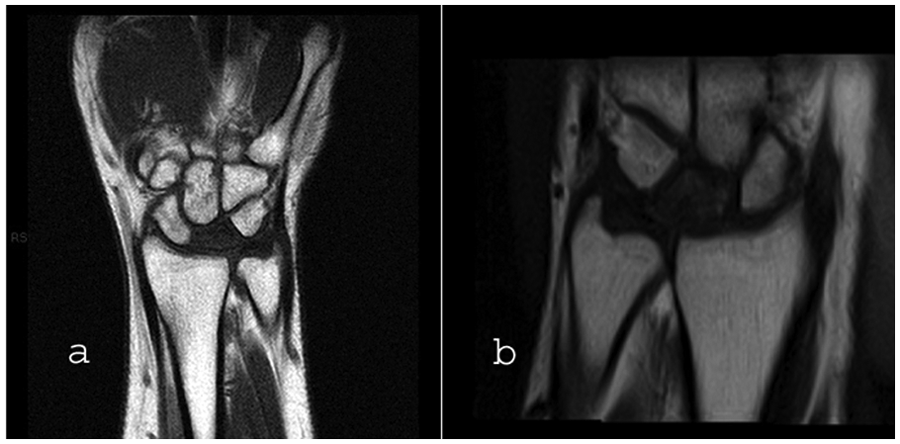
(a) Preoperative T1-weighted coronal MRI scan of the right wrist, suggestive of stage IIIa Kienböck’s disease. (b) Pre-treatment short tau inversion recovery (STIR) sequence coronal scan of the left wrist, indicative of stage I Kienböck’s disease.
She underwent a revascularization procedure to the right lunate (using a reverse-flow pedicled bone graft based on the 4/5 extensor compartment artery). The left hand was managed conservatively in a plaster cast for 3 months. Treatment resulted in improvement in vascularity on MRI scan and resolution of pain.
Several cases of Kienböck’s disease have been described in patients with systemic conditions such as systemic lupus erythematosus (SLE), rheumatoid arthritis, or Crohn’s disease, and the association has generally been attributed to systemic steroid treatment. Bilateral Kienböck’s disease has been described in several small series (Rasmusen and Schantz, 1987; Taniguchi and Tamaki, 1998; Yazaki et al., 2005) and reported systemic associations are uncommon. Mok et al. (1997) discussed a single case of bilateral Kienböck’s disease in a patient treated with steroids for SLE. In reports of bilateral Kienböck’s disease that contain descriptions of occupations, it has been suggested that repetitive stress associated with manual labour may contribute to the development of the disease (but no case-control study has been performed to confirm or refute this). There are no previous reports of Kienböck’s disease occurring in association with diabetes. The co-existence of the two pathologies may represent a chance occurrence, but because the microangiopathy that occurs in diabetes may be an aetiological factor in the development of Kienböck’s disease, we thought it worthwhile to report this case.
Footnotes
Conflict of interests
None declared.