





Abstract
Dear Sir,
A 63-year-old right-hand dominant woman presented to our clinic with left wrist pain two days after slipping on her kitchen floor and landing on her left side. She reported feeling something ‘give’ in her wrist on falling, and on examination there was swelling and bruising on the volar aspect of her left forearm. She had weakness of wrist flexion but the flexor carpi radialis (FCR) was palpable and intact. The remaining flexor tendons appeared to be intact but she did have pain on making a fist. She described this as having felt similar to an injury she had previously sustained to her right wrist when mopping a floor in 2008, which was diagnosed as a closed FCR tendon rupture and required excision of the proximal stump owing to painful adhesions.
She has a history of bilateral trapeziectomies for osteoarthritis with ligament reconstruction on the right side in 1995 and left side in 2001, and a previous right distal radius fracture in 2005 resulting in slight instability of the distal radio–ulnar joint. Aside from this, she has an extensive history of osteoarthritis in other joints from an early age, has had numerous steroid injections to both thumbs prior to the trapeziectomies and to her right elbow, and has been tested for rheumatoid disease and found to be negative. Her medications history includes anti-inflammatories, Ranitidine, Mebeverine, Detrusito and Bendroflumethiazide.
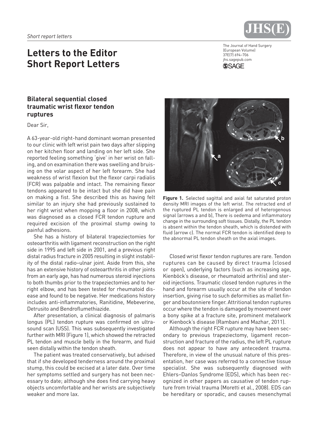
After presentation, a clinical diagnosis of palmaris longus (PL) tendon rupture was confirmed on ultrasound scan (USS). This was subsequently investigated further with MRI (Figure 1), which showed the retracted PL tendon and muscle belly in the forearm, and fluid seen distally within the tendon sheath.

Selected sagittal and axial fat saturated proton density MRI images of the left wrist. The retracted end of the ruptured PL tendon is enlarged and of heterogenous signal (arrows a and b), There is oedema and inflammatory change in the surrounding soft tissues. Distally, the PL tendon is absent within the tendon sheath, which is distended with fluid (arrow c). The normal FCR tendon is identified deep to the abnormal PL tendon sheath on the axial images.
The patient was treated conservatively, but advised that if she developed tenderness around the proximal stump, this could be excised at a later date. Over time her symptoms settled and surgery has not been necessary to date; although she does find carrying heavy objects uncomfortable and her wrists are subjectively weaker and more lax.
Closed wrist flexor tendon ruptures are rare. Tendon ruptures can be caused by direct trauma (closed or open), underlying factors (such as increasing age, Kienböck’s disease, or rheumatoid arthritis) and steroid injections. Traumatic closed tendon ruptures in the hand and forearm usually occur at the site of tendon insertion, giving rise to such deformities as mallet finger and boutonniere finger. Attritional tendon ruptures occur where the tendon is damaged by movement over a bony spike at a fracture site, prominent metalwork or Kienbock’s disease (Rambani and Mazhar, 2011).
Although the right FCR rupture may have been secondary to previous trapeziectomy, ligament reconstruction and fracture of the radius, the left PL rupture does not appear to have any antecedent trauma. Therefore, in view of the unusual nature of this presentation, her case was referred to a connective tissue specialist. She was subsequently diagnosed with Ehlers–Danlos Syndrome (EDS), which has been recognized in other papers as causative of tendon rupture from trivial trauma (Moretti et al., 2008). EDS can be hereditary or sporadic, and causes mesenchymal tissue to be abnormal, resulting in hyperelasticity of connective tissue and increased susceptibility to trauma (Pálvölgyi et al., 1979)
The patient has since been referred for specialist physiotherapy, and although she does not think this condition affected any of her ancestors, her daughter has since also been diagnosed with EDS.
Footnotes
Conflict of interests
None declared.