






Abstract
We present a prospective follow-up of 39 Elektra prostheses in 37 patients (32 women and five men), with a mean age of 56.5 (range 46–71) years; 34 patients had osteoarthritis and three had rheumatoid arthritis. Patients were followed using clinical examination, including measurement of pain on a visual analogue scale, mobility, and strength, after 6, 12, 26, and 52 weeks, and annually thereafter. Radiological examination was done preoperatively and after 6, 26, and 52 weeks, and annually thereafter. The mean follow-up time was 48 (range 3–91) months. Although we observed a fast recovery, including maintenance of mobility and a gradual increase in grip strength, there was a revision rate of 7/38 (24%) after 36 months, increasing to 17/38 (44%) after 72 months. The main reason for revision was loosening of the trapezial component, and biomechanical properties of the trapezial fixation may be the key problem in treating trapeziometacarpal osteoarthritis using a total prosthesis.
Keywords
Introduction
The results of a suspension arthroplasty in treating osteoarthritis of the trapeziometacarpal (TMC) joint are generally good, leaving 80–90% of patients pain free and with good function. However, the disadvantage of this method is a long recovery time of at least 3 to 6 months (Vadstrup et al., 2009). For this reason, the development of a well-functioning joint prosthesis has been attractive. Since de la Caffinière published the first report of a TMC joint prosthesis, many different ball-and-socket designs have been developed (Alnot and Saint Laurent, 1985; Comtet and Rumelhart, 2001; de la Caffinière, 1973; Hannula and Nahigian, 1999).
The Elektra prosthesis (Small Bone Innovations Inc., Morrisville, Pennsylvania, USA; formerly Fixano, Péronnas, France), a cementless ball and socket prosthesis with metal-on-metal articulating surfaces, was introduced in 1996 (Regnard, 2006). The theoretical advantages of this prosthesis were the hydroxyapatite-coated components to facilitate osteointegration. Ten years after the introduction of the Elektra prosthesis, early follow-up results of 100 implants were encouraging, with a fast recovery of 2–3 weeks, good mobility and grip strength, and a revision rate due to aseptic loosening of 15% after 53 months (Regnard, 2006). However, 2 years later Hansen and Snerum (2008) published their experience with 17 implants and found aseptic loosening in 5/17 (29%) and a total revision rate of 7/17 (41%) after 35 months. Hernández-Cortés et al. (2012) published a 2 year follow-up and found radiographic osteolysis in nine of 19 implants, and four underwent surgical revision.
The longer term results of this prosthesis are still not established. For this reason, we wish to contribute with our clinical results in a prospective, long-term follow-up of 39 Elektra prostheses implanted in the TMC joint.
Methods
Between January 2003 and October 2009, 39 TMC joints in 37 patients were treated with the second generation of the Elektra prosthesis and followed prospectively. In total, 34 patients were diagnosed with symptomatic and radiological TMC osteoarthritis (Eaton–Littler stage I–III) (Eaton and Littler, 1973), and three patients had rheumatoid arthritis. Patients with concomitant scaphotrapeziotrapezoid (STT) osteoarthritis or known severe osteoporosis were not recommended for treatment with the prosthesis. The sex, age, hand dominance, and type of employment were recorded preoperatively for all patients.
All operations were done by two consultant hand surgeons using the procedure described by Regnard (2006). Postoperatively, the hand was immobilized in a cast for 3 weeks followed by 3 weeks in a removable splint. After 6 weeks, all patients were given occupational therapy with progressively more load on the hand during the following 2–3 weeks.
Pain, strength and mobility were recorded preoperatively and at 6, 12, 26, and 52 weeks postoperatively. After that patients were seen annually or until revision. Pain was measured using a visual analogue scale (VAS) and expressed in mm, 0 being “no pain” and 100 “maximal pain” (Clarke and Spear, 1964). Grip and pinch strengths were measured using the Jamar dynamometer and expressed in kgf. Mobility was measured as follows: active abduction, defined as the angle between the thumb and index metacarpals when moving the thumb radially away from the index finger in a plane parallel to the palm; active palmar abduction, defined as the angle between thumb and index metacarpal when the thumb rises in a plane perpendicular to the palm; and the distance in mm from the tip of the thumb to the tip and base of the small finger, respectively.
Preoperatively, radiographs were taken of the TMC joint in anteroposterior, lateral projection, and a Bett’s view, and assessed for osteoarthritis (Dela Rosa et al., 2004). Postoperative radiographs were taken after 6 and 26 weeks, and 1 year. Thereafter, radiographs were taken annually. Postoperative radiographs were assessed for implant position, fractures, radiolucent zones, or migration as a sign of implant loosening.
Indications for reoperation were radiological signs of loosening, implant failure, or luxation combined with pain unacceptable to the patient. We decided not to attempt reimplantation of the cup, but instead to remove the cup and neck and convert to suspension arthroplasty. In all except one case, the stem was not prominent in the TMC-cavity and was left in place.
Statistical methods
A one-sample t-test was used to calculate a p-value for the comparison of means. Survival rate was calculated and presented as a Kaplan–Meier plot.
Results
A total of 39 Elektra prostheses were implanted in 37 patients (32 women and five men). Mean age was 56.5 (range 46–71) years. Fifteen prostheses were implanted in the dominant and 24 in the non-dominant hand. Two patients had prostheses in both hands. Mean follow-up was 48 (range 3–91) months. One patient was lost to follow-up after 3 months and censored in the survival analysis. Twenty-two patients were still employed and 15 had retired. Five patients were known to engage in heavy manual work (two nursing assistants, one kitchen assistant, one caretaker, and one nursery school teacher). Mean time before returning to work was 10 (range 2–28) weeks.
The distribution of pre- and postoperative VAS scores are shown in Figure 1. VAS-score decreased significantly after 6 weeks compared with the preoperative level (p < 0.001) and continued to be lower than the preoperative score in patients not revised. One VAS score was missed preoperatively.

Visual analogue scale (VAS) score results and standard deviations. There was a significant (*) reduction in VAS score at all postoperative follow-up times compared with the preoperative level. There were no significant changes in VAS scores between 6 weeks to 3 years. Data for patients who had 4 years follow-up or longer are not shown because of small numbers.
Grip and pinch strength measurements are shown in Figure 2. Grip strength did not significantly change until 26 weeks after the operation compared with the preoperative level (p < 0.001). Grip strength continued to increase until 2 years after operation in patients not revised; a decrease in grip strength was then seen. No significant change was found in pinch strength. Preoperative strength measurement was missed in three patients.
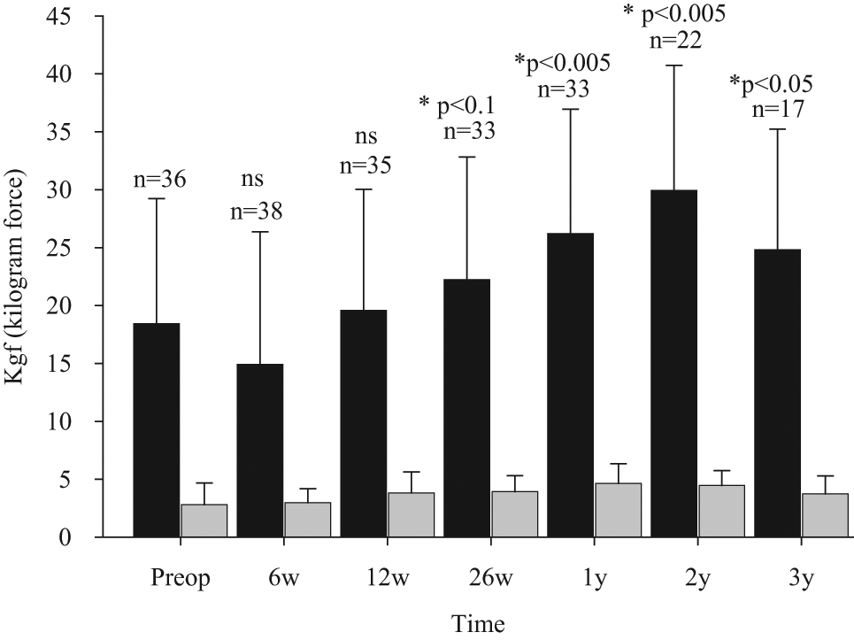
Grip and pinch strength results and standard deviations. Compared with preoperative values, grip strength (black) was unchanged until 26 weeks after the operation. After that grip strength significantly (*) improved compared with the preoperative value. Pinch strength (grey) remained unchanged compared with the preoperative level at all times.
Abduction and palmar abduction did not significantly change compared with preoperative measurements. The distance from the tip of the thumb to the tip of the small finger was 0 mm preoperatively and continued to be so at all follow-up visits. Mean distance from the tip of the thumb to the base of the small finger was 5 mm preoperatively, and no significant change was found.
Peroperatively, there were four cases of a crack fracture in the trapezium and one case of crack fracture in the base of the thumb metacarpal. These patients were given prolonged immobilization in a cast for 6 weeks, followed by a removable splint for 3 weeks. Only one of these has later been revised. In one case, the stem could not be completely inserted. This was solved by the use of a shorter neck.
Eight patients had postoperative complications. One patient developed a superficial infection after 2 weeks; this was treated with oral dicloxacillin for 2 weeks and had no further complications. Four patients developed de Quervain’s disease after 3, 6, 6, and 36 months, respectively. Only one needed surgical treatment. Three patients developed triggering of the thumb after 3, 4, and 17 months, respectively. Two were treated with corticosteroid injection only, and one had surgical release of the A1 pulley.
A total of 17 patients had the prosthesis removed (15 women and two men). Mean age was 58 (range 42–71) years and mean survival of the prosthesis was 37 (range 8–72) months. Of the 20 patients with a follow-up longer than 4 years, 13/20 (65%) prostheses were removed. Four of the reoperated patients — two women and two men — were known to carry out heavy manual work. The probability of implant survival is shown in Figure 3.
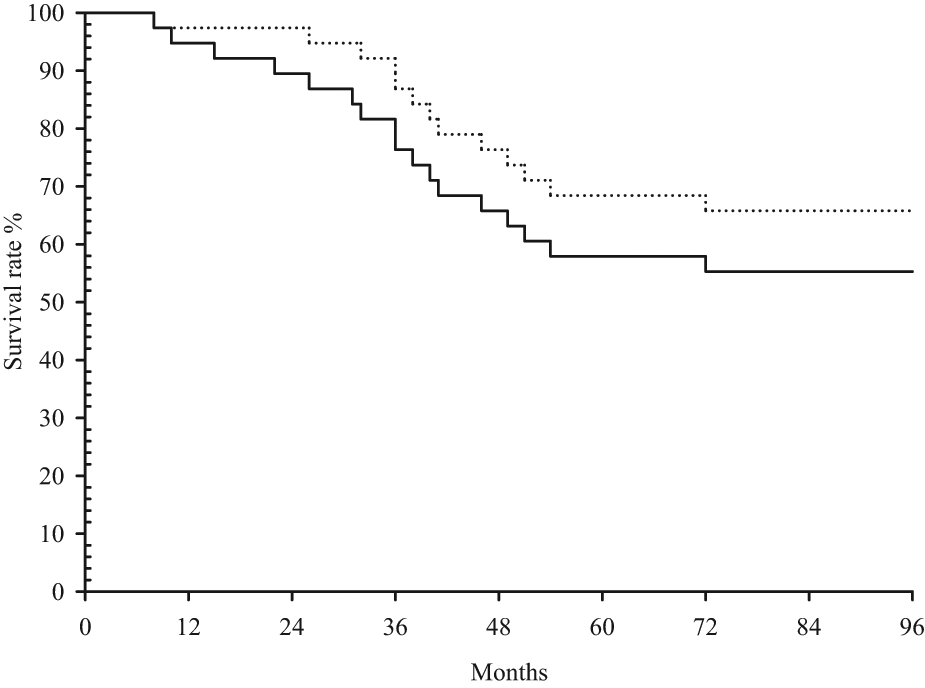
Kaplan–Meier plot showing survival for the Elektra prosthesis in 38 patients. One patient was lost to follow-up after 3 months and is not included in the survival analysis. A total of 17 patients were revised (solid line). Thirteen patients were revised because of aseptic loosening (dotted line).
Thirteen of the 17 prostheses were removed because of aseptic loosening of the cup (Figure 3). In revised patients, radiological assessment showed radiolucent zones in four patients and migration of the cup in nine patients. Radiological assessment of the prostheses in nonrevised patients showed no signs of loosening.
One prosthesis was removed because of a painful dislocation. This was seen on radiographs after 18 months and became symptomatic 9 months later (Figure 4).
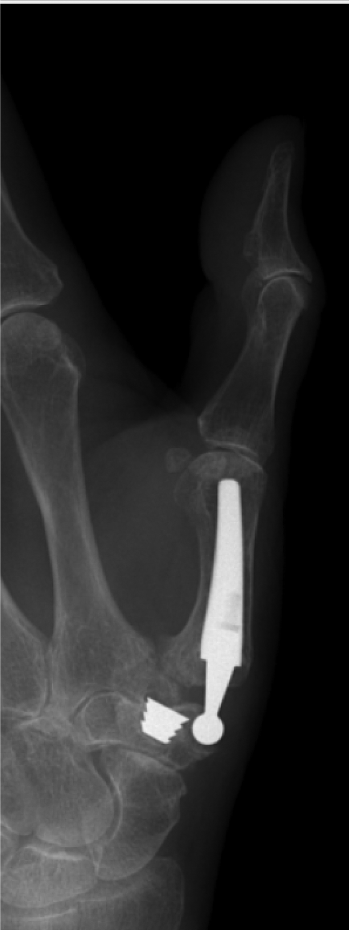
One patient had a symptomatic dislocation and was revised because of instability and pain.
In one patient, the prosthesis was removed because of STT-joint osteoarthritis found during exploration of the prosthesis after 15 months because of persistent pain. STT osteoarthritis was not seen on radiographs and the prosthesis itself had not loosened.
Two patients had the prosthesis removed because of swelling and tenderness on the radial side of the hand, which developed after 5 and 10 months, respectively. In both patients, metallosis was observed. These prostheses were found to be in place and were not loose. Cultures were negative. One of these patients had known hypersensitivity to nickel.
No implant failure or peroperative crack fractures were seen on radiographs. Loosening of the stem was never seen.
Discussion
We have treated a series of patients with the Elektra prosthesis, and although we found fast pain relief, maintenance of mobility, and a gradual increase in grip strength, we found an increasing need for revision. The revision rate was 7/38 (24%) after 36 months, which is close to the results of Hansen et al. (2011) who reported a revision rate of 5/17 (29%) after 35 months. However, with longer follow-up we found an increasing revision rate of 13/38 (34%) after 48 months and 17/38 (44%) after 72 months.
Relatively few studies of long-term follow-up of ball and socket implants for the TMC joint have been published and the results are varying. Wachtl et al. (1998), who reported a follow-up of the cemented de la Caffinière prosthesis, found a survival rate of only 66% after 68 months. Søndergaard et al. (1991) found that 18/22 de la Caffinière prostheses were still in place after 10 years, but with three prostheses revised. Skyttä et al. (2005) even reported a survival of 92% after 10 years for the de la Caffinière prostheses, based on revisions in 49 patients who had rheumatoid arthritis. Lemoine et al. (2009) did a retrospective follow-up of 72 patients treated with the cemented second-generation Guepar prosthesis. After a mean follow-up of 50 months, lucent lines were found in 33% of patients, and loosening in 3% of the trapezial components and 3% of the metacarpal stems. The survival rate was not reported.
To our knowledge, no long-term follow-up studies of uncemented implants other than the Elektra prostheses have been published. Results of the first cementless implant for the TMC joint, the Ledoux prosthesis, were disappointing. Wachtl et al. (1998) found survival of only 28/45 (62%) implants after 16 months.
Authors report failures such as loosening and dislocation in different ways, and the indications for revision may also differ; this makes it difficult to compare the published series. Furthermore, treatment groups may differ in age, gender, occupation, and stage of osteoarthritis, which causes further difficulty in comparing the revision rates.
For both cemented and uncemented implants, the main problem mentioned by the authors of follow-up studies has been the fixation of the cup component in the trapezium. However, the first results published by Regnard (2006) for the Elektra prosthesis showed a revision rate due to aseptic loosening of only 15% after 53 months, indicating that a possible solution for the fixation of the trapezial component had been found.
In the present study, we found a revision rate of 16/38 (42%) after 54 months. A total of 17 prostheses were removed; 13 (34%) were removed because of aseptic loosening of the trapezial component. The Kaplan–Meier plot for prostheses revised because of aseptic loosening shows an increasing failure rate after just 2 years. Furthermore, the survival plot shows a continuous decreasing tendency, indicating that more revisions must be expected in the future. For these reasons, we still consider the bony fixation of the trapezial component to be the biggest challenge in the treatment of TMC osteoarthritis by total arthroplasty.
Several factors are thought to contribute to this problem. Hansen and Snerum (2008) reported a follow-up of 17 Elektra prostheses and mention metallosis and forceful threading before insertion of the cup as possible causes of failure of fixation. Later, this group compared the primary press fit fixation of two different, uncemented cup designs — MOTEC and Elektra — in a pig model and found no difference. However, they found that the threading of the bone before insertion of the Elektra screw cup did weaken the primary fixation (Hansen et al., 2011).
Biomechanical aspects may also play a role. Pinch forces at the pulp of the thumb are magnified 10 to 13 times at the trapeziometacarpal level, leading to significant stress on the implant and increasing the risk of loosening. In the normal TMC joint of the thumb, there is no fixed, single centre of rotation and, as a consequence, the motion in a spherical prosthesis construction may be deleterious for cup fixation or cause exaggerated wear and tear, and perhaps even dislocation (Bozentka, 2010).
Last but not least, the trapezium mainly consists of frail cancellous bone, especially in postmenopausal women. Stress shielding during the years after implantation may increase this problem.
A third-generation Elektra has been designed, in which threading for cup fixation is avoided and a titanium cup is used. Time will show to what extent this will increase the durability of cup fixation. However, as a consequence of our experience with the poor durability of the Elektra prosthesis, our primary method of treating trapeziometacarpal osteoarthritis is once again resection of trapezium and suspension arthroplasty.
Footnotes
Conflict of interests
None declared.
Funding
This research received no specific grant from any funding agency in the public, commercial, or not-for-profit sectors.