






Abstract
Arthroplasty is one of several options for treating symptomatic osteoarthritis of the thumb carpometacarpal joint. There are various hemi- and total arthroplasties available on the market. We report our experience of treating 12 patients with the Moje Acamo CMC1 prosthesis. We reviewed all the patients at a mean of 50 months postoperatively. All patients presented with loosening, migration or tilting of one or both implant components. Nine patients were symptomatic enough to warrant revision surgery with removal of the implant leaving a pseudarthrosis. At the last follow-up, five patients (42%) had already received revision surgery. All patients still possessing an implant showed progressive signs of implant loosening, although some of them had no symptoms. In order to assess their overall disadvantage compared with patients treated by primary trapeziectomy, the outcomes of these two groups were compared (matched pairs). The outcomes after revision surgery were comparable with those of primary trapeziectomy. We no longer use the Moje Acamo CMC1 implant and recommend that patients who have received this implant should be monitored carefully both clinically and radiologically.
Introduction
Arthroplasty is one of several options for treating symptomatic osteoarthritis of the thumb carpometacarpal (CMC) joint. It aims to preserve the length of the thumb (Luria et al., 2007), which is important for maintaining balance of the soft tissues (Cooney and Chao, 1977). In this way, ulnar and axial displacement and metacarpal angulation can be significantly reduced compared with all types of trapeziectomy (either trapezial excision alone or supplemented by a tendon interposition, ligament reconstruction or both) (Luria et al., 2007). There are various hemi- and total arthroplasty implants available on the market. A number of these have been associated with high complication rates, including aseptic loosening and subsidence, subluxation and dislocation, periprosthetic fracture, and infection (Chakrabarti et al., 1997; Hansen and Homilius, 2010; Hernandez-Cortes et al., 2012; Pendse et al., 2009; Perez-Ubeda et al., 2003; Wachtl et al., 1998). Good long-term outcomes have been reported with the de la Caffiniere implant (Johnston et al., 2011).
The implant components for thumb CMC joint arthroplasty can be cemented in place or held with a press fit, which is anticipated to become a stable bone implant interface with/without osseo-integration. Stability at the bone–implant interface depends on a number of factors, including implant material characteristics: elasticity, porosity, chemical composition, and particle release (Branemark et al., 2001; Mavrogenis et al., 2009). Other factors will include bone stock and surgical skill. The relative contribution of each is often unclear.
In the last decade, novel zircon dioxide ceramic implants were introduced for total arthroplasty of the thumb CMC joint; the Moje Acamo CMC1. The implants are fixed using a press-fit technique, without applying cement. The coating of the implants is called Bioverit-1®, a glass–ceramic layer, which is designed to provide a long lasting low friction joint interface. The roughened surfaces of the implant stems are designed to encourage osseo-integration (Ignatius et al., 2005). There have been reports of implantation in the hallux metatarsophalangeal (MTP) joint (Barwick and Talkhani, 2008; Brewster et al., 2010; Kulshreshtha et al., 2009), and the proximal interphalangeal joint (Pettersson et al., 2006; Wesemann et al., 2008). In the thumb CMC joint there are no reports of clinical success and the only two reports are from Hansen et al. reporting a high rate of complications (Hansen and Homilius, 2010; Hansen and Vainorius, 2008).
The aims of this study were to record the outcomes of our implantation of the Moje Acamo CMC1 arthroplasty and compare the outcomes of the patients who had revision surgery with implant removal and trapeziectomy to a similar cohort of patients who had primary trapeziectomy.
Patients and methods
Patients
Between March 2006 and July 2007 we treated 12 patients with symptomatic thumb CMC joint osteoarthritis with Moje Acamo CMC1 arthroplasties. We adhered to the exclusion criteria as advised by the manufacturer (severe subluxation, trapezial height < 8 mm, osteoporosis, algodystrophy, rheumatic diseases, heavy physical work or sports, and scapho-trapezio-trapezoid arthritis). At review, five patients had already undergone revision with removal of the implants and creation of a pseudarthrosis with complete (two patients) or distal excision (three patients) of the trapezium (Figure 1). In order to assess their outcomes more fully we chose to compare their outcome data with age (within ten years) and sex matched patients who had undergone primary trapeziectomies at the same time as the revision surgery (within one year but with a minimum follow-up of one year post-operatively).

(a) Radiograph of ceramic prosthesis with proximal migration, tilting, and loosening, while the distal component shows intense loosening 3.9 y after the implantation. (b) and (c) Radiographs after implant removal and partial trapeziectomy.
Surgical technique
The operations were all performed by one senior surgeon, according to the manufacturer’s manual (see the Online Supplement, II. Surgical Procedure). Before clinical use, the surgeon (a hand surgeon with 30-years experience) had cadaver training for implanting the prosthesis.
The postoperative treatment consisted of pain management, splinting for three weeks with a thumb spica cast, and subsequent physiotherapy.
All the patients were seen regularly for postoperative routine follow up at two and six weeks, three months, and subsequently at one- to two-yearly intervals. The data shown in this study were collected at the last clinical assessment for each patient.
Assessment
Objective. The clinical follow-up assessment included: Kapandji’s opposition score of the thumb (0–10) (Adams et al., 2009), maximal abduction of the thumb CMC joint in the palmar plane (radial abduction), maximal abduction of the thumb CMC joint at 90° opposition (palmar abduction), grip strength (in PSI, measured with a standardized hand bulb dynamometer from North Coast Medical Inc., Morgan Hill, CA, USA), and key pinch strength (in lb, measured with a mechanical pinch gauge from North Coast, Type "0–30 lb"). Both strength measurements were also measured on the contralateral side to calculate relative strengths.
At each follow-up appointment radiographs were taken of the hand in two planes (anteroposterior and oblique) to assess any implant loosening or migration. Two senior orthopaedic hand surgeons (current authors W.D. and M.J.) blindly evaluated these radiographs according to the following criteria.
Implant loosening (criterion: radiolucent line(s) around the stem of the proximal and/or distal device component).
Signs of implant migration (criterion: axial migration of any implant relative to the original postoperative radiographs).
Component tilting (criterion: progressive axis deviation of any implant relative to the original postoperative radiographs).
Perforation of the cortical bone, i.e. perforation or periprosthetic fracture (criterion: continuity of the cortical bone disrupted by the tip of the implant).
Subluxation of the distal on the proximal component (criterion: abnormal alignment of the device components sufficient to cause functional limitation).
Other complications.
The severity of all of these phenomena were classified subjectively on a 0–3 ordinal scale: 0 = not present, 1 = slight, 2 = moderate, and 3 = severe.
Subjective. The self-assessment of the patients at the follow-up examination consisted of the following components:
Visual analogue scores (VAS, 0–10) of pain at rest and pain on maximal loading.
Pain with activity was recorded as the least activity giving pain: no pain, pain on high demand use, pain at normal demand, and pain at rest.
The load-bearing capacity of the hand was assessed on the following scale: high, medium, and low.
Disabilities of the arm shoulder and hand (DASH) score (scale: 0–100, where 0 is the best and 100 is the worst result).
A satisfaction score on the following scale: satisfied, acceptable, and dissatisfied.
Statistical analysis
There were insufficient data for meaningful statistical analysis.
Results
There were 12 patients; three men and nine women. The mean age was 64 (SD 10, range: 50–81) years at the time of surgery. All five patients in whom revision surgery had already been performed had suffered pain and implant migration or loosening. The pain was usually accompanied by swelling. In all of the remaining seven patients there was implant migration or loosening. One was asymptomatic, but the other patients reported swelling, and pain: two at rest, and four at high demand.
Objective and subjective outcome parameters
The objective and subjective outcomes of the three study groups are listed in Table 1. The Kapandji score was either 9 or 10 in all patients.
Clinical outcome. Group IMP: patients still possessing an implant; Group REV: patients followed up after revision; Group PT: patients followed up after primary trapeziectomy.
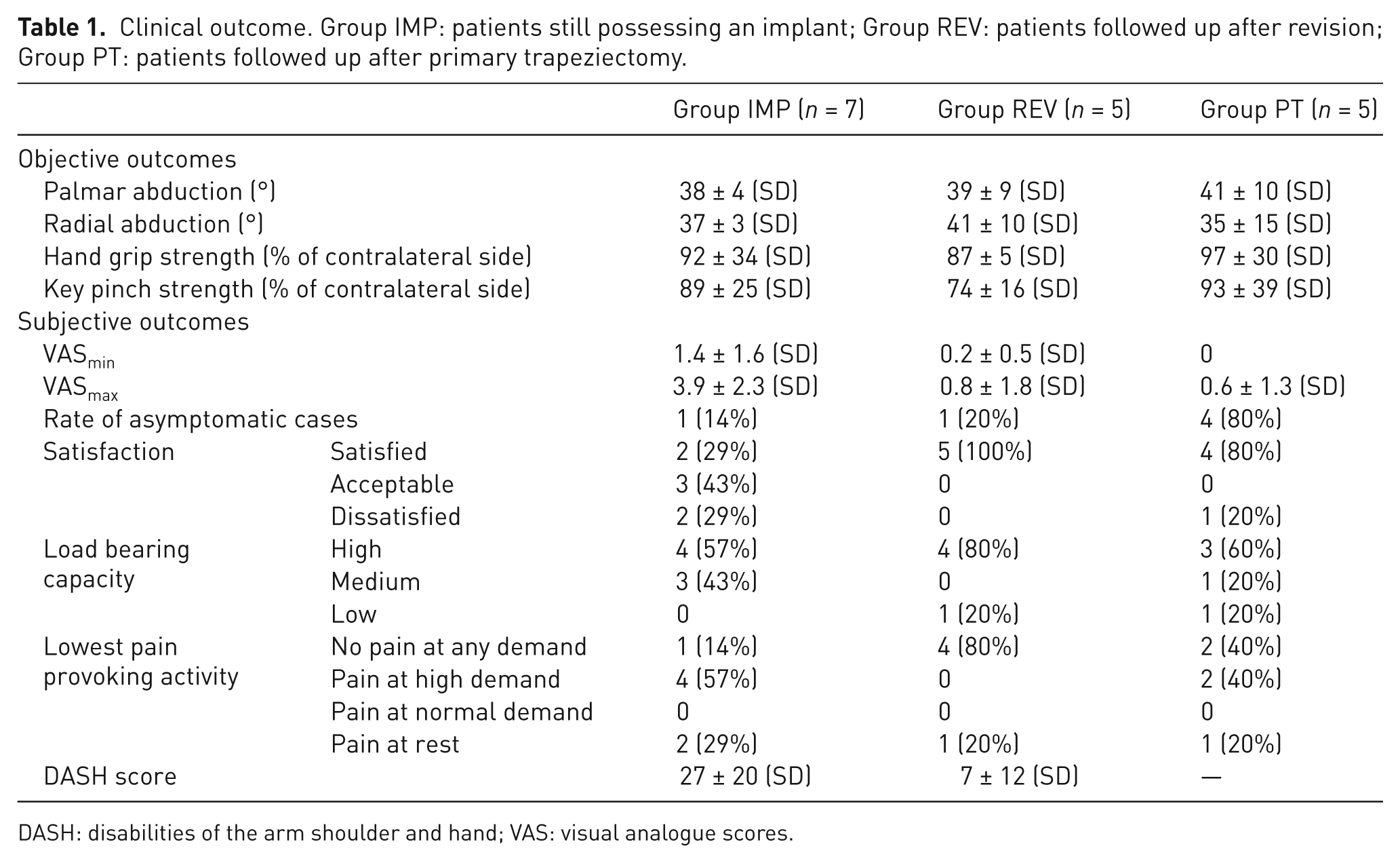
DASH: disabilities of the arm shoulder and hand; VAS: visual analogue scores.
Radiographic analysis
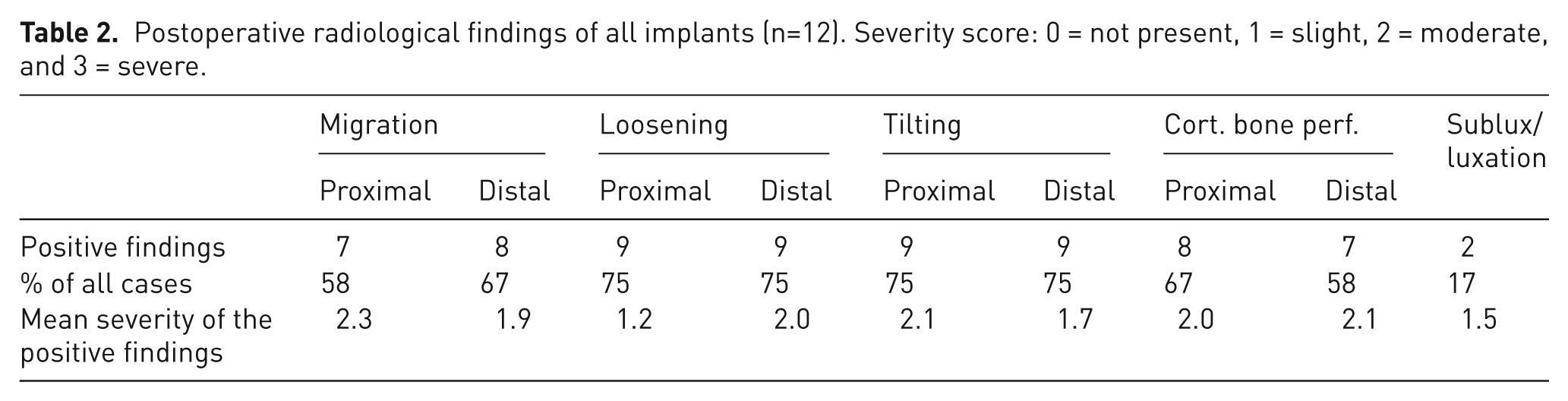
There were signs of migration in at least one component in 83% and tilting in 92%. Only 25% of the implants did not show obvious radiolucent lines around the implant. However, migration or tilting without a clear radiolucent line of at least one component had occurred in these cases as well (Figure 2). A summary of the radiological findings is presented in Table 2.
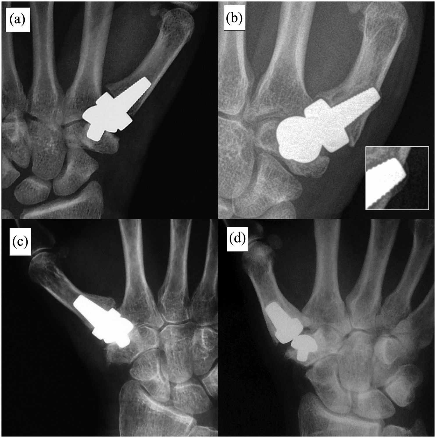
Characteristic complications. Each row contains the radiographs of one particular patient. The radiodraphs ((a) and (c)) in the first column are immediately following surgery. The radiographs in the second column ((b) and (d)) are at most recent follow-up. (b) Shows migration of the proximal and tilting of the distal device component 4.6 y after implantation. The tip of the distal component has penetrated the dorsal cortical bone of the first metacarpal, which has provoked a periosteal reaction. The tip of the proximal component almost reaches the scaphotrapezial joint. (d) Shows migration of the distal component and tilting of the proximal component 4.4 y after implantation. The malalignment of the components causes joint instability.
Postoperative radiological findings of all implants (n=12). Severity score: 0 = not present, 1 = slight, 2 = moderate, and 3 = severe.
Discussion
The mainstay of treatment for thumb CMC joint osteoarthritis is trapezectomy (Salem and Davis, 2012). Most patients do well, but typically there is loss of some length of the first ray with concomitant weakness. In an attempt to improve the outcome various versions of implant artroplasty have been developed, starting with the Swanson silastic trapezial replacement (Swanson and de Groot Swanson, 1985). Soft materials have not proven to be durable so various harder materials, including metals, pyrocarbon and ceramic, have been used. Ceramic spacer balls have been used but have a high failure rate (Adams et al., 2009). They reported on a series of 50 cases and found a high rate (94%) of adverse radiographic findings at three years. This was primarily subsidence into the trapezium, often associated with fracture of the trapezium. Other studies have also been reported to have high failure rates of the Elektra and Pyrocarbon P2 implants (Hernandez-Cortes et al., 2012; Klahn et al., 2012; Maru et al., 2012).
Different types of Moje implants are available for different joints (wrist, MTP and proximal interphalangeal (PIP)). Several studies dealing with this material match our adverse experience. In the MTP joint, failure rates of 6%–24% have been reported (Barwick and Talkhani, 2008; Brewster et al., 2010; Kulshreshtha et al., 2009; McGraw et al., 2010). In the PIP joint, failure rates of 0%–42% have been reported (Pettersson et al., 2006; Wesemann et al., 2008).
In the only previous report of the use of the thumb CMC joint Moje Acamo arthroplasty Hansen and Vainorius (2008) reported on nine cases. They had had to revise three within one year and the remaining six implants showed radiographic signs of loosening. Five showed implant migration and one had a radiolucent line around it. The preoperative mean DASH score of all patients (n = 9) was 57 (range 30–88), while 12 months postoperatively the mean score of the non-revised patients (n = 6) was 40 (range 20–75). There were no other clinical outcome data. In this series, the mean of the available preoperative DASH scores (n = 8) was 52.4 (range 35–56), while 50 months postoperatively the mean score of the non-revised patients (n = 7) was 27.3 (range 0–51). The initial values are similar in the two studies and the more pronounced decline of the mean DASH score in our study was probably influenced by the much longer follow-up period.
Our revision rate (42%) was higher than that of Hansen and Vainorius (2008), but as our follow-up interval is longer, this is also to be expected. Our revision rate is expected to rise further to at least 75%. The radiographic outcomes in this study are very similar to those of the report of Hansen and Vainorius (2008).
The outcome in the group of patients still with an implant gives an inconsistent impression. While the abduction of the thumb and the relative strength results were good (37°–8° and 89%–98%, respectively), the subjective outcomes were poor. The most severe problem was pain with use: it reached a mean of 3.9 on the 0–10 VAS scale. Only one of the seven patients denied having any pain during effort and two patients reported regular pain at rest. Although four of the seven patients reported high load-bearing capacity of the operated hand, only two were ultimately satisfied with the result.
Failure of fixation of the trapezial implant into the bone might be the main cause of failure. The exact reasons are not clear: the mechanical characteristics of the implant material, its surface properties, and the geometrical design need to be considered. For a detailed discussion of our theoretical reasoning of the possible causes of the complications, please see the Online Supplement of this publication (I. Possible Factors of the Observed Complications – A Theoretical Reasoning); they are briefly outlined in the next paragraph.
Improper implant material elasticity may result in stress shielding of the adjacent bone, while a lack of biocompatibility of the implant surface may play a potentially important role in implant loosening. The abrasion of the implant’s coating may cause passive (mechanical) and/or active (biological) degradation of the adjacent tissue. The geometrical design of the implant may influence the micromovements of the device: the circular design of the stem cross-section could potentiate the risk of the rotational shift. Prosthesis malpositioning at implantation may disturb the biomechanics of the joint and also contribute to later loosening.
Revision outcomes
The outcomes of revision surgery were generally satisfactory. Both radial and palmar abduction were better in patients who had been revised than in those still with their implant. The improvement in palmar abduction was only 1°, and radial abduction 4°. The improved joint motility may be explained by both the reduced pain and the reduced tension of the stabilizing soft tissue. The reduced stability of the joint may also explain the observation that, although effort pain was reduced significantly, the hand-grip and key-pinch strength results were slightly reduced after explantation (NS). The DASH score decreased significantly after the revision surgery, suggesting a general improvement in everyday abilities. All of the patients who underwent revision surgery were satisfied with the outcome compared with only one-third of those patients with the device still implanted.
To compare the patients who received revision surgery with a primary trapeziectomy group, a matched-pair comparison was performed. The mean palmar abduction was lower, while the mean radial abduction was higher in the revised patient group than in the primary trapeziectomy group, but neither was significant. Although all the relative strength results were superior in the primary trapeziectomy group, these were not significant. This could be owing to the small sample size of the matched-pair comparison (total n = 10, five pairs). Generally, the comparisons between the two groups only represent observations and are not conclusive owing to the sample size; nonetheless, the statistics have been included for guidance. Overall the final outcomes of the revised implantations and the primary trapeziectomies are very similar.
Failure rates of implants in the hand are not exclusive to the thumb CMC joint. Even well-established implants, such as the Pyrocarbon PIP joint arthroplasty, do not always have consistent results (Mashhadi et al., 2012, McGuire et al., 2011). These probably reflect factors that are, as yet, not understood. Thumb CMC joint arthroplasties seem to be very prone to high failure rates.
Owing to the high rate of failure, we no longer use this implant and we caution other surgeons considering its use. For patients who have already received a Moje thumb CMC replacement, we recommend regular radiographic follow-up as radiological failure often proceeds symptoms. Finally, this implant illustrates how preclinical testing may not translate into clinical practice, so all new implants should only be introduced after large and long enough clinical trials.
Footnotes
Acknowledgements
We thank Simone Gantz for her support in the statistical analysis.
Funding
This research received no specific grant from any funding agency in the public, commercial, or not-for-profit sectors.
Conflict of interests
None declared.