
Abstract

Dear Sir,
Carpal bone and joint injuries in children, especially those involving the midcarpal joint, are very uncommon. Diagnosis of these injuries is often missed or delayed (Gerard, 1980).
We report on the case of an 8-year-old, right-handed girl, who consulted us 70 days after suffering a car accident. She complained of pain, deformity and decreased motion in her right wrist, symptoms that began following the accident. She had consulted at two other institutions, where she was given a diagnosis of wrist sprain.
Physical examination revealed dorsal wrist swelling and deformity and a very limited range of motion. Radiographs and magnetic resonance imaging showed a volar midcarpal dislocation with bony avulsion of the anterior margin of the lunate, without evidence of injury to the scapholunate or lunotriquetal ligaments (Figure 1).
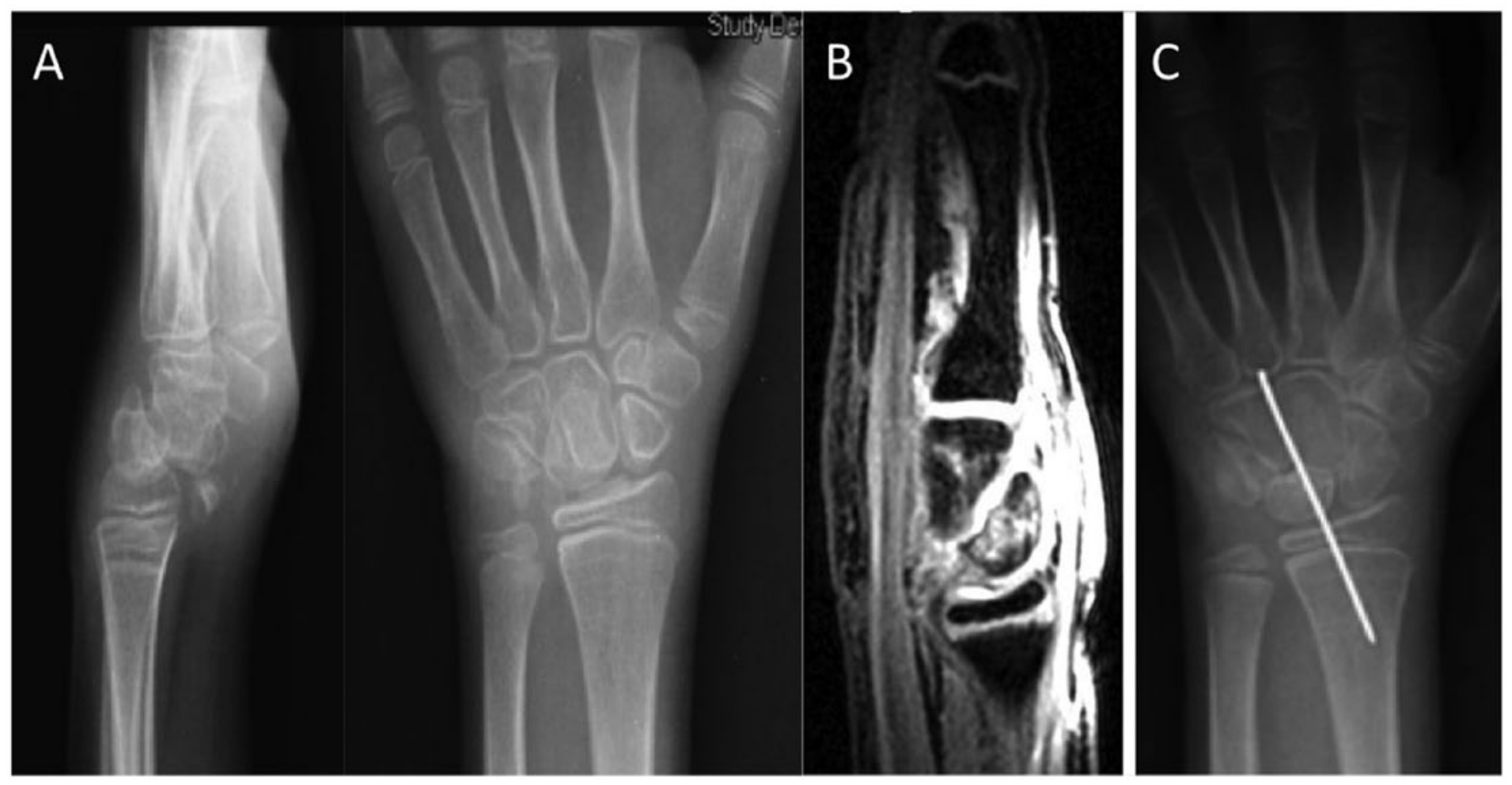
Antero-posterior (AP) and lateral radiographs (A) and magnetic resonance imaging (B) showing palmar midcarpal dislocation with avulsion of the anterior margin of the lunate. (C) Postoperative radiograph showing joint reduction and pin fixation.
An open reduction and temporary K-wire fixation were performed through a double dorsal and palmar approach (Figure 1). A major impact injury to the cartilage of the head of the capitate bone was evident. All scar tissue was removed, preserving the remnants of the midcarpal ligaments. Once the midcarpal joint was reduced, stabilization was performed with a 1.4 mm K-wire. The remnants of the midcarpal volar and dorsal (radiolunotriquetal) ligaments were sutured and a dorsal capsulodesis was also performed.
Following surgery her wrist was immobilized for 6 weeks in a long arm cast.
At 4 years follow-up, the patient reported no pain. Passive wrist range of motion was almost symmetrical (flexion 80°, extension 70°, ulnar deviation 30° and radial deviation 15°) (Figure 2). Key pinch strength measured 5 kgf for her right hand and 6 kgf for the left, while grasp strength was 12 kgf for both sides. Final radiographs showed moderate midcarpal joint narrowing and lunate enlargement in the sagittal plane (Figure 2).
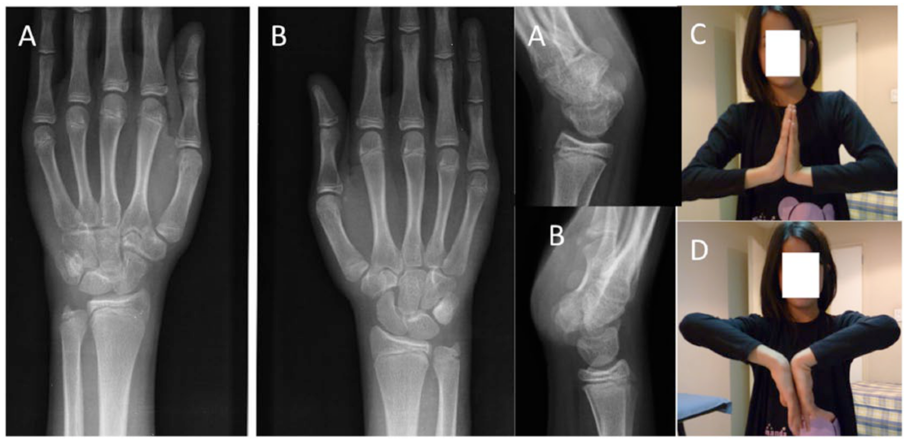
Result 4 years after treatment. Right wrist radiographs (A) demonstrated correct joint alignment, with slight joint narrowing and lunate enlargement in a sagittal plane as compared with the left wrist (B). Wrist range of motion was slightly decreased as compared with the contralateral side (C, D).
The most common form of midcarpal disorder in children is instability, which might be related to general joint laxity, or secondary to trauma (Chou et al., 2010; Craigen, 1996; Gerard, 1980).
Only a single case of midcarpal dislocation has been reported (Graham and Jacobson, 1999); this was in a 15-year-old girl with a non-traumatic midcarpal dislocation lasting 3 years, treated with an arthrodesis. Whether this case was an actual midcarpal dislocation or the result of a long-term midcarpal instability will remain unknown.
To our knowledge, our report is the first report of posttraumatic midcarpal dislocation in a child. Midcarpal dislocation in children, even when treated late, may benefit from joint reconstruction rather than joint fusion.
Footnotes
Declaration of conflicting interest
The authors declared no potential conflicts of interest with respect to the research, authorship, and/or publication of this article.
Ethical approval
The local ethics committee approved the protocol of the study. Patients provided written informed consent before participation, in accordance with the Declaration of Helsinki guiding biomedical research involving human subjects.