
Abstract

Dear Sir,
The cutaneous innervation of the dorsum of the hand is derived from dorsal sensory nerves originating from the superficial radial and ulnar nerve, and by dorsal branches (DBs) originating from the proper digital nerves (PDNs). There is little detail in available literature with regards to the anatomy of the DBs, but informal intra-operative observation suggests that they are sometimes unexpectedly substantial in diameter. We therefore undertook a cadaveric study to analyse the origin, distribution and diameter of the DBs.
Dissection was performed on five matched pairs of fresh-frozen female cadaveric hands. Bruner incisions were made in each web space, and the common digital nerves, PDNs and DBs were identified. Under 3× loupe magnification, the presence/absence of a DB was noted on each side of the digits (see Figure 1), and its point of origin documented relative to the first annular (A1) pulley. After meticulous dissection of the adventitial tissue, the nerves were sectioned, and the diameters of the PDNs, and their respective DBs, were measured immediately proximal and distal to the DB origin using an eyepiece graticule (×25 magnification). All measurements were taken as a mean of three readings, measured in millimetres, using a surgical ruler. The intra-observer variation was assessed by calculating the coefficient of variation for each digit.
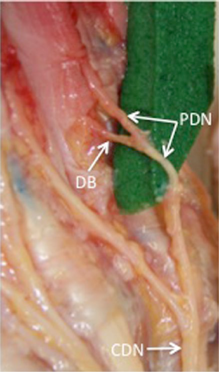
Cadaveric dissection to show the ulnar proper digital nerve (PDN) and DB of the index finger in the second web space of a right hand. The green background material was used to transfer the transected nerve segment to microscope graticule for diameter measurement.
DBs originated from the majority of PDNs dissected (92% of 100 PDNs) and were absent in a minority (8%), most commonly on the radial side of the thumb (n = 3). The DBs originated alongside, or proximal to, the A1 pulley in 58% of cases, and distal to the A1 pulley in 34% of cases, most commonly in the third web space. Diameter measurements demonstrated that DBs were consistently greater than two-thirds of the diameter of their corresponding PDNs, and in some, almost equal (Table 1). This was particularly apparent on the radial side of the middle finger (1:0.94) and the radial side of little finger (1:0.93). Analysis of intra-observer error using paired t-tests was not statistically significant. (Coefficients of variation recorded for the radial side of the digits, from thumb through to little finger, 17.7, 6.4, 8.4, 13.1 and 7.8, respectively. For the ulnar side, from the thumb to the little finger, they were 10.6, 8.7, 11.4, 14.4 and 10.3).
Diameter of the DBs and their parent digital nerves.
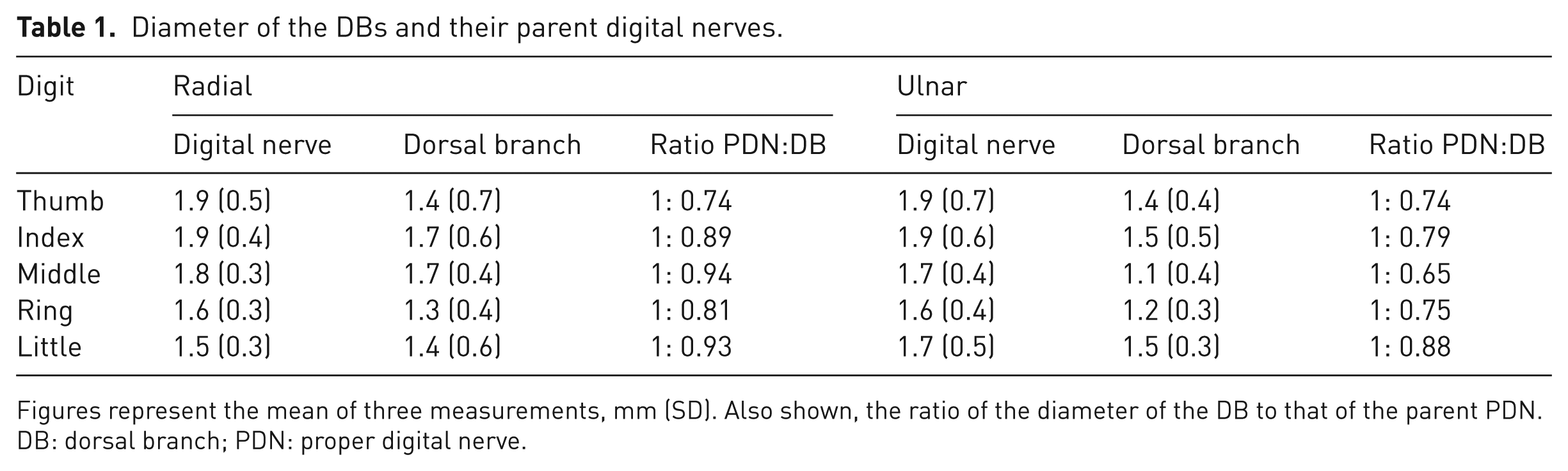
Figures represent the mean of three measurements, mm (SD). Also shown, the ratio of the diameter of the DB to that of the parent PDN.
DB: dorsal branch; PDN: proper digital nerve.
Our study confirms the findings of other previous studies, as to a fairly constant presence of the DBs, including in the thumb. In particular, Bas and Kleinert (1999) found DBs in 76% of cases; they were frequently absent in the thumb or the ulnar side of the little finger. We particularly wish to highlight that the relative diameter of the DBs may be greater than previously documented. Previous studies, limited solely to the radial side of one ring finger (Bas and Kleinert, 1999), or to both sides of one single index finger (Wallace and Coupland, 1975), have suggested a relative diameter of half the size. In our considerably larger study, albeit performed without the benefit of histological nerve staining, we found the DB:PDN diameter ratio to be greater, particularly in the radial side of the middle and little fingers, where the two nerves were almost equal in size (Figure 1).
Although we present a relatively small series, we hope this will raise an awareness of the anatomy and diameter of the DBs and PDNs, which will help to guide safe operative dissection in the webspaces, for example when the anatomy is distorted by scar tissue in recurrent Dupuytren’s disease. We also hope it may inform choice and design of reconstructions that depend on the DBs for cutaneous innervation, such as the Foucher flap (Foucher and Braun, 1979) and the bilaterally innervated sensory cross-finger flap (Lassner et al., 2002).
Footnotes
Acknowledgements
The authors wish to thank Dr Rob Elton for his statistical guidance and to Charles Booth, Cristina Gatti and Justine Jaka who provided technical assistance for the completion of this report. We also gratefully acknowledge the support received from Professor Gordon Findlater, Iain Campbell and Susan Bond from the Department of Anatomy, University of Edinburgh.
Declaration of Conflicting Interests
The authors declared no potential conflicts of interest with respect to the research, authorship, and/or publication of this article.
Informed consent
Informed consent was obtained for all cadaveric specimens used and all dissections or images used comply with the Anatomy Act 1984.