
Abstract
Congenital hypoplasia of the extensor tendon central slip is a rare entity. This article describes the clinical characteristics in a series of 22 fingers in 16 patients (mean age: 10 months), and the outcomes of conservative treatment. Nine of 22 fingers were classified as slender or hypoplastic. Treatment with bracing was successful in 21 digits, resulting in full active extension of the proximal interphalangeal joint at a mean of 8.5 months after treatment. Bracing was unsuccessful in one digit, in which operative treatment resulted in a successful outcome. Some residual deformity was observed in ten fingers after a mean follow-up period of 2 years and 1 month. Congenital hypoplasia of the central slip can be treated successfully by the conservative hand bracing when worn with full compliance. Treatment time is extended by the infrequent application of the hand brace or in the case of hypoplastic slender fingers.
Introduction
Flexion deformity of the proximal interphalangeal (PIP) finger joint is an occasional observation in children. Among these deformities, camptodactyly is relatively common, and is defined generally as non-traumatic congenital flexion contracture of the PIP joint that cannot be corrected passively to full extension (Foucher et al., 2006; Smith and Kaplan, 1968). By contrast, patients have rarely been observed with an extension lag of the PIP joint that can be corrected passively to almost full extension. This condition has been mentioned briefly as congenital hypoplasia of the extensor tendon central slip in very few publications in the English literature (Carneiro, 1993; Kim et al., 2014; Lin and Chiu, 2004), and the detail of pathology and treatment progress for congenital hypoplasia of the central slip remains unclear. We have managed patients with congenital hypoplasia of the central slip conservatively by extension brace treatment as the first-line choice, and used operative intervention only for those patients who failed brace treatment. This article describes the clinical characteristics of the condition and the effectiveness of conservative treatment of congenital hypoplasia of the extensor tendon central slip. The intraoperative anatomical findings of the extensor tendon apparatus in the single case of failure are described.
Methods
We performed a retrospective review of 22 consecutively treated digits in 16 patients with congenital hypoplasia of the extensor tendon central slip. The patient demographic data and treatment progress were reviewed retrospectively from medical records. Our institutional review board approved the study protocol.
Congenital hypoplasia of the central slip was defined as an infant with an extension lag of the PIP joint alone, who could not extend the finger PIP joint actively since birth, but in whom the joint could be passively extended to full, or almost full, extension due to a secondary minor joint contracture (Carneiro, 1993). The extension lag displayed by these patients was large enough for the patients’ mothers to recognize easily (60°–80°), although they could not be measured exactly because the patients were very young infants. There was neither pain nor swelling around the finger joint, and both active and passive flexion of the involved metacarpophalangeal (MP) and distal interphalangeal joints was intact. No nodules could be found in the palmar fascia, and the skin was not thickened or dimpled. Patients with camptodactyly, arthrogryposis, and trigger finger were carefully excluded, as were patients with pre-existing neuromuscular conditions. Camptodactyly was diagnosed as a contracture of the PIP joint without an extension lag, which was uncorrectable both actively and passively to full extension (Foucher et al., 2006). Trigger finger was diagnosed as snapping during active extension of PIP joint in spite of usual posture of flexion of PIP joint and a palpable nodule around the A1 pulley due to thickening of the flexor tendon (Shah and Bae, 2012). Associated congenital anomalies were evaluated.
The characteristics of affected fingers were evaluated, including their distribution, whether they were more slender visually than the unaffected fingers, and whether their PIP joint could be extended passively to full extension with or without a secondary minor joint contracture of less than 30°. If the affected finger was visually determined to be more slender than the same, unaffected finger in the contralateral hand, it was defined as a hypoplastic slender finger.
Conservative treatment was commenced in all patients, using a static hand brace that could maintain the PIP joint in a position of extension (Figure 1). We recommended the hand brace be worn 24 h a day. If full compliance could not be achieved (for example if the patients were removing the splint themselves), we recommended that they wear it during the day or night as much as possible. Surgical treatment was indicated only for patients who did not respond to conservative brace treatment or for those who could not wear the brace equipment due to PIP joint instability.
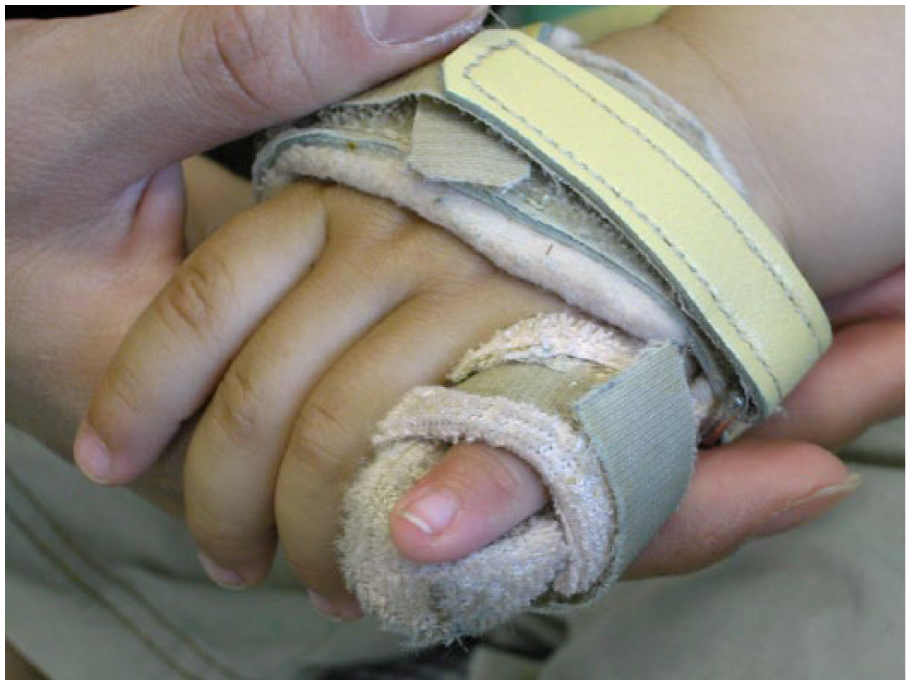
Representative figure of the static hand brace.
Clinical outcomes were evaluated with measurement of the residual lack of full active extension of the PIP joint, and remaining deformity of the affected fingers at final follow-up. The time period over which patients wore the hand brace was recorded. The patients were followed-up by either the first or second authors every few months. We asked the patients’ mothers how long the hand brace was worn at every outpatient visit. Although we could not assess the exact amount of time the patient spent in the brace, parent-reported information regarding the estimated wear time, defined in the general terms ‘frequent’, ‘occasional’ (daytime or night time) or ‘seldom’, could be obtained adequately. The patients were divided into two groups: those who wore the brace frequently were designated as group F, and the patients whose brace use was defined as ‘occasional’ or ‘seldom’ were designated as group O. The time to achieve active full extension of the PIP joint was compared between group F and O.
Data analysis was carried out with StatView 5 (SAS Institute Inc., Cary, NC). Differences between the two groups were assessed using the Mann–Whitney U-test. A p value <0.05 was considered statistically significant.
Results
Patient demographic data and results are listed in Table 1. Patients included five girls and 11 boys, mean age 10.4 months (range 0–34) at presentation. Six patients had associated congenital anomalies: one hypoplastic toe, one clinodactyly, one symphalangism of the small finger, one cerebral palsy, one clasped thumb and one intrauterine growth retardation. Ten patients had unilateral isolated digital involvement, and six had bilateral involvement (with the same digits affected bilaterally). In total, congenital hypoplasia of the central slip was found in two index, 15 middle, one ring and four little fingers. Nine of 22 fingers were hypoplastic and more slender than the contralateral unaffected fingers. At presentation, 15 of 22 fingers were correctable passively to full extension, seven had a secondary minor (less than 30°) restriction of passive PIP joint extension with an extension lag. This PIP joint restriction did not increase with passive MP joint extension; the absence of the dynamic tenodesis effect indicated the presence of joint contracture.
Patient characteristics, treatment methods, and results.
+: present; −: absent; PIP: proximal interphalangeal.
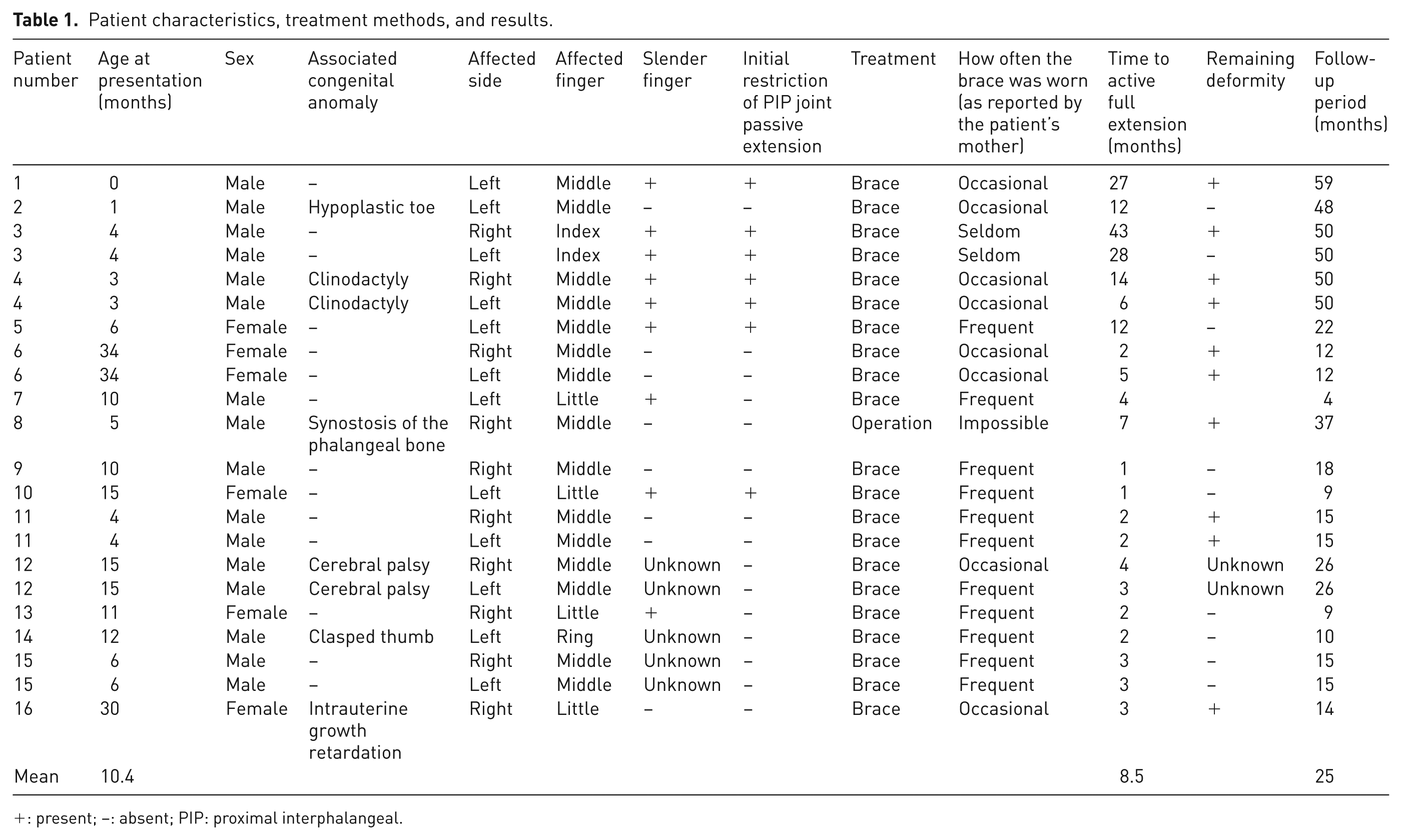
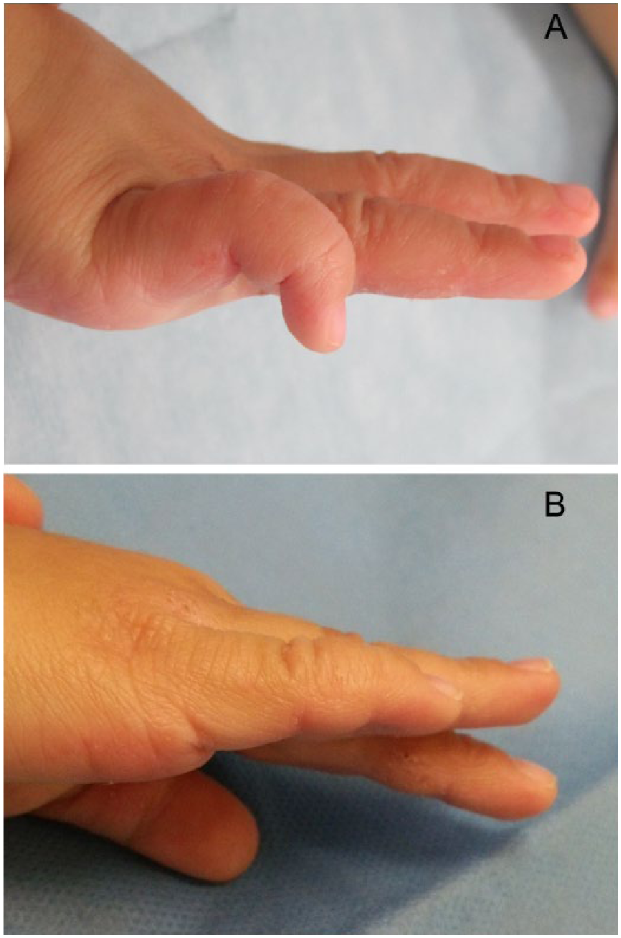
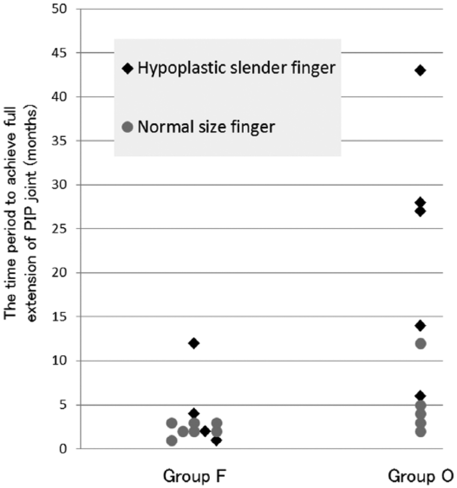
Mean follow-up was 2 years and 1 month (range 4 months–4 years and 11 months). Conservative hand brace treatment was performed in 21 fingers and operative treatment was performed in one finger. The hand brace was worn frequently for 11 fingers (group F), with occasional or seldom use in ten fingers (group O); two wore the brace during the day, six at night and two rarely. Although all fingers had pretreatment extension lag of the PIP joint, all obtained active full extension of the PIP joint after a mean 8.5 months post-treatment (Figure 2). The mean time to achieve active full extension of the PIP joint after treatment was significantly shorter in group F (3.2 months) than in group O (14.8 months, P = 0.003, Figure 3). The time to achieve active full extension of the PIP joint was significantly shorter in normal sized fingers (3.7 months) than in hypoplastic slender fingers (15.2 months, p = 0.041, Figure 3). Ten fingers had remaining deformity at final follow-up: one had extension lag of the MP joint and one had radial drift deformity of the PIP joint as illustrated in the operative case presented below. Eight patients demonstrated hyperextension of the MP joint with slight flexion of the PIP joint (claw finger deformity, Figure 4(A)), in these cases, hyperextension of the middle finger MP joint disappeared with full extension of the PIP joint when the patient adopted the scissors posture (Figure 4(B)) because extension of the middle finger MP joints was restricted by flexion of the ring and little finger MP joints through the juncturae tendinum. This finding is similar to Bouvier’s sign, where the PIP and distal interphalangeal joint can be extended following the correction of MP joint hyperextension, indicating insufficiency of the intrinsic musculature of the hand (Sammer and Chung, 2009; Sapienza and Green, 2012). Intrinsic insufficiency did not always occur in hypoplastic digits; four of these patients had fingers of normal size.
(A) Initial finding of flexion deformity of the PIP joint in the right little finger of case number 16. (B) Active full extension of the PIP joint in the right little finger 3 months after hand brace treatment.
Comparison of treatment time between group F (frequent use of the hand brace) and group O (occasional or seldom application of the hand brace). The mean time taken to achieve active full extension of the PIP joint after treatment was significantly shorter in group F (3.2 months) than in group O (14.8 months, P = 0.003), and it was significantly shorter in normal sized fingers (3.7 months) than in hypoplastic slender fingers (15.2 months, P = 0.041).
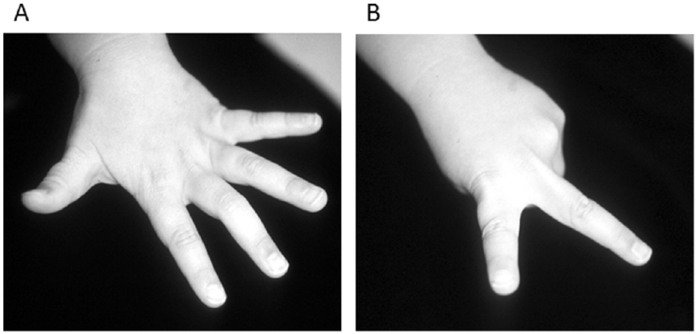
Representative figure of the remaining claw finger deformity after hand brace treatment. (A) Slight flexion deformity of the PIP joint in the middle finger with hyperextension of the MP joint. (B) Full extension of the PIP joint in the middle finger when hyperextension of the MP joint was restricted by flexion of the ring and little finger MP joint in scissoring posture.
Surgery was performed in one middle finger (patient number eight in Table 1) because of failure of bracing due to PIP joint laxity. The PIP joint became hyperextended and displayed a radial deviation when the hand brace was applied to maintain the PIP joint in full extension. It was assumed that the patient also had hypoplasia of the volar plate, collateral ligaments and central slip. Intraoperative findings showed attenuation of the central slip of the extensor tendon, which was thinner than the lateral bands. We could not confirm the status of the volar plate or collateral ligaments. The attenuated central slip was plicated with 3 mm of shortening proximally and the PIP joint was fixed in the extension position with Kirchner wire for 5 weeks. Active full extension and flexion of the PIP joint was achieved 7 months after surgery. The hyperextension instability of the PIP joint was corrected, although radial deviation persisted postoperatively.
Discussion
Congenital hypoplasia of the central slip of the extensor tendon is an extremely rare condition. There have been some reports on congenital hypoplasia of the extrinsic extensor tendon of the hand (Hamanishi et al., 1986; McMurtry and Jochims, 1977; Tsuge, 1975; Tungshusakul et al., 2011; Vartany et al., 1996) but, to our knowledge, there are only three published reports (two case reports and a case series of five patients) on congenital hypoplasia of the central slip in the English literature (Carneiro, 1993; Kim et al., 2014; Lin and Chiu, 2004).
Previous reports on congenital hypoplasia of the central slip have all described surgical treatment. Carneiro described five cases, all treated surgically (Carneiro, 1993). Intraoperative findings showed that the central slip was universally attenuated; it was partially excised, advanced distally and sutured to the insertion of the central slip. Lin and Chiu reported on a 19-year-old man with bilateral laxity of the central slip resulting in Boutonniere deformity. He was treated surgically by non-excisional tightening of the central slip (Lin and Chiu, 2004). Kim et al. reported a 26-year-old man with congenital Boutonniere deformity in both little fingers. At operation, findings were a poorly differentiated extensor mechanism and the absence of the tendinous attachment on the dorsal part of the middle phalanx (Kim et al., 2014). The extensor mechanism was reconstructed.
Although surgical treatment has been performed in all previous cases with satisfactory results, the present study reveals the effectiveness of hand brace treatment for congenital hypoplasia of the central slip in a larger case series of 22 fingers on 16 patients. We think that surgical treatment should be reserved for patients in whom conservative treatment has failed. We believe that if conservative hand brace treatment is initiated from the patient’s early childhood, the attenuated central slip is gradually tightened by the correction force applied by PIP joint extension during growth, reducing the risk of a persistent Boutonniere deformity as described in previous case reports (Kim et al., 2014; Lin and Chiu, 2004).
The mechanism of attenuation of the central slip remains controversial. Carneiro described that the attenuation was caused by an intrauterine traumatic accident rather than a formation disorder occurring during embryogenesis (Carneiro, 1993). In contrast, others believed that attenuation of the central slip occurs due to a developmental error in the formation of the tendon (Kim et al., 2014; Lin and Chiu, 2004). Because hypoplastic slender fingers and a persistent Bouvier’s sign associated with the insufficiency of the intrinsic muscles of the hand was found in about half the cases in the present study, we surmise that attenuation of the central slip is caused by embryonic developmental failures.
In this series, central slip hypoplasia was associated in some cases with intrinsic insufficiency. Unfortunately, the rate of accompanying intrinsic insufficiency in this series is unclear, as we did not perform the Bouvier test at the first medical examination. However, it is likely that congenital intrinsic deficiency was involved in hypoplasia of the central slip, as some patients with slender fingers or with residual clawing deformities were correctable with the Bouvier test at final follow-up.
The present study has some limitations. It was a retrospective study with a relatively small number of cases, no untreated control group and a short follow-up period of about 2 years. It is important to continue monitoring the children successfully treated with a brace until they reach adulthood. It was not feasible to measure the extension lag and the exact range of motion of the fingers precisely because the patients were very young. The correlation between the degree of extension lag and time for correction in splintage is unclear, and further research into this correlation is necessary. The clinical identification of the slender finger was based on visual assessment alone, and not assigned by means of an exact measurement. Radiographic changes around the PIP joint were not evaluated. It remains unclear whether the period to achieve full active extension of the PIP joint is faster after surgical treatment than after hand brace treatment. Future prospective studies are needed to investigate the healing period with conservative hand brace treatment compared with an untreated group or surgical treatment.
In conclusion, congenital hypoplasia of the central slip can be treated successfully by using a static hand brace to keep the PIP joint in the extension position, especially with the frequent application of the hand brace. Successful treatment takes longer when the hand brace is only worn occasionally or rarely, and in the case of hypoplastic slender fingers.
Footnotes
Declaration of Conflicting Interests
The authors declared no potential conflicts of interest with respect to the research, authorship, and/or publication of this article.
Funding
The authors received no financial support for the research, authorship, and/or publication of this article.
Ethical approval
The study was approved by the institutional review board at Osaka City General Hospital.