
Abstract

Dear Sir,
The soft tissue defects with bone or tendon exposure in multiple finger injuries usually needs more than one flap for reconstruction. A number of options exist (Ishiko et al., 2009; Kim et al., 2014; Li et al., 2015). Here we report a patient of three-finger defects using a free first dorsal metatarsal artery perforator flap.
A 41-year-old male patient presented to our hospital 4 hours after having his left index, middle and ring fingers hurt by an electric planer. The defects extended from middle phalanx to distal phalanx in finger pulp; the soft-tissues, including the proper palmar digital arteries and nerves of three fingers, the deep digital flexor tendon of middle finger, were damaged seriously (Figure 1(a)). After debridement, a Kirchner wire was used to fix the middle finger to stabilize the distal interphalangeal joint; the injured fingers were put together, and the wound margin on adjacent fingers sutured, to make the separate wounds integrated as one wound (Figure 1(b)).
(a) a 41-year-old male patient with defects of left index, middle and ring fingers by an electric planer; (b) the wound margin in adjacent fingers were sutured up to be a united wound; (c) a free dorsal metatarsal artery perforator flap designed for reconstruction; (d) dissected flap; (e) after flap transfer.
A free dorsal metatarsal artery perforator flap was outlined on the left instep (Figure 1(c)). Suitable perforators located in advance with the help of Doppler sonography. Then the flap was dissected (Figure 1(d)). The first dorsal metatarsal artery and its digital branch, a digital branch of the first plantar metatarsal artery, three branches of great saphenous vein, the first common digital nerve, a dorsal digital nerve, and the extensor hallucis brevis tendon of the big toe, were dissected and included in the flap.
The flap was transferred to the recipient area, with the distal side of the flap docking to the index finger, and the proximal side docking to the middle and ring fingers (Figure 1(e)). The extensor hallucis brevis tendon was anastomosed to the deep digital flexor tendons of middle finger. For vascularization, two arteries were anastomosed end-to-end to ulnar digital artery of index and middle fingers, three veins were anastomosed end-to-end to plantar and dorsal digital vein of the ring finger, respectively. Two nerves were anastomosed end-to-end to the ulnar digital nerve of the index and middle finger, respectively. Finally, a plaster slab was placed to immobilize the fingers, and the donor site was closed directly.
After surgery, the patient was given anticoagulant therapy for 5 days. Then the finger joint functional exercises were started 10 day post-operation, and the index, middle and ring fingers were separated 21 days after surgery. Nine months post operation, a revision surgery was performed for flap thinning. We followed the patient 3 months after revision surgery and found a functionally and cosmetically good result (Figure 2). The range of motion was 0° to 45° for the distal interphalangeal joint of the index finger, 0° to 35° for the middle finger, and 0° to 45° for the ring finger.
Good cosmetic and functional result was obtained 3 months after revision surgery. (a) Finger extension; (b) finger flexion.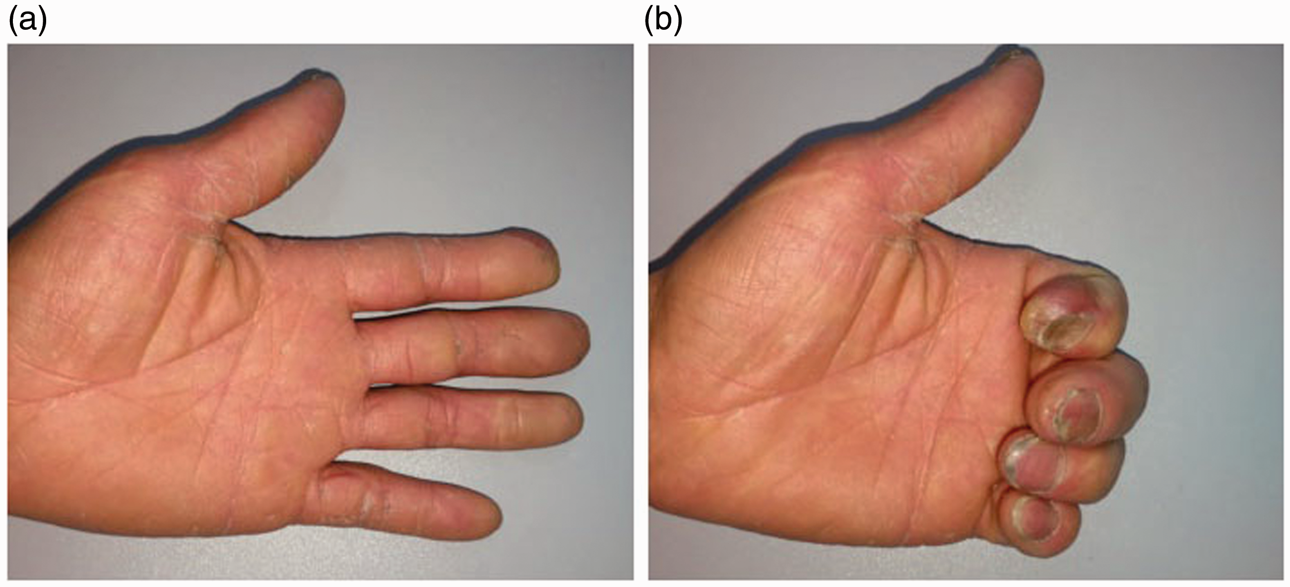
Composite tissue defect of multiple fingers is a difficult problem in hand surgery. The principle of reconstruction is to restore major important tissue using secondary tissue, with a minimum harm. Since the similarity between hand and foot in anatomy, foot is a favourable option for reconstruction of finger defect or toe-to-finger transplantation.
Our case suggests that the first dorsal metatarsal artery perforator flap can reconstruct multiple finger defects, though this procedure needs a secondary procedure for separation of fingers.
Footnotes
Declaration of conflicting interests
The authors declared no potential conflicts of interest with respect to the research, authorship, and/or publication of this article.
Informed consent
Written informed consent was obtained from the patient for the publication of this case report together with the accompanying images.