
Abstract
Dear Editor,
Vascularized bone grafts based on the 1,2 intercompartmental supra-retinacular artery (ICSRA) can be used for treating scaphoid nonunions (Chang et al., 2006). The vascularized bone graft is thought to import osteogenic progenitors to enhance the healing potential of the fracture. Its inherent blood supply improves graft survival rather than depending upon the unreliable vasculature of the scaphoid (Ong et al., 2011). Three-dimensional (3D) high frequency power Doppler ultrasonography is a non-invasive technique highly sensitive to slow and fast flow and can detect neovascularization in vivo fracture healing (Sun et al., 2012). We undertook a study to assess the microvasculature of vascularized bone grafts used for scaphoid nonunion using 3D high frequency power Doppler ultrasonography.
Twenty patients with a mean age of 23 years (range 20–27) who underwent operation for scaphoid nonunion between April 2012 and March 2014 were included. The mean delay from injury to surgery was 209 days (range 182–256). Before operation all patients had radiographs, 3D CT scans of the wrist to define fracture pattern and displacement, and MRI to assess vascularity of the scaphoid. All fractures were at waist level. After making a dorsoradial incision, the 1,2 ICSRA was visualized on the surface of the extensor retinaculum. A bone graft was elevated preserving the 1,2 ICSRA and perforating vessels. The scaphoid nonunion site was freshened, curetted and compressed with a cannulated Herbert screw (Depuy Synthes). The vascularized bone graft was transposed to reach the nonunion site where it was gently overlaid on the dorsal aspect and stabilized with a K-wire that was cut short.
CT scan was carried out in all patients between 12–18 weeks after operation to confirm scaphoid union and bridging trabeculae. At the same time, all received 3D high frequency power Doppler ultrasonography assessment over the dorsal wrist and scaphoid area using a high frequency (centre frequency at 55 MHz) in vivo micro-imaging system (Mindray & General Electricals, China). Doppler spectral analysis was used to assess the 1,2 ICSRA pedicle and bone graft including direction of flow, perfusion, and vessel calibre.
Patients were followed-up for a mean of 2.5 years (range 2.2–3). The scaphoid fracture united in all cases, with a mean time of 188 days (range 168–206). Doppler ultrasonography confirmed pulsatile blood flow signals from the vascularized graft pedicle and vascularity of the callus tissues around the scaphoid suggesting neo-angiogenesis and vascularized graft incorporation across the nonunion site. The origin of 1,2 ICSRA pedicle (mean diameter 0.5 mm) from the radial artery (mean diameter 1.5 mm) with a reverse pattern pulsatile flow was also appreciated in all cases (Figure 1). All grafts remained vascularized with no evidence kinking or thrombosis of the pedicle.
3D high frequency power Doppler ultrasound images. (a) shows the bone graft (BG), (b) confirms the pedicle of the 1,2 ICSRA vascularized bone graft (VAS) and surrounding vascularization of the callous tissues, (c) clearly illustrates the position of bone graft over the scaphoid, reverse flow of small sized (0.5 mm) 1,2 ICSRA pedicle whose orientation, location, and directions are perceptibly different from radial artery and its branches. The pulsatile flow and wave patterns are seen (d).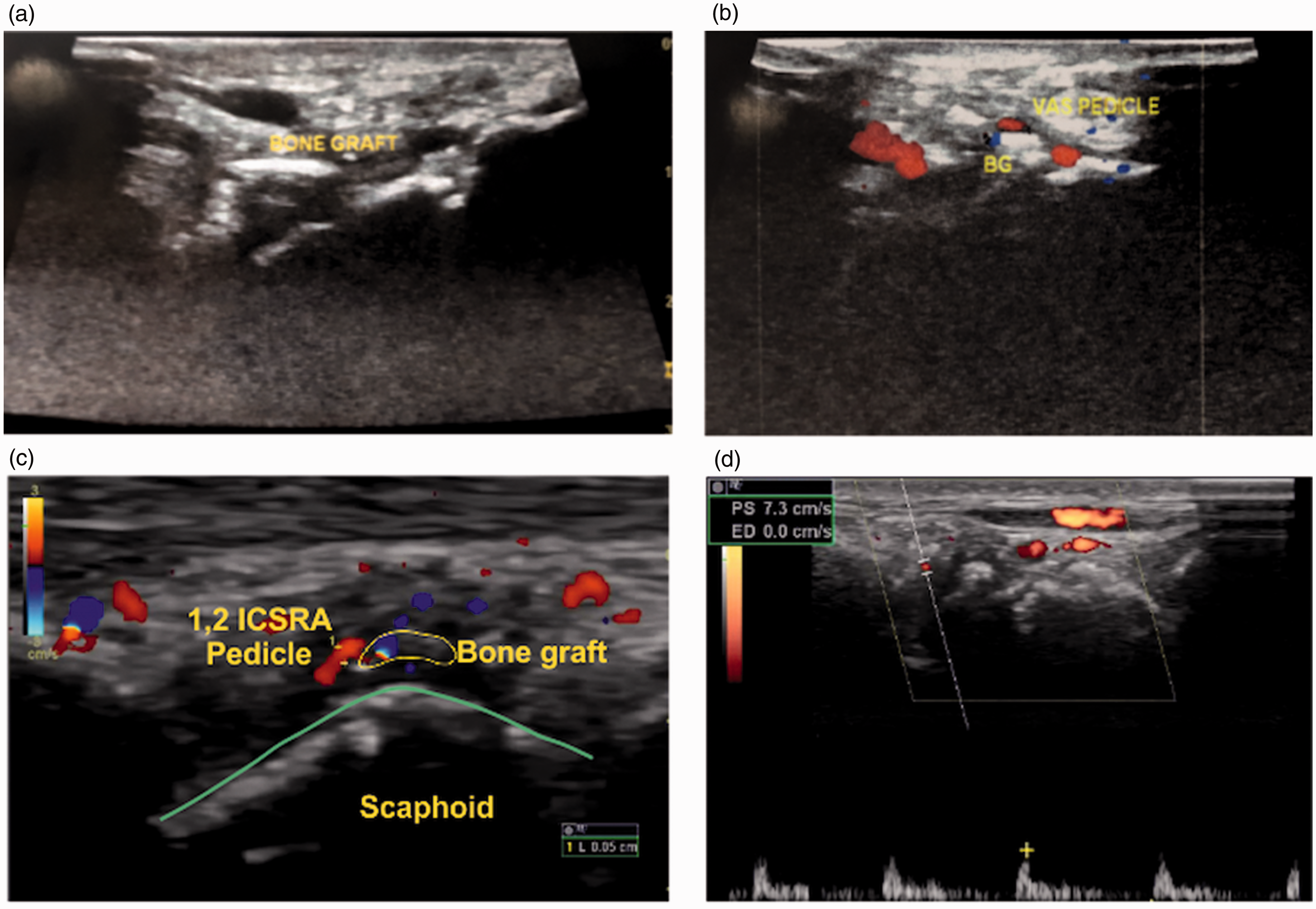
Studies of the biology of fracture healing have demonstrated that angiogenesis is a critical process in the time course of healing. For successful bone healing the injury site must be revascularized. Thus, methods for non-invasive assessment of the presence of vessels and the vascularity of the callus tissues may be valuable for gauging the status of the healing process (Augat et al., 2014). 3D high frequency power Doppler ultrasonography allows discrimination of Doppler signals from different depths. It detects moving interfaces and scatters from within a well-defined sample volume, which then can be positioned anywhere along the axis of the ultrasound beam. In vivo assessment of microvasculature during femoral fracture healing in rats and evaluation of vascularization and blood flow at the fracture site by 3D high frequency power Doppler ultrasonography was comparable with CT-based micro angiography (Sun et al., 2012). It is noninvasive, real-time, and can be used for longitudinal follow-up to quantify both vascularization and blood flow. The technique has limitations. The detection of flow signals is limited by the penetration depth. With the use of higher Doppler frequency, penetration depth would be decreased, so researchers must compromise between accuracy and detection depth.
Identification of 1,2 ICSRA pedicle along with the bone graft located over the scaphoid nonunion site and Doppler spectral analysis with pulsatile wave forms demonstrates the feasibility and reproducibility of 3D high frequency power Doppler ultrasonography for studying microvasculature during scaphoid fracture healing. The technique is likely to be applicable to study of fracture healing at other sites.
Footnotes
Acknowledgements
We thank the patients who have been the backbone of this study and the radiology team who supported us with images and results.
Declaration of conflicting interests
The authors declared no potential conflicts of interest with respect to the research, authorship, and/or publication of this article.
Funding
The authors received no financial support for the research, authorship, and/or publication of this article.
Ethical approval committee
The study was approved by an ethical committee: 01/2017. Informed and duly signed consent was obtained from all patients.