






Abstract
The aim of our study was to compare the medium-term clinical and functional outcomes of early repair of a complete Zone 2B or 2C transection of the flexor digitorum profundus tendon associated with either the repair of the two slips of the flexor digitorum superficialis tendon, only one slip repaired (the other slip was resected) or no slips repaired (both slips were resected). The outcomes of these three repair techniques were compared based on the total active motion, residual flexion deformity of the proximal interphalangeal joint, Strickland’s formula and Tang classification, Disability of Arm Shoulder and Hand score and complications. The study reviewed 61 fingers (51 patients) at a mean follow-up of 89.5 months. There was no significant difference in finger mobility comparing the three techniques for management of flexor digitorum superficialis. All but one patient returned to work and the DASH score (mean 6.9) suggests that function does not appear to be greatly affected.
Level of evidence
III
Keywords
Introduction
When dealing with Zone 2 complete flexor tendon lacerations, there is still uncertainty about how to best manage the flexor digitorum superficialis (FDS). Tang (1994) concluded that it was preferable to repair only the flexor digitorum profundus (FDP) in Zone 2C lacerations. However, Sadek (2020) recently published a retrospective study of 53 patients (64 fingers) comparing FDS repair in Zone 2B and suggested that it was preferable to repair both tendons since they found a small gain in grip strength; however, the decision to repair the FDS was made intraoperatively based on the ease of repair and gliding of the FDP tendon. Some authors advise against simply resecting the FDS as it eliminates the vascular contribution of the vincula to the FDP and instead recommend resecting a single slip if the suture is too bulky (Bellemère and Ardouin, 2014; Dy and Daluiski, 2014; Giesen et al., 2017). This approach is based on animal and cadaver studies showing that repairing the two tendons in Zone 2C caused more adhesions, worse tendon gliding and likely an increased risk of secondary rupture (Hwang et al., 2009; Paillard et al., 2002; Tang et al., 2007, 2003; Zhao et al., 2002). The debate is far from being settled. Gibson et al. (2017), in a survey of hand surgery practices in the United States, found that 8% of surgeons repaired only the FDP, 65% repaired both tendons and 27% repaired only one of the two FDS slips. Repairing a single FDS slip is a compromise that many surgeons appear to have adopted, although few clinical studies have compared this technique to the others.
The primary aim of our study was to compare these three FDS management techniques when combined with FDP repair. We were specifically interested in Zone 2B/2C injuries as it is here the tendon interaction occurs between the FDP and the two FDS slips, away from their insertions. The secondary aim was to evaluate the quality of life and return to work in a population of patients who suffered complete flexor tendons laceration in Zone 2B/2C. We hypothesized that early management of Zone 2B/2C flexor tendon lacerations by suture repair of the FDP tendon and suture repair of a single FDS slip with removal of the other slip will produce better functional outcomes in the medium term than the two other techniques described.
Methods
This was a single-centre retrospective study involving patients who suffered a complete FDP and FDS laceration in the finger(s), potentially combined with collateral neurovascular bundle damage in Zone 2B/2C between 1 January 2007 and 1 January 2019 and who underwent emergency surgical treatment (<24 hours) in a European Federation of Hand Emergency Centre (FESUM Centre).
Exclusion criteria were the presence of a skin defect requiring a flap procedure, amputation or preoperative devascularization, associated injury to the extensor mechanism, presence of infection, joint perforation or associated fracture. Patients were contacted and invited to attend for clinical and functional evaluation. All patients received clear information about the study’s aims and provided written consent before being included. Patients who did not consent to participating or who were unable to provide written consent along with patients under 18 years of age at the time of the injury event were excluded.
Surgical treatment
Surgical treatment was performed by senior hand surgeons in the operating suite under regional anaesthesia with a tourniquet inflated at 250 mmHg. Their expertise level for tendon repair were: level 4 (six repairs), level 3 (38 repairs) and level 2 (17 repairs) (Tang and Giddins, 2016). A complete injury assessment was done after Bruner extension of the wound. Complete transection of the FDP and two FDS slips was confirmed. Transection of one or more pulleys was also noted; potential transection of a nerve and/or proper digital collateral artery was recorded.
Then the flexor tendon repair was initiated. The tendon stumps were retrieved in the wound. In some instances, retrieval of the proximal stump required a proximal incision after having located it with a silicone drain.
The FDP was repaired using a four-strand Kessler or Tsuge pattern with 4-0 slowly absorbable (PDS®, Ethicon™, NJ, USA) or nonabsorbable suture (Flexocrin®, B. Braun™, Saint-Cloud, France). A peripheral running 5-0 slowly absorbable suture (PDS®) was applied next to reduce the bulk of the repair (Wu and Tang, 2014). The suture’s tension was estimated using about 1 cm of tendon shortening and restoration of an even digital cascade.
The strategy for managing the FDS varied between surgeons and defined which of the three study groups the patient belonged in.
The two FDS slips were sutured with one Kessler suture in each slip. Only one slip was repaired using a Kessler suture, while the other slip was removed (Figure 1).
Intraoperative photographs of a right hand in which the flexors in the index finger were completely transected in Zone 2B. (a) Suturing of the flexor digitorum profundus (FDP) and one flexor digitorum superficialis (FDS) slip and (b) After the tendons were repaired, the ulnar slip of the FDS was removed. No slips were repaired and the FDS was removed.

The third operating step consisted of verifying the tendon’s bulk within the tendon sheath through passive finger testing. When possible, the A2 and A4 pulleys were preserved to prevent bowstringing. If the tendon repair was too bulky and impinged on the pulleys, the A2 and/or A4 pulley was partially resected from its lateral edge, always preserving at least one-third of the A2 pulley. If tendon gliding was not restored after complete repair of the FDP and the two FDS slips, the decision to resect one of the two slips was taken.
Lastly, any unilateral nerve and vessel damage was repaired using an operating microscope, digital arteries with interrupted 10-0 nonabsorbable suture (Ethilon®, Ethicon™, NJ, USA) and digital nerves with 9-0 nonabsorbable suture.
Postoperatively, all patients were prescribed a protocol of immediate controlled active motion (Elliot et al., 1994; Giesen et al., 2017). Immediately after surgery, the hand was protected in a short forearm-based dorsal splint from the mid-forearm to the tip of the fingers. The wrist and the metacarpophalangeal joints were in 45°–60° of flexion and the interphalangeal joints were fully extended. Two days after surgery, a customized dorsal thermoplastic splint was made and had to be worn day and night. Then rehabilitation started supervised by a hand therapist at least five times a week in sessions of minimum 30 minutes. The first 3 weeks of rehabilitation consisted of passive mobilization of the fingers in flexion with the wrist flexed and extension with the wrist and metacarpophalangeal joint flexed. The aim was to obtain full range of passive motion of the fingers before starting active flexion. When the FDS and FDP were repaired, passive mobilizations alternated between dorsal interphalangeal (DIP) and proximal interphalangeal (PIP) joints. Then the patient had to perform active flexion of the fingers, starting from full extension to 25%, 50% and 100% of flexion, progressively. The aim was to obtain full finger flexion in the third week. From the third week, progressive extension of the wrist was performed during flexion exercises to activate the tenodesis effect. The splint was removed 6 weeks after operation. Passive and active flexion exercises were continued with the addition of progressive loading exercises according to the patient’s progression and pain until 3 months after surgery.
Clinical review
Patients who accepted our invitation were seen by a single independent observer after a minimum follow-up of 24 months, between April and July 2021. Medical data were retrieved from the patient’s records and were confirmed during the clinical review visit. This included information about the wound (side, location, type and number of fingers involved), hand dominance, profession (manual or non-manual work, manual work was defined as a work that implied heavy and repetitive manual tasks), smoking activity, the type of suture repair for each tendon and the presence of associated neurovascular damage.
Bilateral and comparative measurements of active and passive motion of the involved finger(s) were done with a goniometer on the metacarpophalangeal (MCP), PIP and DIP joints. The clinical outcomes were evaluated using:
the total active motion (TAM), TAM percentage relative to the healthy contralateral finger (TAMc) (Natal-Albelo et al., 2020), PIP flexion deformity (degrees), Strickland’s formula (Strickland and Glogovac, 1980) by grouping the ‘excellent’/‘good’ categories and ‘fair’/‘poor’ categories, Tang score (Tang, 2007) by grouping the ‘excellent’/‘good’ categories and ‘fair’/‘poor’/‘failure’ categories.
The patient’s function and quality of life was assessed using the Disability of Arm Shoulder and Hand (DASH) score (Hudak et al., 1996). Return to work after the accident was determined. Lastly, postoperative complications that justified surgical intervention were documented (secondary rupture, stiffness requiring subsequent tenolysis procedure, infection).
Statistical analysis
Qualitative variables were described as counts and percentages and quantitative variables as means and standard deviation or by the median and interquartile interval if the data were not normally distributed (checked graphically and with the Shapiro–Wilk test). The results of the three groups of patients were compared, then additional tests were done after excluding patients who suffered a complication. The comparability of groups was evaluated using the chi-squared test or Fisher’s exact test and with a Kruskal–Wallis test.
The qualitative and quantitative results were compared between the three groups using a generalized linear mixed model. The odds ratio or mean values and their 95% confidence intervals were derived from the model as an effect size. The residual flexion deformity was compared between the three groups using a Kruskal–Wallis test. The threshold for statistical significance was set at 5%.
Results
Study population
While 69 patients suffered a complete transection of the two flexor tendons in Zone 2B or 2C, a full set of data was available from only 51 patients (74%) (61 fingers) (Figure 2). The mean follow-up at the review was 89.5 months (range 31–172). Most of the patients were male (80%) and the median age at the time of injury was 27.5 years (range 18–64). Thirty-one patients (61%) were manual workers at the time of the injury. There was no difference between the three groups in terms of patients’ characteristics, location of the laceration (Zone 2B or 2C) and the need for neurovascular repair (Table 1).
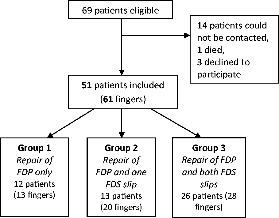
Study flowchart.
Description of the injured fingers by group.
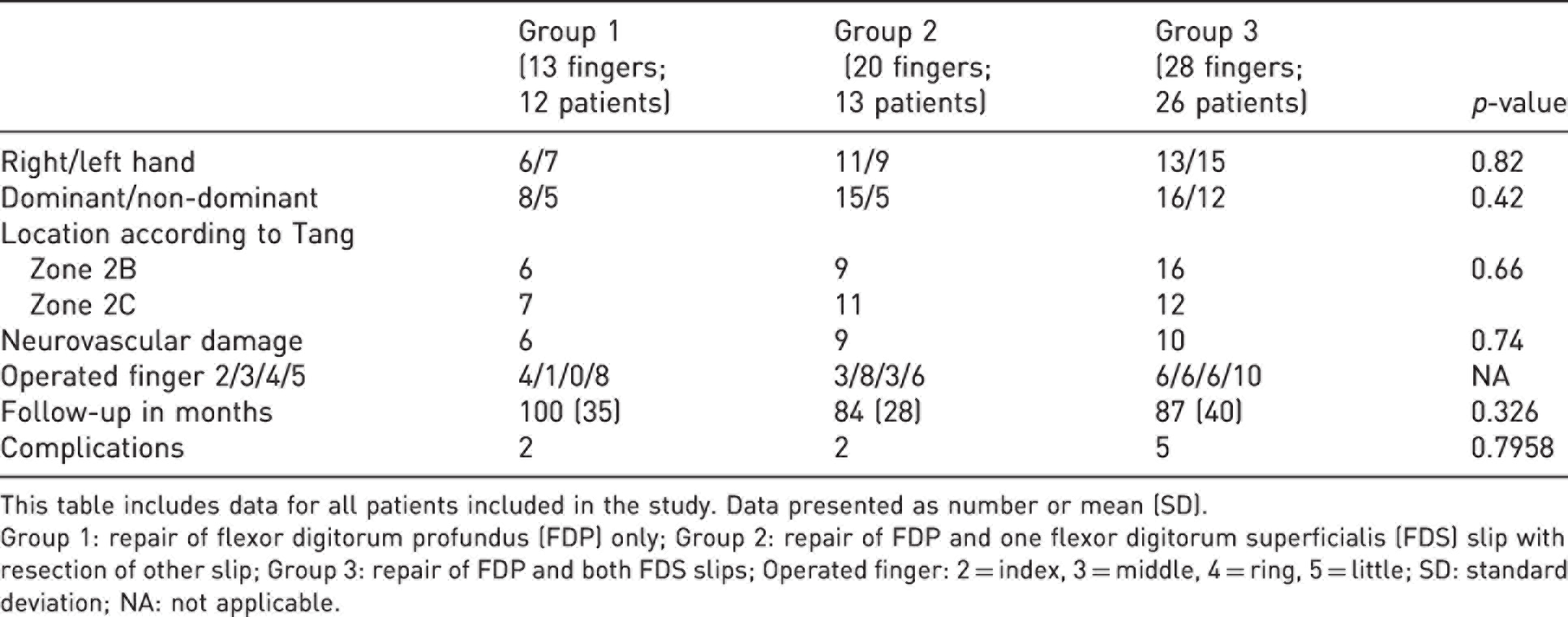
This table includes data for all patients included in the study. Data presented as number or mean (SD).
Group 1: repair of flexor digitorum profundus (FDP) only; Group 2: repair of FDP and one flexor digitorum superficialis (FDS) slip with resection of other slip; Group 3: repair of FDP and both FDS slips; Operated finger: 2 = index, 3 = middle, 4 = ring, 5 = little; SD: standard deviation; NA: not applicable.
Clinical outcomes, functional outcomes and return to work
There was no significant difference in the mean TAM or the mean TAMc values between the three groups (p = 0.1299 and 0.998, respectively). Comparing the outcomes according to Strickland’s formula and the Tang classification did not reveal any significant differences between the three groups. The residual flexion deformity in the PIP joint was larger in Group 2, but this was not significantly different (p = 0.1299) (Table 2). The mean DASH score in the entire patient population was 6.9, with no significant difference between the three groups (p = 0.152). All the patients had returned to work at the time of the clinical review except for one (manual worker) who could not return to work because of his injury.
Postoperative results for each group.
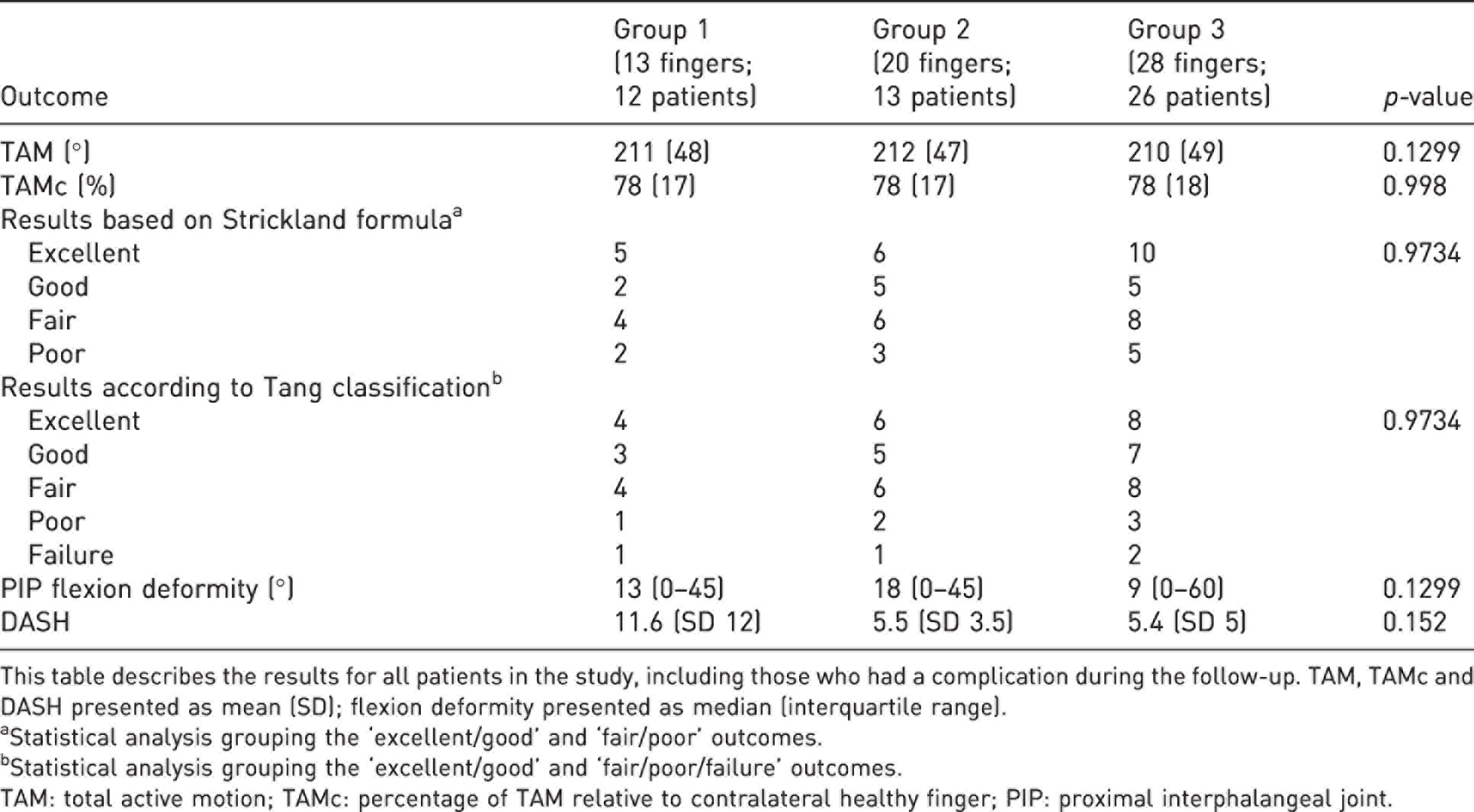
This table describes the results for all patients in the study, including those who had a complication during the follow-up. TAM, TAMc and DASH presented as mean (SD); flexion deformity presented as median (interquartile range).
aStatistical analysis grouping the ‘excellent/good’ and ‘fair/poor’ outcomes.
bStatistical analysis grouping the ‘excellent/good’ and ‘fair/poor/failure’ outcomes.
TAM: total active motion; TAMc: percentage of TAM relative to contralateral healthy finger; PIP: proximal interphalangeal joint.
Complications
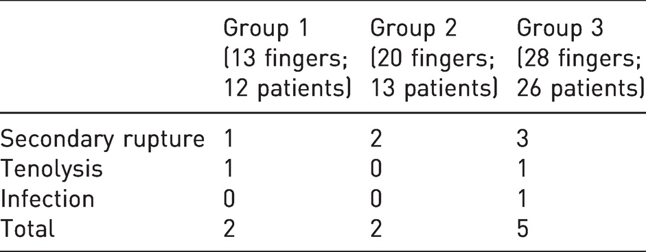
Nine fingers (17%) (nine patients) had a complication that required another surgical procedure (secondary rupture, tenolysis or infection). Three of the six secondary ruptures involved repair of the little finger. None of the three groups had a significantly larger number of complications (p = 0.7958) (Table 3). A subsequent statistical analysis was done after excluding the nine fingers with a complication in order to focus on the outcome of the fingers that were not reoperated. We found no significant difference in the values of the TAM, TAMc and Strickland’s formula. There appeared to be a trend of better outcomes in Group 1 patients based on the Tang classification, without a significant difference. The mean residual flexion deformity was low and not significantly different between the three groups (Table 4).
Complications requiring revision surgery.
Comparisons of outcomes after excluding the fingers that had a complication and required revision surgery.
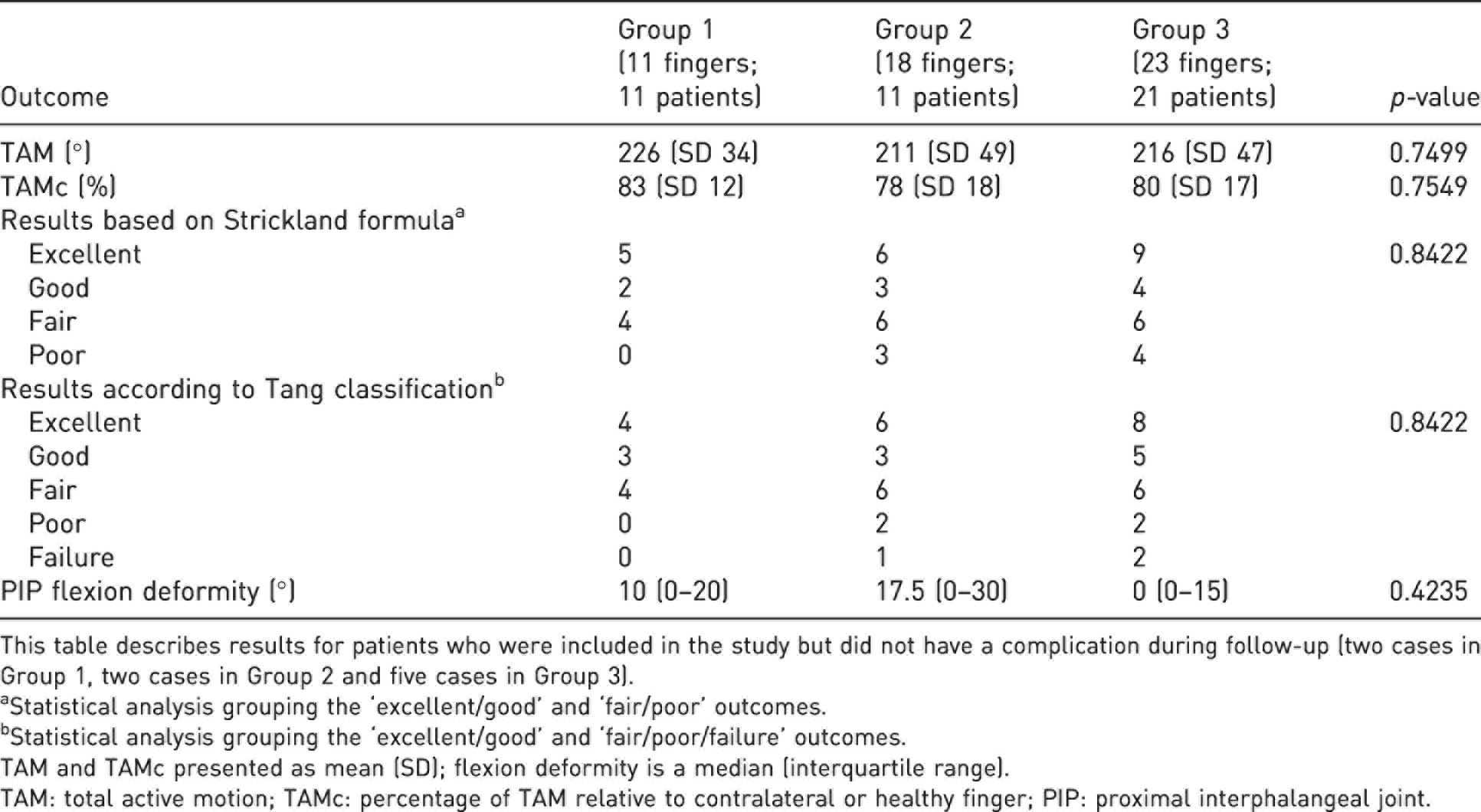
This table describes results for patients who were included in the study but did not have a complication during follow-up (two cases in Group 1, two cases in Group 2 and five cases in Group 3).
aStatistical analysis grouping the ‘excellent/good’ and ‘fair/poor’ outcomes.
bStatistical analysis grouping the ‘excellent/good’ and ‘fair/poor/failure’ outcomes.
TAM and TAMc presented as mean (SD); flexion deformity is a median (interquartile range).
TAM: total active motion; TAMc: percentage of TAM relative to contralateral or healthy finger; PIP: proximal interphalangeal joint.
Discussion
This study presents a lot of details of patients undergoing flexor tendon repair over a period of 12 years. We found no significant differences in the TAM, TAM relative to the contralateral side, Strickland’s formula, Tang classification or residual PIP flexion deformity in complete Zone 2B/2C transections of the finger flexor tendons in which the FDS was managed three different ways. These findings do not allow us to come out in favour of a particular surgical technique. Thus, the management of the FDS will continue to be controversial.
Tang (1994) found a non-significant trend to better TAM when the FDS was resected in Zone 2C. Sadek (2020) found no significant difference in finger mobility after Zone 2B injuries between repairing and not repairing the FDS. Lastly, Natal-Albelo et al. (2020) recently published conclusive findings that FDS excision led to worse short-term functional outcomes (3 months), assessed by TAM, Strickland and DASH, but found no significant differences at 6 months.
To our knowledge, no comparative clinical studies have been done of repairing one slip of the FDS and resecting the other slip. This surgical strategy was based on several biomechanics studies (Hwang et al., 2009; Paillard et al., 2002; Zhao et al., 2002) showing that partial FDS repair significantly reduced the tendon’s gliding resistance compared with repairing both slips. In our study, this strategy did not appear to produce different clinical outcomes compared with the two other techniques used and described in the literature.
The secondary objective of our study was to evaluate the functional outcomes and return to work in this population of patients. Transection of the flexor tendons remains a somewhat rare injury, making up <1% of all hand injuries (Dy and Daluiski, 2014; Hill et al., 1998), and affecting a young, active population having a mean age of 34 to 36 years (Khor et al., 2016; Moriya et al., 2016; Pan et al., 2020; Rigo and Røkkum, 2016). The transection occurs in the context of a work-related injury in 25% of cases (de Jong et al., 2014). Logically, the clinical results of a finger flexor tendon repair will determine the return to work. Most of our patients were manual workers, who undertook heavy and repetitive manual tasks (31 patients, 61%); only one patient had not returned to work, but he had injured all four fingers. While other studies are required, surgical repair of flexor tendon lesions in Zone 2B or 2C do not appear to compromise the return to work, including in manual workers. Similarly, the results of the DASH questionnaire in our population were very good (6.9 points) and not different between the three groups, which seems to confirm that Zone 2B/2C flexor tendon lesions do not appear to be a major disabling condition. The DASH score provides a broad, general view of upper limb use during the patient’s day-to-day life and has been validated in this context (Karjalainen et al., 2019). However, certain limitations could restrict its use for evaluating the outcomes of finger flexor repairs as it does not take into consideration factors such as the number of fingers affected, hand dominance and the type of finger injured. The Michigan Hand Questionnaire (MHQ) is a tool that provides a more comprehensive view of the morbidity of hand pathology (Chung et al., 1998).
In our case series, the tenolysis rate was (two fingers, 3.2%) similar to other published studies. The need for tenolysis can be explained by poor compliance to the postoperative rehabilitation instructions, resulting in secondary stiffening. The most common complication was secondary tendon rupture (10% of our case series). This occurrence was slightly higher than the rate reported in the literature of 4–10% for four-strand sutures (Dowd et al., 2006; Dy et al., 2012; Harris et al., 1999; Venkatramani et al., 2019). Three of the six secondary ruptures occurred after repair of tendons in the little finger, which is known to be more challenging than the other fingers (Giesen et al., 2018; Rigo and Røkkum, 2016).
Our study has several limitations that must be mentioned. First, the grip strength was not measured during the follow-up visits. Some studies (Sadek, 2020; Starnes et al., 2012) have collected this information (grip strength), while others mention a risk of measurement bias due to pain during its use and the risk of secondary rupture during a forceful gripping (Orkar et al., 2012). Other confounding factors are age, sex, occupation, type and number of fingers injured (Libberecht et al., 2006; Silfverskiöld et al., 1993). We recognize that the small patient population also contributed to a lack of statistical power. While the number of patients reviewed appears to be too small to draw any conclusions, the size of our case series was the same or even larger than other published studies on this topic, particularly focused on Zone 2 lacerations. The sample sizes range from 33 patients (37 fingers) (Tang, 1994) to 53 patients (64 fingers) (Sadek, 2020). The low incidence of flexor tendon injury and the large share of patients lost to follow-up in this young, active population contributed to the small sample size. Given the retrospective design of this study another limitation is represented by selection bias. Indeed, Group 1 included more little fingers than the two other groups. The smaller size of the little finger could have influenced the surgeon’s decision to resect the FDS to improve FDP mobility in the tendon sheath. Moreover, there were more multiple finger injuries in Group 2. A larger sample size may have increased the statistical power and increased the probability of finding significant differences between the three techniques. The results of the parameters evaluated in this study were very similar between the three techniques, which means that very large samples would be needed to detect statistically significant differences, although these differences are not likely be clinically relevant.
In repair of lacerations of the finger flexor tendons in Zone 2B or 2C, the medium term clinical and functional outcomes do not seem to differ between resecting one FDS slip, both FDS slips or repairing the FDS. The patient’s quality of life and ability to return to work (even manual work) do not appear to be strongly impacted.
Footnotes
Acknowledgements
The statistical team of the Lille University-Hospital Center who performed the statistical analyses of this study.
Declaration of conflicting interests
The authors declare no potential conflicts of interest with respect to the research, authorship, and/or publication of this article.
Funding
The authors received no financial support for the research, authorship, and/or publication of this article.
Ethical approval
Ethical approval was given by our local Research Ethics Committee based on the retrospective nature of the study, as all the procedures being performed were part of the routine care and follow-up of patients.
Informed consent
Oral and written informed consent was obtained from all individual participants included in the study.
