
Abstract

Dear Editor,
A 20-year-old woman presented 6 months after a motor vehicle accident that caused a right C5-C7 brachial plexus palsy. She had surgery elsewhere prior to her presentation to treat a clavicular fracture. Surgical exploration showed avulsed C5-C7 nerve roots with no potential for grafting. She underwent transfer of the spinal accessory nerve to the suprascapular nerve, long head of triceps motor branch to the teres minor motor branch and to the anterior division of the axillary nerve, and fascicles from the ulnar nerve to the biceps motor branch. At 26-month follow-up she had recovered 70° of shoulder abduction, complete external rotation and full elbow flexion scoring M4 on Medical Research Council (MRC) grading. However, she still had very limited (20°) shoulder flexion and was unable to greet people with a handshake.
One year later, the patient still complained of absent shoulder flexion and so we elected to pursue surgical reconstruction. On clinical examination, the upper clavicular part of the pectoralis major appeared weak and was unsuitable for transfer to the anterior deltoid. Transfer of the sternal portion of the pectoralis major was therefore considered.
Through a deltopectoral approach with extension over the previous incision for clavicular plating, both the deltoid and the upper part of the pectoralis muscles were exposed. Both these muscles appeared atrophic. However, the lower part of the pectoralis major was normal in appearance and the pectoralis minor muscle was functional too. The sternal portion of the pectoralis major muscle was released from the ribs. The neurovascular pedicle then was dissected in the deltopectoral groove and contractions of the muscle flap were confirmed by stimulating the pectoral nerve. The muscle flap was flipped 180° around the neurovascular bundle to make the under surface of the muscle face upwards. With the shoulder adducted, the flipped sternal portion of the pectoralis major was attached to the posterior portion of the acromion, in maximum possible tension. (Figure 1). Additional sutures were placed to anchor the flap on the anterior portions of the acromion and clavicle. The humeral insertion of the pectoralis tendon was left unaltered, as adequate tension to the muscle transfer was obtained.

(a) Schematic illustration of the surgical procedure for transferring the lower portion of the pectoralis major muscle to the deltoid muscle and (b) Intraoperative view of the lower portion of the pectoralis major muscle flipped over the deltoid muscle.
At 18-month follow-up after the anterior deltoid reconstruction, her shoulder abduction was 70°, external rotation was complete, and shoulder flexion was 60° (Figure 2). The DASH score improved from 30 preoperatively to 70 postoperatively. No improvement in shoulder abduction was noted, possibly because the insertion of pectoralis major tendon was not moved laterally, aiming instead to reconstruct shoulder flexion.
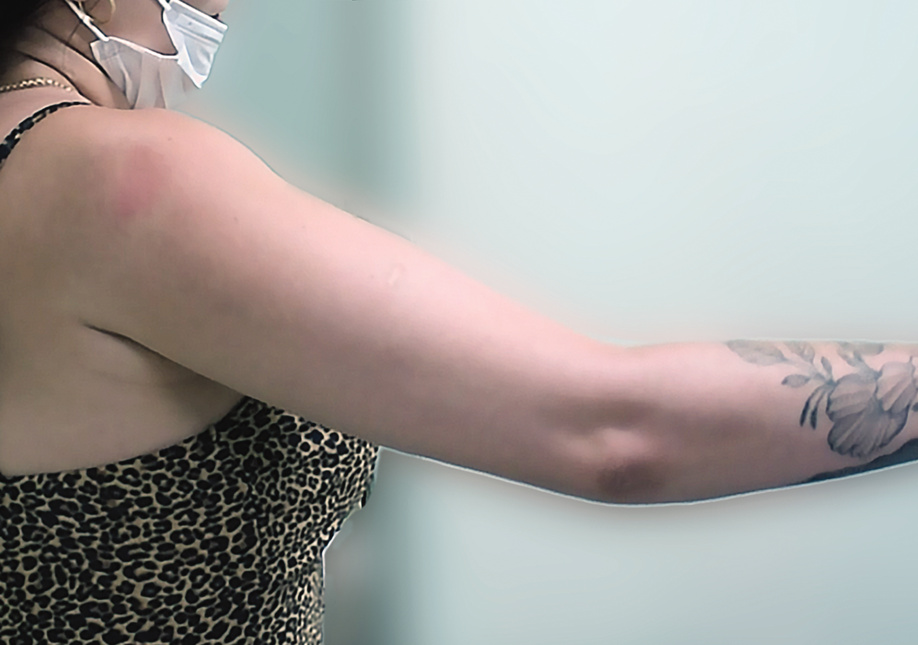
Demonstrating shoulder flexion 18 months after surgery.
Shoulder flexion is largely performed by the anterior deltoid muscle, with some contributions from rotator cuff muscles (Kronberg et al., 1990; Myers et al., 2005; Wattanaprakornkul et al., 2011). In general, after a successful transfer of the spinal accessory to suprascapular nerve, both shoulder abduction and flexion is restored while poor recovery of external rotation is to be expected. Our patient did recover external rotation but not shoulder flexion, and potential options for the reconstruction of shoulder flexion were limited.
The trapezius, long head of triceps, the long head combined with the short head of the biceps, latissimus dorsi and upper pectoralis major muscles have all been proposed for deltoid function reconstruction (Resch et al., 2008). Although in our patient the upper trapezius was available, the poor results reported from trapezius muscle transfers often with less than 20° of forward flexion made us look at other options (Rühmann et al., 1998). The biceps tendon was not an option, because it was a reinnervated muscle. We also avoided using the latissimus dorsi muscle due to our past experience of poor outcomes on occasions following transfer of the thoracodorsal nerve to the anterior division of the axillary nerve. The long head of triceps was unsuitable for transfer because its donor nerve was used to reinnervate the axillary nerve. The upper pectoralis major was paralyzed in our patient and hence was not an option. In addition, only 30° of shoulder flexion was restored using this transfer in a previous study (Resch et al., 2008). The suboptimal outcomes were possibly due to partial denervation of the upper pectoralis muscle that commonly occurs with brachial plexus injuries. For all these reasons, a lower pectoralis major transfer to the anterior deltoid was considered.
This transfer could be considered as a salvage procedure to achieve shoulder flexion in patients with partial brachial plexus injuries.
Footnotes
Declaration of conflicting interests
The authors declare no potential conflicts of interest with respect to the research, authorship, and/or publication of this article.
Funding
The authors received no financial support for the research, authorship, and/or publication of this article.
Informed consent
Written informed consent was obtained from the patient(s) for their anonymized information to be published in this article.