
Abstract
This article reviews and highlights complications of flexor tendon repairs. Although the outcomes of flexor tendon repairs have improved over the years, fair or poor functional outcomes are seen, especially in patients whose trauma involves multiple structures of the hand and in zone 5 with multiple tendon lacerations. Rupture of the flexor tendon after repair is no longer a major problem if current repair principles are carefully adhered to. Different degrees of adhesion formations and interphalangeal (IP) joint stiffness still occur in a few patients. Early active postoperative mobilization and use of a shorter splint with sparing of the wrist are effective measures to prevent adhesion formation and IP joint stiffness. Given the overall poor results and high rate of complications with flexor digitorum profundus (FDP) repairs in zone 1, a direct repair of the FDP tendon to any short remnant of the distal insertion with 10-strand or even stronger core suture repair is adopted by many units.
Introduction
The outcomes of flexor tendon repairs have remarkably improved due to more detailed understanding of flexor tendon anatomy and biomechanics, its repair techniques and rehabilitation protocols (Elliot and Giesen, 2013; Giesen et al., 2017; Lalonde and Martin, 2013; Moriya et al., 2017; Tang, 2007, 2013, 2018a, 2018b).
Adherence to crucial steps, such as strong tendon repair, the release of critical pulleys, an intraoperative extension-flexion test to assess the quality of the tendon repair (Tang et al., 2017) and the rehabilitation protocol of early active motion, have ensured good to excellent outcomes in more than 85% to 90% of adult patients after zone 2 flexor tendon repair (Giesen et al., 2009, 2018; Moriya et al., 2017; Pan et al., 2020; Zhou et al., 2017). However, despite these advances, some fair and poor functional outcomes are still seen, especially in patients whose trauma involves multiple structures of the hand or multiple tendon lacerations in flexor zone 5.
Many factors involving pre-, intra-, and postoperative management can influence functional outcomes. Despite the best efforts of surgeons, there are still some outside factors having adverse effects, such as the type of trauma and compliance of patients. The reoperation rates reported previously were between 6% (Dy et al., 2012) and 13% (Rigo and Røkkum, 2016). The purpose of this review is to present and highlight the complications still encountered after flexor tendon repair and provide tips for the prevention and management from the personal experience of the authors who have dealt with a significant number of these problems.
General complications
Adhesion formation
Adhesion formation is the most common complication encountered after surgical repair of tendon injuries (Momeni et al., 2010; Pulos and Bozentka, 2015), mainly resulting from the imbalance between intrinsic and extrinsic healing factors. Adhesion formation represents secondary extrinsic tendon healing and cannot be entirely avoided. Various factors, such as the location of flexor tendon injury (Svingen et al., 2022), associated pulley injury, involvement of flexor digitorum superficialis (FDS) tendon, and concomitant vascular or nerve injuries (Demers et al., 2022), are all known to considerably influence the severity of tendon adhesion. Extensor tethering by fibrin-oedema and reduced passive finger flexion are the most common reasons for loss of flexion after flexor tendon surgery. It is typically seen in patients with complications or patients who fail to go through focused postoperative hand therapy.
Tendon adhesions are frequently observed after crush injuries, and adhesion formation has been found to be proportional to the extent of soft tissue trauma (Strickland, 2000). Early active motion is currently the most effective way to prevent or decrease adhesion formation. A strong tendon repair is one of the prerequisites for this early active motion regimen.
The release of tendon adhesions is the most frequent reason for reoperations after flexor tendon repair. Previous reviews have identified the overall rate of adhesion formation after primary repair of flexor tendons requiring tenolysis to be between 4% and 10% (Pulos and Bozentka, 2015). Karalezli (2019) reported a lower tenolysis rate after finger flexor tendon repair in zones 1, 3 and 5 than in zone 2. However, the tenolysis rate reported over the past decade varies widely from 0% to 23% (Giesen et al., 2009, 2018; Karalezli, 2019; Pan et al., 2019; Svingen et al., 2022). Different trauma mechanisms, location of injuries, surgical methods and rehabilitation protocols are the most important impacting factors. Variations in indications for tenolysis as well as in individual patient willingness or demand for further treatment might explain the wide range of tenolysis rates reported in literature. Results from a large registry study showed that no patients underwent tenolysis in the low-income group, compared with 5.4% in the middle-income group and 8.7% in the high-income group (Svingen et al., 2022).
Karalezli (2019) reported a tenolysis rate of 23% after zone 2 flexor tendon repairs using a modified Kessler method (two-strand) and modified Duran passive motion protocol for postoperative rehabilitation. Civan et al. (2020) evaluated the tenolysis rates of zone 2 flexor digitorum profundus (FDP) with FDS tendon repairs using a four-strand technique and early passive motion exercises. A total of 23 out of 149 patients and 28 out of 194 fingers (14.4%) required tenolysis. However, Moriya et al. (2019), reporting on the outcomes of the flexor tendon repairs of zone 2 in 102 fingers using a six-strand core suture and early active motion protocol, noted tenolysis was needed in 5 (5%) fingers. Sadek (2020) also reported similar rates with three fingers needing tenolysis out of 64 flexor tendons repairs in zone 2B using the six-strand repair method and a combination of active and passive motion postoperative protocol. Pan et al. (2019, 2020) reported a tenolysis rate of 2% after zone 1 and 2 primary tendon repairs with early active motion protocol. Adhesions may be more common among patients treated with weak tendon repairs and passive motion postoperative rehabilitation protocol.
The tenolysis rate varies among the digits. Compared with the flexor tendon repair of the fingers, there are fewer reports on adhesion after flexor pollicis longus (FPL) repair. With the use of strong tendon repair and early active motion, many authors report no tenolysis (Giesen et al., 2009; Pan et al., 2017; Moriya et al., 2021). Fewer satisfactory outcomes are often reported in the little finger compared to the other fingers in zones 1 and 2. Orkar et al. (2012) compared the outcome of flexor tendon repair in the index and little fingers and found tenolysis in 1 (1.7%) patient in the index finger group and in 8 (7.4%) patients in the little finger group.
In our experience, the tenolysis rate after repairs in zone 2 without digital bony injury was similar to that of Pan et al. (2019, 2020), most likely due to the identical clinical and rehabilitation protocols followed in the two units (Tang et al., 2017). The tenolysis rate after tendon repair in zone 5 is higher, at about 15% to 20%.
Despite these, some patients end up with a limited range of motion from adhesions that may require tenolysis. In general, tenolysis is recommended if the injured fingers cannot recover at least 40% to 50% of the range of interphalangeal (IP) joint active motion postoperatively. Tenolysis should be deferred for at least 3 months after the initial surgery, because the digital function usually continues to improve dramatically in the first few months. In our experience, we have seen patient outcomes continuing to improve even after 6 months to 1 year after a tendon repair. The timing of tenolysis, hence, should be judged clinically and should be delayed until there is no further progression in the range of movement of the digit.
Our results of flexor tendon repair using the modern principles of the six-strand core repair method, partial or entire venting of critical pulley, intraoperative extension-flexion test and early active motion protocol have shown good outcomes. In the last 3 years, we have performed one tenolysis out of 43 digits after repair in zone 1. The index finger of this patient had unsatisfactory flexion (10°) of the distal IP joint with normal extension after primary repair (rated as fair according to Tang criteria). We performed tenolysis 3 months after the primary repair (Figure 1). None of our patients needed tenolysis after zone 2 repair. Four patients with severe hand injury (two wrist amputations, one extensive crush injury and one ‘spaghetti wrist’) underwent tenolysis after zone 3, 4 and 5 tendon repairs (Figure 2).
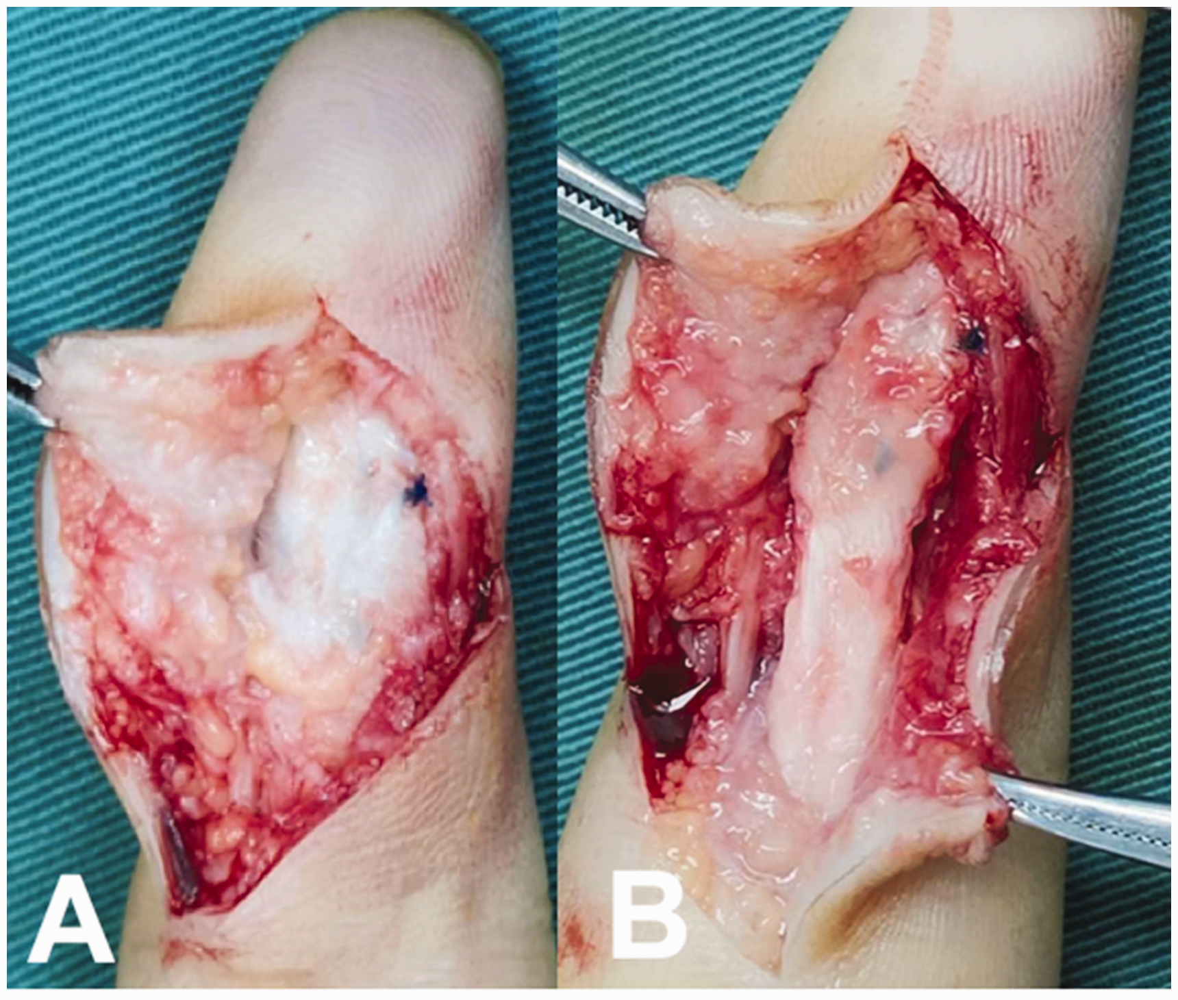
Index finger in a 48-year old man 3 months after zone 1 tendon repair. (a) Adhesion formation shown surrounding the tendon and (b) The adhesion tissue was resected and healed tendon seen.
Hand of a 52-year old man six months after replantation following traumatic amputation at palm level. (a) Preoperative image demonstrating limited active flexion of fingers. (b) Severe adhesion formation seen on exploration and (c) Tendons after tenolysis and resection of the adhesions surrounding the tendons.
Interphalangeal joint stiffness
IP joint stiffness is the most frequent late complication after a flexor tendon repair (Momeni et al., 2010; Pulos and Bozentka, 2015). Flexion contractures of the proximal IP (PIP) joint may be a consequence of collateral ligament and volar plate contracture, fracture of a phalanx, scarring of the skin or flexor tendon adhesions. The incidence of joint stiffness is high, especially in patients experiencing severe hand injuries involving multiple structures.
The incidence of IP joint stiffness reported in flexor tendon injuries is 17% (Momeni et al., 2010; Pulos and Bozentka, 2015). The most common cause of joint stiffness is the postoperative protective splinting of the finger. Seiler (2011) states that joint contracture may be more common among patients treated with dynamic flexion splinting with elastic traction. The rates of flexion contracture of more than 15° in the PIP joint were reported to be in the range of 29% to 40% with the Kleinert technique, 13% to 28% with early passive motion, and 10% to 24% with early active motion. Güntürk et al. (2018), reporting on the results of repair of zone 2 flexor tendon lacerations using the four-strand modified Kessler core suture and modified Kleinert postoperative protocol, have noted PIP contractures in 59%, of which 10% of the contractures were more than 20°. That was the main reason for the Kleinert postoperative rehabilitation method falling into disrepute worldwide.
The incidence of joint stiffness can be reduced by continuous joint mobilization exercises and correct postoperative splinting. In our practice, we maintain the wrist in 20° flexion, metacarpophalangeal joint in 30° flexion and IP joints in full extension after flexor tendon repair using a splint. PIP joint release combined with flexor tenolysis is a valid solution for recalcitrant contractures. Zuo et al. (2020) achieved excellent functional outcomes of 84% according to the modified Strickland criteria in 15 digits with combined PIP joint release and flexor tenolysis.
Rupture of tendon repair
Factors contributing to rupture after a flexor tendon repair include weak suture material, poor surgical technique with gapping, repair too weak to sufficiently undergo aggressive postoperative rehabilitation protocols and poor patient compliance. With a better understanding of the concept of flexor tendon repair and improvement in surgical and postoperative rehabilitation, the rate of rupture after flexor tendon repair reduced considerably to almost zero.
Although early active motion has been shown to reduce the occurrence of adhesions, the repair should be sufficiently strong to allow this. Otherwise, the risk of tendon rupture will increase. Peck et al. (1998) reported a rupture rate of 46% in zone 2 flexor tendon repair with rehabilitation using controlled active motion. In contrast, the rupture rate was 4% in a study by Elliot et al. (1994). The most likely reason for such a significant difference in the rupture rate would be the repair technique. Even though a two-strand core suture was used in both these studies, the rupture rate of 46% was probably due to the use of a weaker modified Kessler core suture popularized by Kleinert, while the rupture rate of 4% was associated with a more original version of Kessler’s, which Elliot described as Kessler/Kirchmayer (Savage, 2012). In a review by Momeni et al. (2010), the rate of rupture of flexor tendon repair was reported to be in the range of 4% to 30%. The strength of a tendon repair using a two-strand core suture technique is not sufficient to tolerate the postoperative early active mobilization rehabilitation. This is the prime reason for the high rupture rate reported in the earlier publications. Biomechanical studies have shown that six-strand core suture repair has better strength to tolerate the early active motion (Wu and Tang, 2021). The rupture rate reported was 3% in a study by Savage and Risitano (1989) using a six-strand core suture for zone 2 tendon injuries. Currently, multi-strand core suture has become the accepted practice. Svingen et al. (2022) identified risk factors for reoperations after zone 1 and 2 flexor tendon repairs in a registry study and found that a modified two-strand Kessler core suture technique had a higher association to rupture compared with a four-strand loop suture.
The rupture rate after flexor tendon repair has reduced considerably since the principles of stronger repair, pulley venting and early rehabilitation have been popularized and adopted widely. Recent studies in the last 5 years have shown rupture rates in the range of 0% to 5% in zones 1 and 2 using a six-strand core suture combined with early active mobilization (Chen et al., 2021; Giesen et al., 2018; Moriya et al., 2017; Pan et al., 2019; Sadek, 2020; Zhou et al., 2017).
Historically, the FPL tendon had a higher rate of tendon rupture after repair compared to the fingers. Among the other fingers, the little finger is reported to be more prone for rupture (Dowd et al., 2006). In a study by Orkar et al. (2012), 3 (5%) tendons ruptured in the index finger group while 12 (11%) failed in the little finger group. However, this appears to have changed in the recent past, with no ruptures reported in many publications, and 77% to 91% of patients noted to have excellent and good outcomes (Baer et al., 2003; Giesen et al., 2009; Pan et al., 2017). Moriya et al. (2020) reported three FPL tendon ruptures in 17 patients, but all three ruptures were in patients treated by surgeons who were inexperienced in tendon repair surgery.
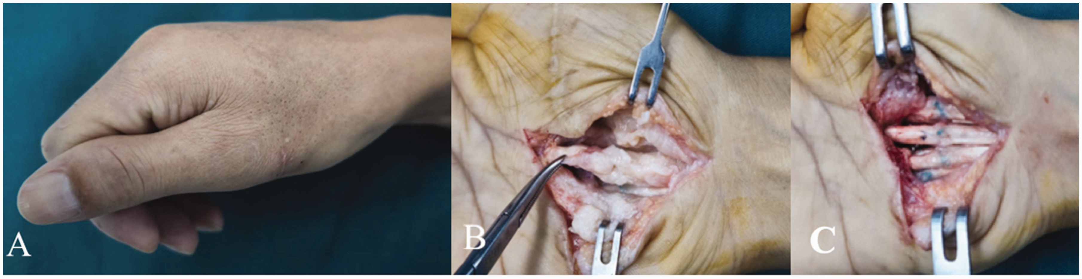
Most tendon ruptures are reported to occur around day 10 after a tendon repair. Other studies, however, report ruptures as late as 6 to 7 weeks postoperatively (Momeni et al., 2010). Rupture of a flexor tendon after repair can occur any day in the first few weeks, often because of inappropriate use of the hand by the patient. Once the diagnosis is confirmed, immediate exploration and re-repair is the preferred treatment (Figure 3). Generally, if a rupture occurs more than 4 to 6 weeks after the initial repair, tendon grafting or staged tendon reconstruction may be required.

(a) Rupture of FDP tendon after a 2-strand modified Kessler repair. (b) Repair using a 6-strand M-Tang technique 3 weeks after the initial repair. (c) and (d). Clinical pictures showing full flexion and extension of the finger 6 months after surgery.
We recommend a six-strand core suture to repair flexor tendon in zones 2 to 5, while a four-strand core suture is reserved for smaller tendons, such as the FDS or FDP of the little finger. This is combined with a safer rehabilitation protocol of early active and passive motion in all cases. We have not noticed any tendon ruptures in our series of more than 100 flexor tendon repairs within the last 3 years (Tang, 2005, 2013).
Infection and wound problems
Other complications including infection, disruption of surgical wound and additional further surgical procedures caused by soft tissue injuries have also been reported (Giesen et al., 2018; Moriya et al., 2019). Demers et al. (2022), reporting on the overall incidence of complications after flexor tendon repair in a registry study, noted the rate of infection as 3.1% and disruption of the surgical wound as 1.8%. The most common reason found was a significant degree of wound contamination. Serious infection after flexor tendon repair is rare but devastating. Most cases of infection encountered in our department were superficial inflammation of soft tissue caused by foreign body-type reactions to suture materials 3 to 6 months after the initial repair. We had only one patient who developed a severe infection caused by pseudomonas aeruginosa following multiple tendon repairs in zone 5.
Tendon triggering and bowstringing
Triggering and bowstringing of the flexor tendon after repair have been reported (Momeni et al., 2010; Pulos and Bozentka, 2015) and are associated with the treatment of flexor pulleys during flexor tendon surgery. The bowstringing is more often observed in severe hand injuries associated with the loss of most of or entire pulleys of the digits. Giesen et al. (2017, 2018) noted that when the bulkiness at the repair site was 150% of that of the normal tendons, active gliding of the repaired tendons was impeded. Venting of the A2 or A4 flexor tendon pulleys was once considered aggressive (Tang, 2018, 2019). However, this consideration has changed in the past decade. Proper venting of either a part of the A2 pulley or the entire A4 pulley is a key step in attaining an optimal outcome to allow the repaired tendons to glide smoothly. Recently, the concern has not been bowstringing caused by venting of the pulley, but insufficient gliding of the tendon as a result of inadequate venting. Moriya et al. (2016a, 2016b) and Giesen et al. (2018) described more aggressive approaches including venting the entire A2 pulley or venting the A4 and A3 pulleys together during flexor tendon repair surgery. They reported no clinically dysfunctional bowstringing in either of those two studies. We do not encourage this aggressive approach but do recommend judicious venting of these important pulleys whenever indicated due to catching of the repair site. With the application of the concept of pulley venting and intraoperative extension-flexion tests, tendon triggering and bowstringing can be avoided.
Site-specific complications
Zone 1
Achieving satisfactory outcomes after zone 1 flexor tendon repair is challenging and some loss of motion is anticipated (Huq et al., 2013). The mechanism of injuries in zone 1 involves a heterogeneous collection of injury patterns primarily due to direct laceration or avulsion of the tendon.
A length of tendon stump of more than 1 cm is sufficient to allow direct tendon repair using a combination of core and epitendinous sutures. The repair method and rehabilitation protocol are the same as that after flexor tendon repair in zone 2 (Tang, 2019). The functional outcomes and complications of tenolysis and rupture of flexor tendon repairs in zone 1 are often reported and analysed together with zone 2 tendon repairs in the published literature. None of the patients required tenolysis or surgery for rupture after zone 1 tendon repairs in recent publications (Giesen et al., 2018; Pan et al., 2020).
Excellent or good outcomes are not consistently achieved in cases where the distal tendon stump is less than 1 cm long. A myriad of techniques has been described for treating the tendon-bone interface including tie-over button, bone anchor and suture anchors (Huq et al., 2013). No single technique has emerged to be superior to others. McCallister et al. (2006) performed a study comparing flexor tendon repairs of zone 1 using the micro anchor technique with the standard pullout method. They reported no differences in outcomes for range of motion of the distal IP joint between the two groups.
A wide range of complications were reported including failure of the repair, nail deformity, repeat surgery and wound infection using pullout and all-inside suture fixation. Kang et al. (2008) reported an infection rate of 22% and a rate of abnormal nail growth of 35% using a button pullout technique with polypropylene sutures. The occurrence rate of complications, including skin infection, osteomyelitis, chronic draining granuloma and nail deformity, was in the range of 24% to 63% using a transosseous or non-transosseous all-inside technique (Geary et al., 2020; Li and Hammert, 2022). Although these complications, to a certain extent, can be avoided using a bone anchor, the main disadvantage of a bone anchor compared to the other techniques is the cost of the implant. In addition, a foreign body-type reaction to the suture material and osteomyelitis have been reported in isolated cases (Huq et al., 2013).
Compton et al. (2022) reported similar outcomes and fewer complications in patients with zone 1 FDP avulsions treated without tendon repair compared to those who underwent acute tendon repair. Overall poor results with FDP repairs in zone 1 is widely recognized and the complications with these techniques are many. Therefore, we do not recommend a pullout suture technique and our current preference is doing a direct repair of the FDP tendon to any short remnant of the distal insertion of the FDP tendon with a 10-strand or even stronger core suture (Calcagni et al., 2022; Tang, 2022).
Zone 2
Although, historically, zone 2 flexor tendon repairs have been associated with a poor outcome, with the advances in surgical techniques and postoperative rehabilitation of early mobilization, the repair of flexor tendons in this zone no longer poses a major problem. The rupture rate after primary repair has considerably reduced with the adoption of strong repair methods using a six-strand core suture and pulley venting (Chen et al., 2021; Giesen et al., 2018; Moriya et al., 2017; Pan et al., 2019; Sadek, 2020; Zhou et al., 2017).
Despite advances in the surgical and rehabilitation protocol of flexor tendon repairs, adhesion formation continues to occur in 6% of the cases undergoing early active motion (Starr et al., 2013), with a tenolysis rate of 5% to 10%. Injury characteristics including both the FDS and FDP laceration, concomitant vascular injury and nerve injury, and the extent of soft tissue damage was associated with an increased risk of requiring tenolysis (Demers et al., 2022; Svingen et al., 2022). The outcomes of zone 2 tendon repairs are likely less satisfactory when the injuries are more severe (such as with fractures or with extensive avulsion of skin and soft tissue). More optimized rehabilitation protocols may reduce the density of adhesion formation, which is caused by inadequate differential tendon motion between FDS and FDP during the initial healing phase (Chinchalkar et al., 2016). Combining synergistic wrist motion and active mid-range digit flexion could improve the differential tendon excursion without overloading the healing tendons (Chinchalkar et al., 2016). The use of an extension splint keeping the repaired finger in more extension at the MP joint, to allow a larger IP joint flexion, is very useful in treating adhesions. This splint can be used during the first 1 to 2 months after repair, when the adhesions can still be remodelled.
Zones 3 to 5
Since the flexor tendon repair methods and rehabilitation protocols in zones 3 to 5 are similar to that of zone 2, there are few separate reports on the functional outcomes of flexor tendon repairs specifically in zones 3 to 5, and most of them are reported in combination with those of zones 1 and 2. As with the repairs in zone 2, strong repair methods using a standard four- or six-strand core suture have made active early mobilization possible, providing better outcomes and a low rupture rate in these zones as well. However, tendon injury in zones 3 to 5 often involves multiple tendons with concomitant nerve and vascular injuries or even extensive skin and soft tissue injuries. Despite strong repairs and early active motion, there is still a high adhesion rate in these zones. The complication rate after tendon repairs in zones 3 to 5 is impacted by many factors including mechanism of injury, structures involved, treatment protocol and compliance of patients.
Yii et al. (1998), reporting on 52 patients with flexor repairs in zone 5, noted no tendon ruptures. Wilhelmi et al. (2005) reported tenolysis in one patient and a second surgery for rupture in three patients after zone 5 flexor tendon repairs. Yazdanshenas et al. (2016) noted tenolysis in seven out of 153 patients, and combined tenolysis and neurolysis in three patients. Demirdover et al. (2018), reporting on the functional outcome in wrist injuries between the distal wrist crease and flexor musculotendinous junction (spaghetti wrist), noted eight out of 156 patients needed revision and reoperation. Three of these had tendon adhesions, three had neuromas and adhesions simultaneously, one had an ulnar nerve neuroma and one patient had two neuromas on the median and the ulnar nerves and a rupture of the FDS tendon to the little finger. In multiple tendon injuries caused by clean sharp lacerations, adhesion formation can be avoided through a combination of meticulous surgical technique, early active motion and patient compliance. However, in multicomponent soft tissue crush injuries of the wrist, the ends of tendon and nerves can be ‘horsetail’-shaped. The adhesion formation may be inevitable in these situations because of severe oedema, poor repair quality of tendons and poor nerve function.
Summary
With the advances in repair techniques and rehabilitation strategies for a flexor tendon repair, the rupture rate has substantially reduced, whereas tendon adhesion still remains the most common and difficult problem. Early active motion postoperatively is an effective measure to prevent adhesion formation. Reliable surgical repairs and exact venting of the pulleys is a prerequisite to decrease resistance to gliding and reduce the risk of rupture. A shorter and free wrist positioning splint can be used to make finger joints fully extendable to prevent PIP joint stiffness. For compliant patients, out-of-splint passive and active finger motion after a strong tendon repair has been proven to be safe (Pan et al., 2020). Appropriate rehabilitation protocols can be adjusted according to the preferences of the surgeon and therapist and the characteristics of individual patients.
Meticulous operative technique and appropriate rehabilitation protocols remain the most effective measures to prevent complications in clean tendon injuries. However, adhesions and joint contractures are unavoidable in severe hand injuries involving multiple structures because of restrictions in allowing early active motion. A comprehensive analysis of the biomechanics, healing ability and healing speed of involved structures are required. Publications of basic research in recent years show promising results in achieving scarless healing with biologic modulators (Yang et al., 2022; Zhou et al., 2018).
Footnotes
Declaration of conflicting interests
The authors declared no potential conflicts of interest with respect to the research, authorship, and/or publication of this article.
Funding
The authors received no financial support for the research, authorship, and/or publication of this article.