
Abstract
We present two cases of isolated post-traumatic osteoarthritis in the middle carpometacarpal joint.
Keywords
Carpometacarpal joint (CMCJ) osteoarthritis is rare but is more frequent in the little finger ray CMCJ (Cobb et al., 2018; Gehrmann et al., 2015). We present two cases of isolated middle ray CMCJ osteoarthritis after hand injuries.
A 34-year-old deliveryman injured his left hand in a traffic accident. He complained of dorsal pain and swelling and sought acupuncture and rehabilitation. He visited our clinic 10 months after the injury because of worsening pain and weakness that was affecting his ability to work. Isolated middle ray CMCJ osteoarthritis was seen on radiographs and a computed tomography (CT) scan and no other CMCJs were injured. Capitometacarpal arthrodesis was carried out (Figure 1). Six months later, he was able to return to delivery work, experiencing no pain. His grip strength improved from 31.0 kg preoperatively to 47.9 kg (compared 54.9 kg on the healthy side) 1 year postoperatively.
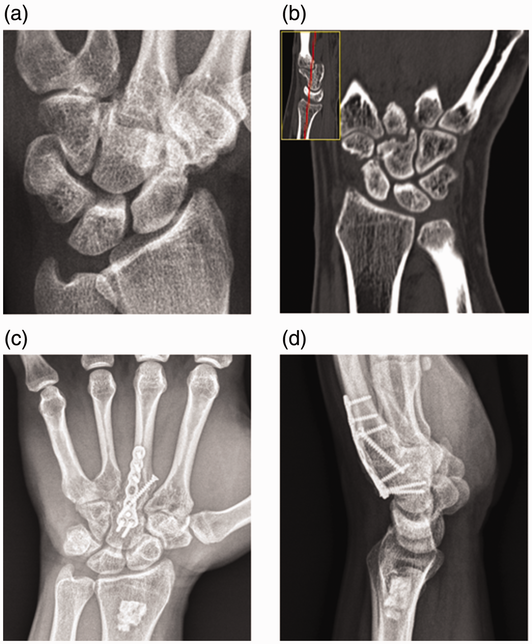
(a, b) A 34-year-old man: radiographs of the left wrist. Computed tomography scan with sagittal cuts of the CMCJs of the (c) index, (d) middle and (e) ring finger rays. (f) 3-D reconstruction with the arrow indicating isolated middle ray CMCJ osteoarthritis. (g) Coronal cut of the CMCJs and (h, i) radiographs of middle ray CMCJ arthrodesis 12 months postoperatively. CMCJ: carpometacarpal joint.
A 53-year-old stock trader, had a fall from a height of 1.5 metres, striking his right hand on the ground. He attended our clinic 6 months later as the symptoms of pain, swelling and grip weakness had persisted. Radiographs and a CT scan revealed isolated osteoarthritis in the right middle ray CMCJ, primarily affecting the dorsal half of the joint. He was treated with capitometacarpal arthrodesis (Figure 2). His grip strength improved from 20.9 kg preoperatively to 29 kg (compared to 42.2 kg on the healthy contralateral side) at the final 26-month follow-up, with intermittent pain measuring 1 on a visual analogue scale (VAS) of 1 to 10.
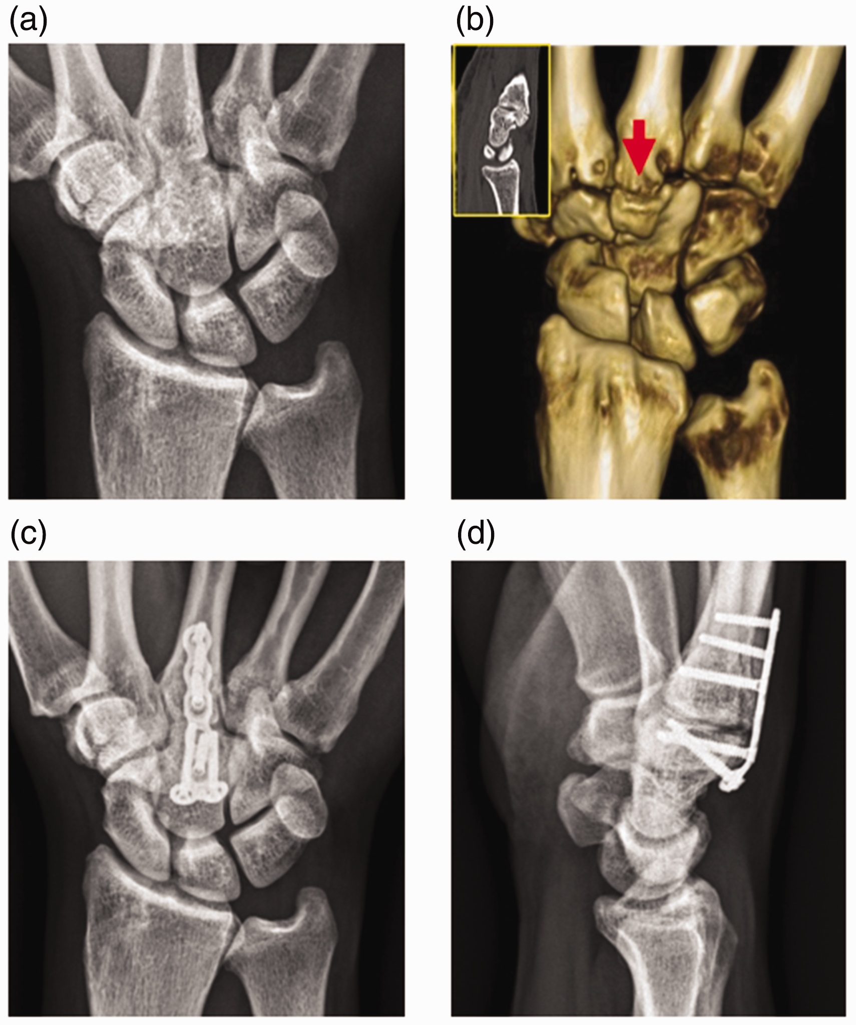
(a, b) A 53-year-old man: radiographs of his right wrist. Computed tomography scan with sagittal cuts of the CMCJs of the (c) index, (d) middle and (e) ring finger rays. (f) 3-D reconstruction with the arrow indicating isolated middle ray CMCJ osteoarthritis. (g) Coronal cut of the CMCJs. (h, i) Radiographs of middle ray CMCJ arthrodesis 26 months postoperatively. CMCJ: carpometacarpal joint.
The middle ray CMCJ serves as the keystone owing to its central and recessed location compared to the other CMCJs (Bhardwaj et al., 2020) and the ray is the central pillar of the hand (Flatt, 1959). Although osteoarthritis is seen in carpal boss syndrome, it has not been reported after trauma. CMCJ fracture-dislocations occur most commonly in the ring and little CMCJs because of their relative mobility. In our two cases, axial loading is the likely mechanism of injury as no dislocation occurred.
The middle ray CMCJ is a rigid joint, so arthrodesis is a suitable option. In these two cases, arthrodesis resulted in satisfactory functional outcomes.
Footnotes
Declaration of conflicting interests
The authors declared no potential conflicts of interest with respect to the research, authorship, and/or publication of this article.
Funding
The authors received no financial support for the research, authorship, and/or publication of this article.
Ethical approval
Ethical approval was granted by the Institutional Review Board of Ditmanson Medical Foundation Chia-Yi Christian Hospital (IRB2023096).