
Abstract
We compared the healing quality of Bruner and McCash incisions for limited fasciectomy in a randomized clinical trial involving 12 women and 60 men with Dupuytren’s disease. None had had previous surgery on the affected hand, and all were treated between May 2017 and November 2018. Patients were graded according to the Tubiana classification and randomized to Bruner or McCash groups. Scar assessment was performed 6 months postoperatively using four methods: visual analogue scale score, Vancouver Scar Scale score, and Patient and Observer Scar Assessment Scale. The mean age of the patients was 61 years, and 72% were Caucasian. The mean time since disease onset was 6 years. Compared with the McCash group, the Bruner group had more concerning postoperative complications including wound dehiscence, pain and hypertrophic scarring. The McCash incision technique had more satisfactory results in terms of fewer postoperative complications, better quality of healing and higher patient satisfaction.
Keywords
Introduction
Surgery may be indicated for contractures occurring in Dupuytren’s disease (DD) that are greater than 30° at the metacarpophalangeal joint, of any magnitude at the proximal interphalangeal joint, or include painful nodules (Strickland and Leibovic, 1991). It also may be indicated in cases where the patient suffers from mild contractures or nodules during certain activities (Ruettermann et al., 2021). However, complications such as skin necrosis; vascular, neurovascular and tendon injuries; haematoma; infection; and disease and contracture recurrence can occur after these procedures (Rayan, 2007).
Wound healing problems after surgery have been associated with the severity of the contracture, rate of disease progression, comorbidities, proximal interphalangeal joint involvement and treatment provided (Eberlin and Mudgal, 2018). The most commonly discussed techniques for treating DD are limited fasciectomy, dermofasciectomy and percutaneous needle fasciotomy (Alser et al., 2020). Limited fasciectomy is the most commonly used treatment because it helps patients regain normal hand function, and most patients are satisfied with the outcome (Ruettermann et al., 2021). However, the procedure may be associated with scarring and a long recovery period, which can interfere with daily activities. Patients accept the treatment despite the inconvenience because it results in satisfactory aesthetic improvement (Poelstra et al., 2020). Limited fasciectomy may be performed via a Bruner incision, which is a longitudinal zig-zag design along the entire length of the digit where disease is visualized or palpated. Alternatively, it can be via a McCash incision where wounds (usually transverse) are left open to heal by secondary intention. This study compares the final aesthetic outcome of limited fasciectomies using either the Bruner or the McCash incision technique, as the type of incision may affect wound healing and aesthetic outcome (Bruner, 1967; McCash, 1964).
Methods
Study design
We conducted a randomized clinical trial between 23 May 2017 and 11 November 2018. The trial was designed using the CONSORT group checklist. Patients were included if they met the following eligibility criteria: patients of either sex with a clinical diagnosis of DD and no previous surgery or trauma to the affected hand. Patients with amputations and those who were clinically unfit for outpatient surgery were excluded. Contractures were classified according to Tubiana, a system that assesses the severity of DD by observing the flexion contracture of each ray, assigning values for the metacarpophalangeal, proximal and distal interphalangeal joints, and classifying the variation of DD into four different stages: I (deficit less than or equal to 45°), II (46–90°), III (91–135°) and IV (above 135°) (Tubiana, 1986). The operations and follow-up occurred at Casa da Mão/Escola Paulista de Medicina, Universidade Federal de São Paulo.
The Research Ethics Committee of the Federal University of São Paulo, Escola Paulista de Medicina approved this study under protocol number 1814239. In addition, the Brazilian Registry of Clinical Trials approved this study under protocol number RBR- 2fcsnhq.
Determination of sample size
Sample size calculations were performed to support the number of patients in the trial. For the two-tailed Student t-test, 33 patients per group were required, considering a power of 90% and p < 0.05. The standard deviation for the difference between the scores of the questionnaires was calculated (based on the known sick population and based on the literature) with a confidence level of 90% and a power of 80%.
Randomization and blinding criteria
Randomization was performed by generating random numbers using a randomization program prior to the start of the study. The patients were divided into two groups, Bruner and McCash, in a 1:1 ratio. The envelopes were opaque and sealed to ensure concealment randomization. They were opened in the operating theatre after the patient was ready for surgery, minutes before the induction of anaesthesia.
Surgical procedures and follow-up
All patients underwent limited fasciectomy with no difference in surgical technique other than the incision. Three surgeons (one level 3 and two level 4, according to Tang and Giddins, 2016) performed the operations, with an equal distribution of patients and techniques between them. Procedures were performed under brachial plexus block anaesthesia. All patients received similar dressings, non-steroidal anti-inflammatory drugs (every 12 hours for a period of 5 days) and cephalexin 500 mg, every 6 hours for 7 days as part of the postoperative protocol developed at the institution’s Department of Infectious Disease. Patients returned on postoperative days 7, 14 and 21 for dressing changes and then monthly for clinical assessment for the first 6 months. The following were considered postoperative complications: wound dehiscence, pain, haematoma, infection, neurovascular injury, hypertrophic scarring and skin necrosis.
Scar evaluation
We assessed scar quality and the patient’s scar assessment at six months postoperatively using three methods: a visual analogue scale (VAS) score, the Vancouver Scar Scale (VSS) and the Patient and Observer Scar Assessment Scale (POSAS).
The VAS method assesses the healing progress from the patient's perspective, allowing for self-evaluation of dissatisfaction. The scale ranges from 0 to 10 and explicitly evaluates the appearance of the skin. A score of 0 indicates complete satisfaction, representing normal skin with no visible scars; a score of 5 reflects moderate alteration, indicating that the appearance is not significantly different from the average; and a score of 10 represents complete dissatisfaction, indicating a severely altered appearance. Results are classified using a ruler-shaped figure divided into three sections based on the scores. Scores below four are categorized as ‘good’, which indicates mild changes. Scores ranging from 4 to 6 are considered ‘fair’, reflecting moderate alterations, while scores above 6 are labeled ‘poor’, signifying significant dissatisfaction. A lower score represents a better appearance of the scar (Brokelman et al. 2012; Voutilainen et al. 2016).
The VSS assesses the functional and aesthetic aspects of the scar. It consists of items related to the pigmentation, vascularization, malleability and height of the scar. The final score ranges from 0 to 13, with the lowest score corresponding to the best outcome (Santos et al., 2014; Brunelli et al., 2020).
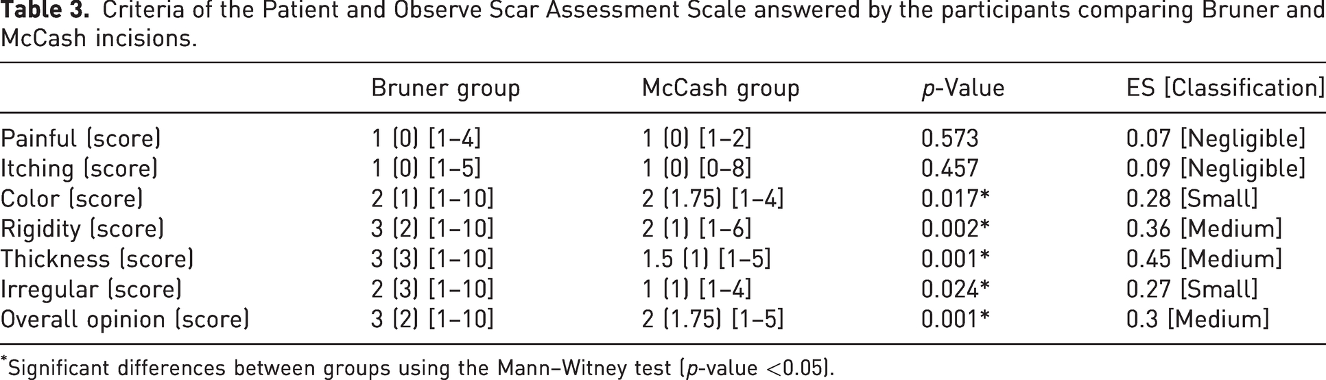
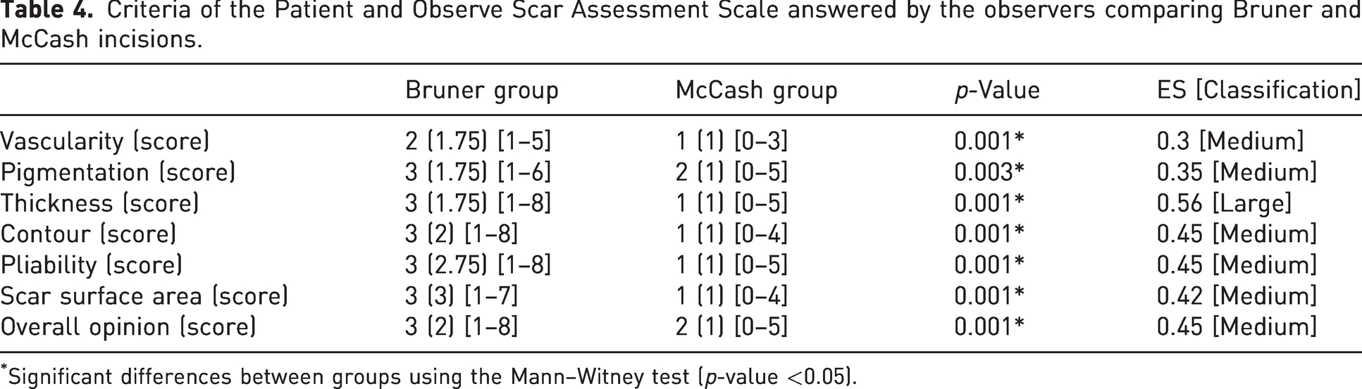
The POSAS (second version) (Draaijers et al., 2004; Lenzi et al., 2019), translated, validated and culturally adapted for Brazil, assesses the quality of the scar according to both the observer and the patient. It consists of two numerical scales, one for the patient (POSAS-P) and one for the observer (POSAS-O). The patient scale assesses pain, itching, colour, stiffness, thickness and irregularity. The observer scale assesses vascularity, pigmentation, thickness, contour relief, pliability and scar surface area. A score was also given for the overall opinion of both the patient and the observer.
Statistical analysis
The normality and homogeneity of the data were assessed using the Shapiro–Wilk and Levene tests. Given the discrete nature of the data and the non-normal distribution of the VAS scores, descriptive statistics were calculated using the median (interquartile range) and minimum and maximum values. In addition, to characterize the participants in each group, the frequencies of several variables were presented, including sex, ethnicity, dominant hand, type of work, factors affected with DD, the number of affected fingers in each hand and Tubiana score.
The results of POSAS-P, POSAS-O, VSS and VAS scores were compared between the groups using the Mann–Whitney U-test. Effect size (ES) values were calculated for each comparison and categorized as follows: 0.5 (large), 0.49–0.3 (medium), 0.29–0.1 (small) and ≤0.1 (negligible) (Fritz et al., 2012). All inferences were made at a significance level of p ≤ 0.05.
Results
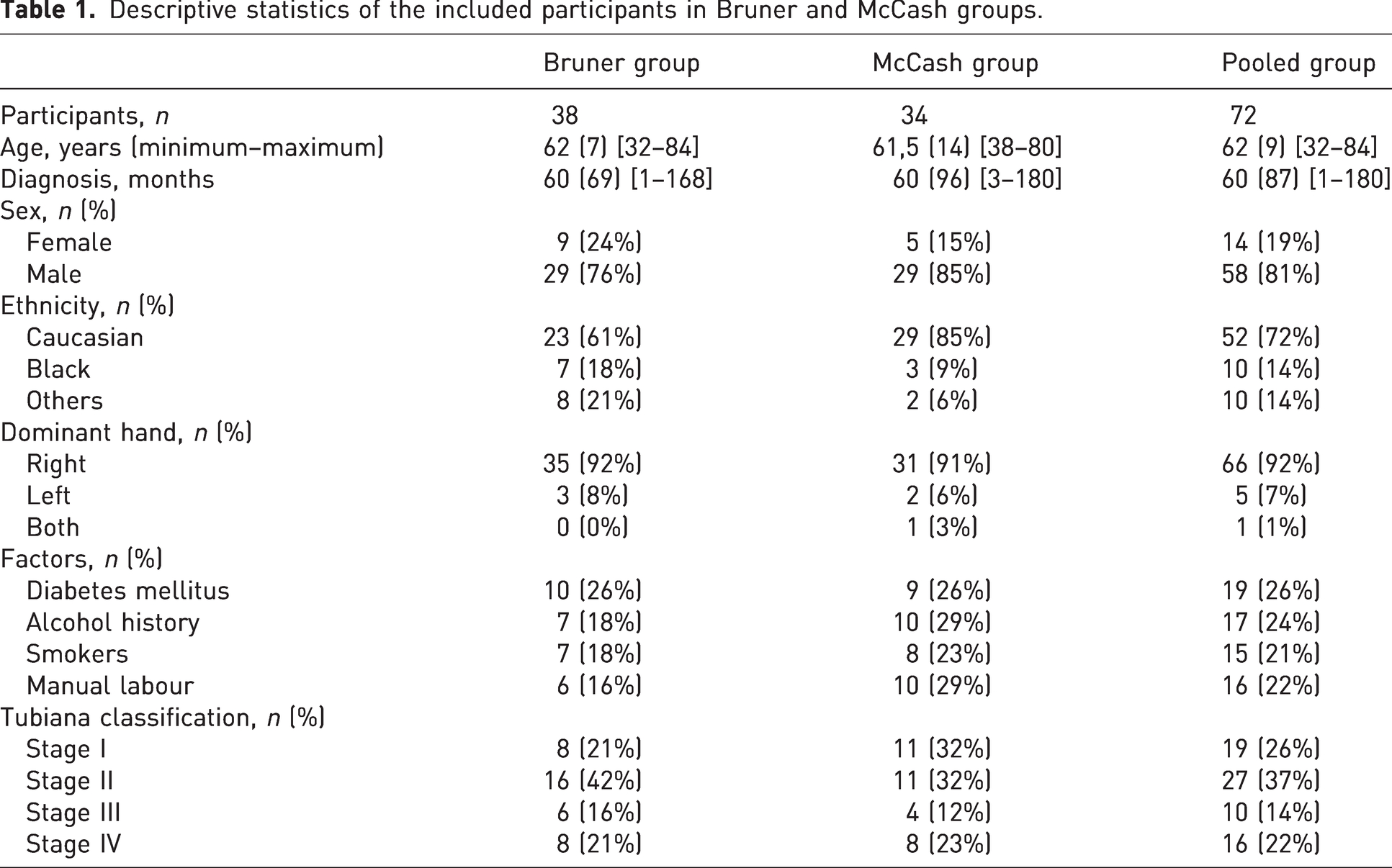
The study included 72 patients (12 women and 60 men), 38 patients in the Bruner group (nine women and 29 men) and 34 patients in the McCash group (five women and 29 men) (Figure 1). The mean age of the patients was 62 years, and 72% were Caucasian. Diabetes mellitus was the most common comorbidity (26%). Seventeen patients had a history of alcohol abuse and 15 were smokers. The operated side was the right dominant hand in 66 patients. Sixteen reported performing manual labour. According to the Tubiana classification, 19 patients were in stage I, 27 in stage II, 10 in stage III and 16 in stage IV (Table 1). There were no major differences between the two groups.
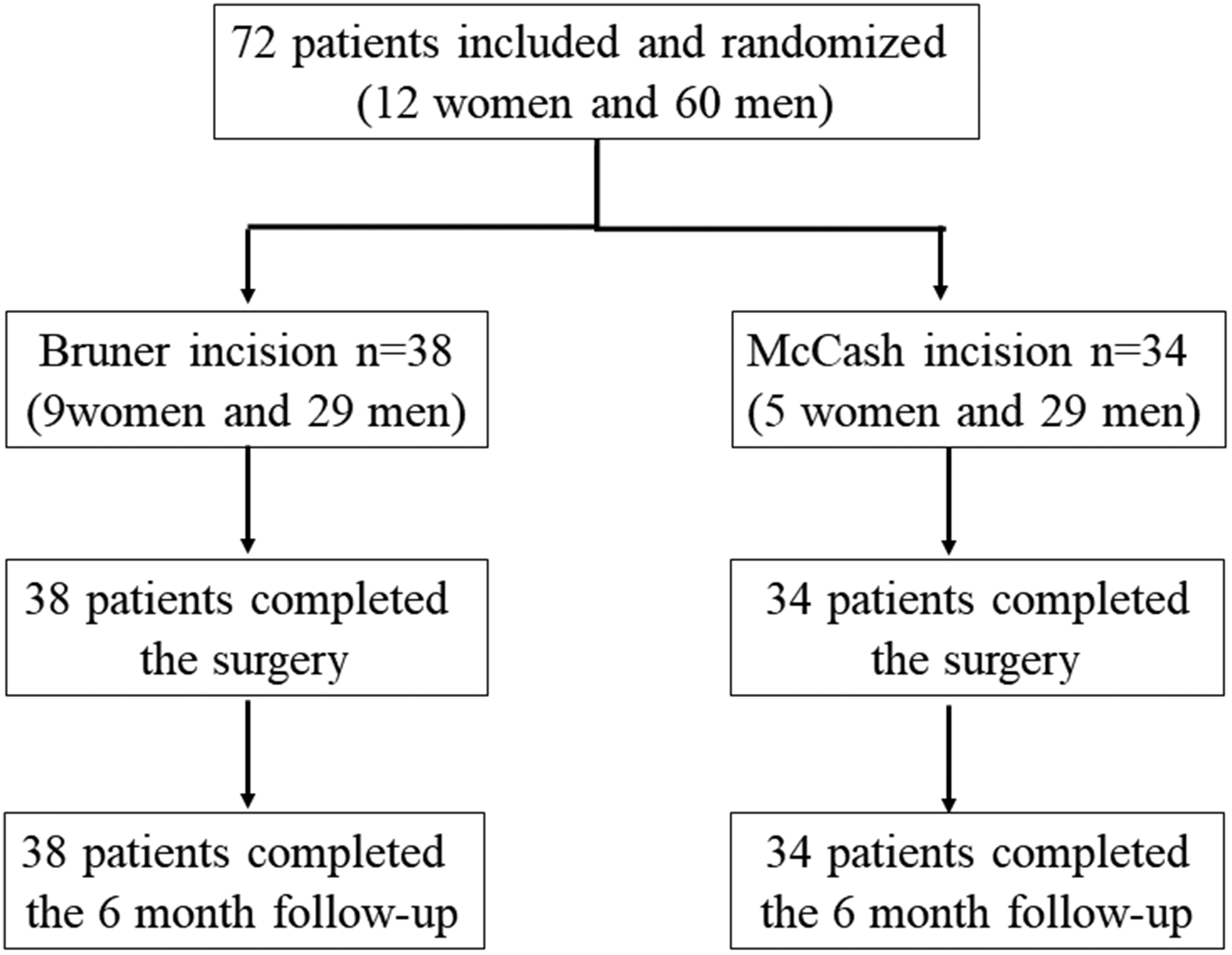
Consolidated standards of reporting trails (CONSORT) patient flow diagram.
Descriptive statistics of the included participants in Bruner and McCash groups.
The results of the scale analysis (VAS) showed that the Bruner group showed a higher score in all criteria assessed, resulting in worse results being seen by the patients. The average healing time for patients undergoing the Bruner technique was 14 days, and for patients undergoing McCash surgery, it was 30 days. Possible complications, including dehiscence, hematoma, infection and skin necrosis, were separated by group. In the Bruner group, dehiscence occurred in five patients, two reported pain, six had hematoma, one had infection and 12 had skin necrosis. The only complication present in the McCash group was pain, reported by six patients, which lasted an average of 21 days.
Figure 2 shows that the VAS scores were significantly higher in the Bruner group (p = 0.007; ES = 0.32 (mean)). Figure 3 shows photographs of the hands of patients operated on using the Bruner and McCash techniques at various postoperative intervals. In addition, the VSS scores were significantly higher in the Bruner group for all criteria (p < 0.045) with small and large effect sizes (Table 2), as shown in Figure 4.
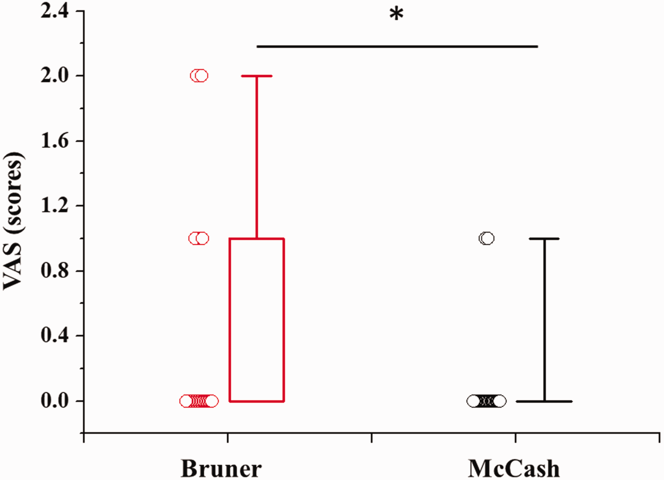
Visual analogue scale scores for the Bruner and McCash incisions.

Photos of the hands of patients operated with Bruner and McCash incision. Photos were taken immediately before surgery (a/f) and at 7 days (b/g), at 14 days (c/h), at 30 days (d/i) and at 6 months (e/j) after surgery.
Criteria of the Vancouver Scar Scale comparing Bruner and McCash incisions.
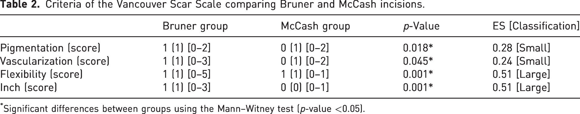
Significant differences between groups using the Mann–Witney test (p-value <0.05).
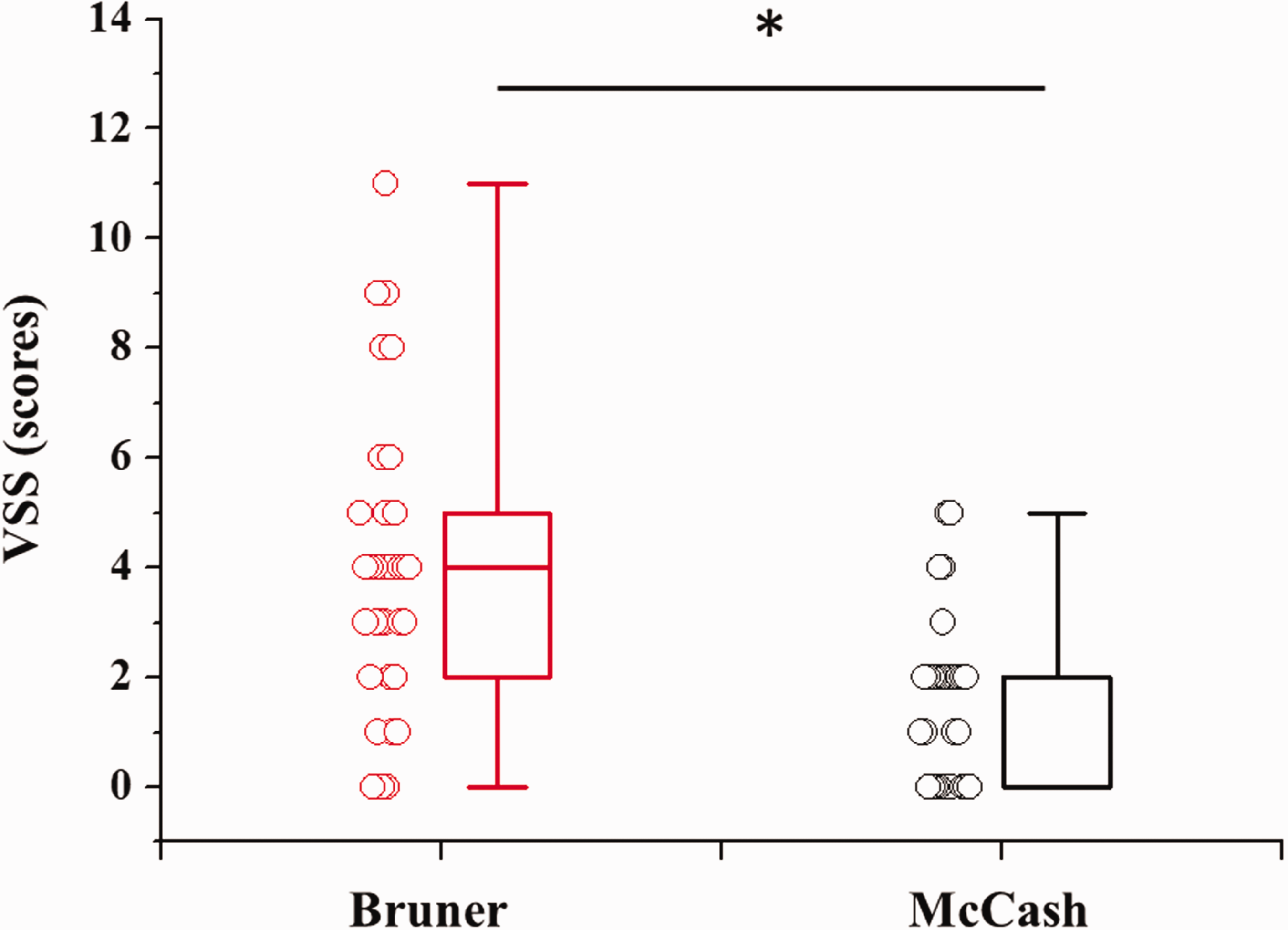
Scores obtained for the Vancouver Scar Scale using Bruner and McCash incisions.
Table 3 shows the participants’ responses to the POSAS analysis. The Bruner group had higher scores for colour, rigidity, thickness, irregularity and general opinion (p < 0.024) with small to medium effect sizes. Consequently, the overall index was significantly higher for the Bruner group (p = 0.002; ES = 0.37 (medium)), as shown in Figure 5.
Criteria of the Patient and Observe Scar Assessment Scale answered by the participants comparing Bruner and McCash incisions.
Significant differences between groups using the Mann–Witney test (p-value <0.05).
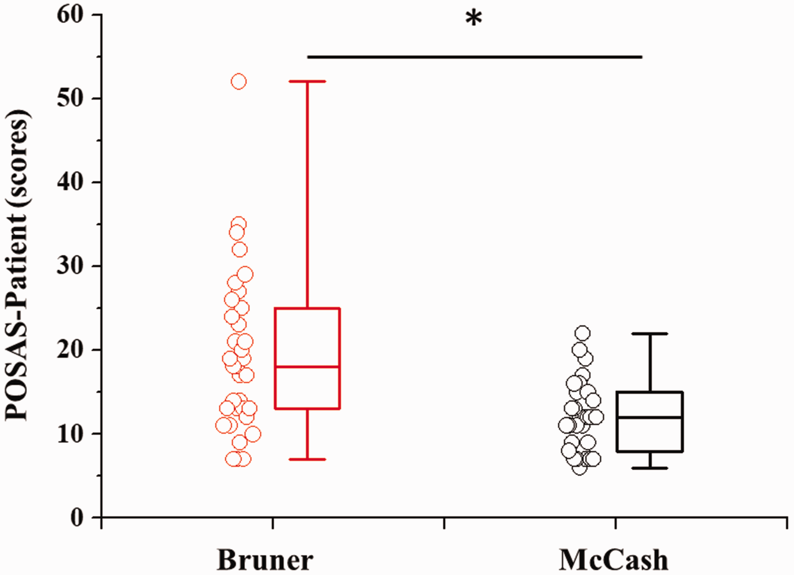
Quality of the scar perceived by the participants using the Patient and Observer Scar Assessment Scale using the Bruner and McCash incisions.
Furthermore, the observers’ responses indicated that the Bruner group also had high scores in all criteria (p < 0.003) (Table 4) with medium to large effect sizes. Therefore, based on the observers’ responses, the overall index was significantly higher for the Bruner group (p = 0.001; ES = 0.50 (medium)), as shown in Figure 6.
Criteria of the Patient and Observe Scar Assessment Scale answered by the observers comparing Bruner and McCash incisions.
Significant differences between groups using the Mann–Witney test (p-value <0.05).
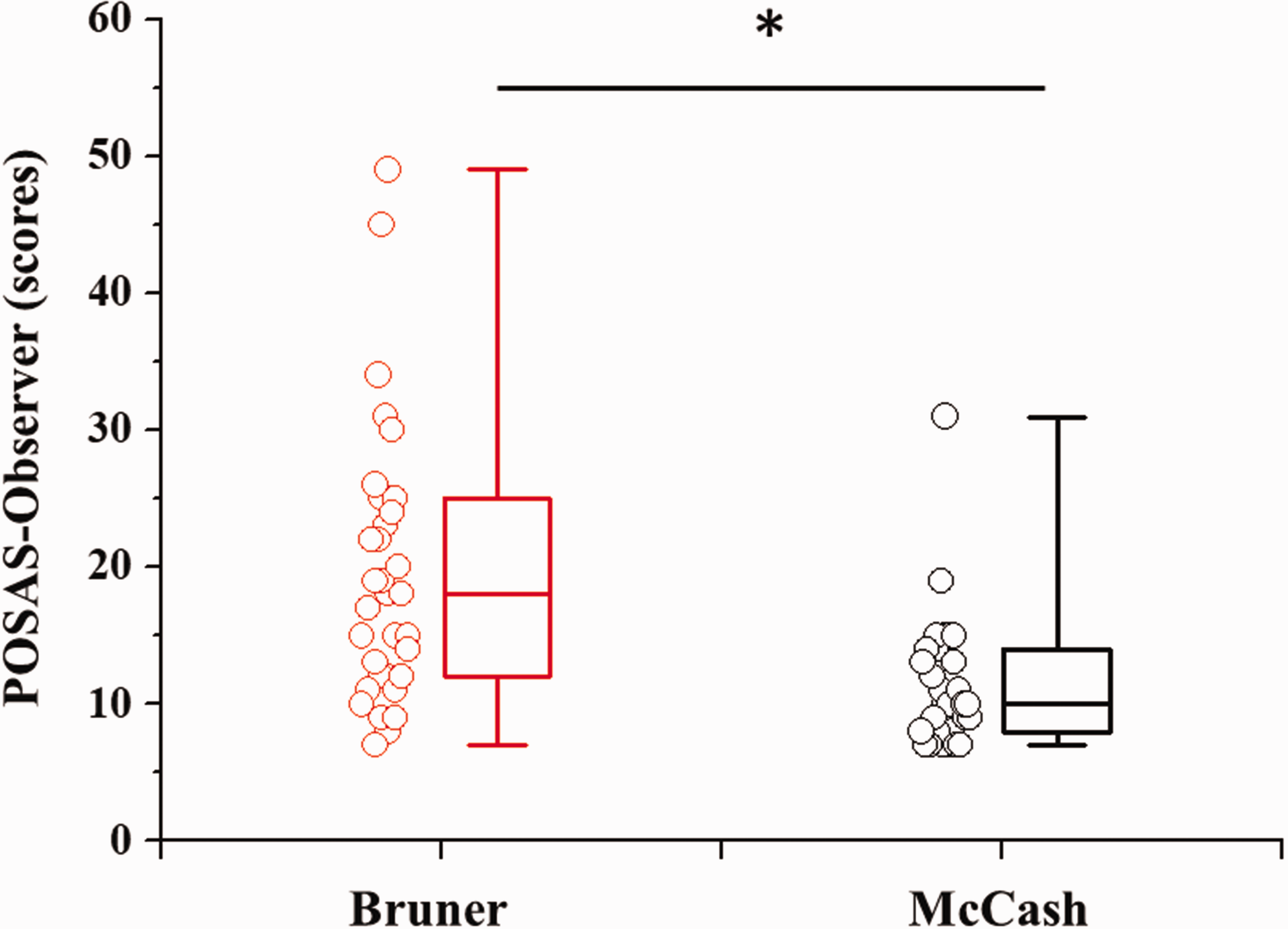
Quality of the scar perceived by the observers using the Patient and Observer Scar Assessment Scale using the Bruner and McCash incisions.
Discussion
Our results indicate that the McCash incision outperforms the Bruner incision in all categories analyzed, including postoperative complications, healing quality and patient satisfaction. The open approach has advantages and disadvantages, but the McCash incision has proved to be a reliable and important technique in the management of DD (Lesiak et al., 2017). A study of 59 patients showed favourable results in correcting deformities and restoring hand function by using the McCash technique to treat different stages of DD (Silva et al., 2023). Another study showed that most patients who underwent this technique did not require postoperative hand therapy (Lesiak et al., 2017). In a study with extended follow-up, patients were analysed for 7 and 21 years after open palm surgery. The authors concluded that the McCash technique resulted in fewer complications and superior outcomes in terms of skin closure (Roulet et al., 2018).
Both techniques analysed require a long recovery process with swelling during immobilization, which can lead to stiffness (Denkler et al., 2022). However, the open palm technique can probably avoid complications such as haematoma, skin ischaemia and necrosis, tension and pain (Guilhen et al., 2014). Although the McCash technique may not offer the same level of coverage for the surgical wound as the Bruner technique, patient satisfaction is not affected in the short or long term (Nann et al., 2023). The results obtained in terms of vascularization, mobility and pain showed that the McCash incision was better in all aspects, supporting the existing literature for improved outcomes with use of the McCash technique.
Our study had limitations. Patients were from a single institution, which may have influenced sample selection. The study lacked a universally accepted standard for assessing the characteristics of scars, despite the existence of various assessment methods. A review by Park et al. (2022) highlighted the complementary nature of existing scar assessment scales. To date, the most widely used and validated method is the VSS, while the POSAS scale offers a novel approach by considering both patient and observer perspectives, and the VAS is the only method that quantifies patients’ subjective assessment of their scars (Park et al., 2022).
The results of this study are relevant because currently there are no specific methods available to assess healing after surgical treatment for DD. Nann et al. (2023) highlighted that patient satisfaction may be uncertain in the early stages of healing and improve over time.
Footnotes
Acknowledgement
None.
Contributorship
All surgical procedures were performed by the orthopedic surgeons LGSL, JBGS and FF. All authors contributed to the research design, conception, data analysis and interpretation. LGSL, RPC, HBN and FF contributed to the writing and critical revision of the manuscript. All authors reviewed and edited the manuscript and approved the final version of the manuscript.
Declaration of conflicting interests
The authors declare no conflict of interest with respect to the research, authorship, and/or publication of this article.
Ethical approval
This work was approved by the Ethics and Research Committee of the Federal University of São Paulo under number 1.814.245, and duly registered with the World Health Organization – ReBEC under numbers 2fcsnhq and UNT U1111-1273-2903.
Funding
This work was supported by UNIFESP/SP/Brazil and Evaluation of Graduate Education (Coordenação de Aperfeiçoamento de Pessoal de Nível Superior, Brazil (CAPES), contract 88887.514571/2020-00) for financial support and scholarships.
Informed consent
Written informed consent was obtained from the patient(s) for their anonymized information to be published in this article.