
Abstract
Introduction:
This study investigated the association between capitate morphology and the success rates of non-vascularized bone graft (NVBG) and vascularized bone graft (VBG) operations for scaphoid nonunion.
Methods:
We retrospectively analysed 213 patients with established scaphoid non-union who underwent either non-vascularized bone graft (n = 75) or vascularized bone graft (n = 138). We analysed risk factors, including demographics, medical history, implant type, previous surgery, bone graft donor site and capitate morphology for their association with treatment outcome. We also assessed the relationship between capitate and scaphoid morphology in the uninjured wrists of 31 patients.
Results:
The overall success rate was 81% for NVBG and 86% for VBG, with no significant difference. For NVBG, Type II capitate, age 40 or older and tobacco use were significant risk factors for failure. For VBG, only previous surgery was a significant risk factor. In uninjured wrists, a Type II capitate was associated with a significantly thinner scaphoid waist and a lower waist index than a Type I capitate.
Conclusion:
Our study introduces a new radiographic classification of capitate morphology and establishes its significant association with scaphoid morphology. Beyond traditional factors, capitate morphology can help to assess scaphoid morphology and potentially infer vascularity, aiding the selection of the most appropriate bone graft type for treating scaphoid non-union.
Level of evidence:
III
Introduction
Scaphoid fractures are the most common carpal bone injuries, with reported incidence rates ranging from 1.4 to 29 per 100,000 person-years (Dy et al., 2018; Wells et al., 2021). Despite various treatments, scaphoid non-union (SNU) occurs in 5–50% of cases, leading to progressive reductions in wrist range of motion and grip strength (Dodds et al., 2006; McCallister et al., 2003). Untreated SNU can lead to wrist arthritis and functional disability. Given the frequent presence of bone defects and humpback deformity in established scaphoid non-union, bone grafting with internal fixation is widely recognized as the standard surgical treatment (Duncumb et al., 2022; Miller and Huang, 2024; Ross et al., 2020; Zondervan et al., 2024).
Although vascularized bone graft (VBG) is often considered beneficial for scaphoid non-union at risk of avascular necrosis (AVN) or with compromised vascularity (Higgins and Giladi, 2021; Pulos et al., 2018; Sgromolo and Rhee, 2019; Waitayawinyu et al., 2009), its efficacy is debated. This stems from the unreliable detection of AVN or scaphoid vascularity by MRI, radiographs, intraoperative assessment of punctate bleeding and histology. Consequently, some comparative studies of VBG and non-vascularized bone graft (NVBG) techniques have shown no statistically significant difference in outcomes (Duncumb et al., 2022; Fujihara et al., 2023; Guria et al., 2025; Ross et al., 2020), questioning the benefit and necessity of VBG in routine SNU management.
The correlation between scaphoid vascular anatomy and scaphoid morphology was first described by Morsy et al. (2019). They proposed that scaphoid shape was associated with differing vascular supply patterns which could influence the predisposition to non-union, AVN and Preiser disease. Specifically, Type II (slender) scaphoids possess a less robust vascular network than Type I, featuring only a single dorsal ridge vessel, an absence of anterior waist vessels and a lack of anastomoses between proximal and distal vascular networks, and no vessels entering at the scapholunate region. These characteristics increase the risk of insufficient blood supply, potentially hindering bone healing. However, a conclusive clinical correlation between Type II scaphoid morphology and increased non-union or treatment failures has not been established, primarily owing to the difficulty in scaphoid classification caused by morphological alterations during the process of fracture non-union.
The capitate, with its extensive articulation with the scaphoid, may play a role in determining scaphoid type. Unlike the scaphoid, whose joint line and length are significantly influenced by different degrees of scaphoid flexion or fracture on posteroanterior (PA) radiographs, the joint lines on the capitate are more readily observed and measured. We use the trans-section angle and the ratio of the capitotrapezoid (CT) joint line to the scaphocapitate (SC) joint line to classify capitate morphology into Type I and Type II (Figure 1). This study investigated the association between capitate and scaphoid morphology and its effect on the failure of VBG and NVBG surgery for scaphoid non-union.
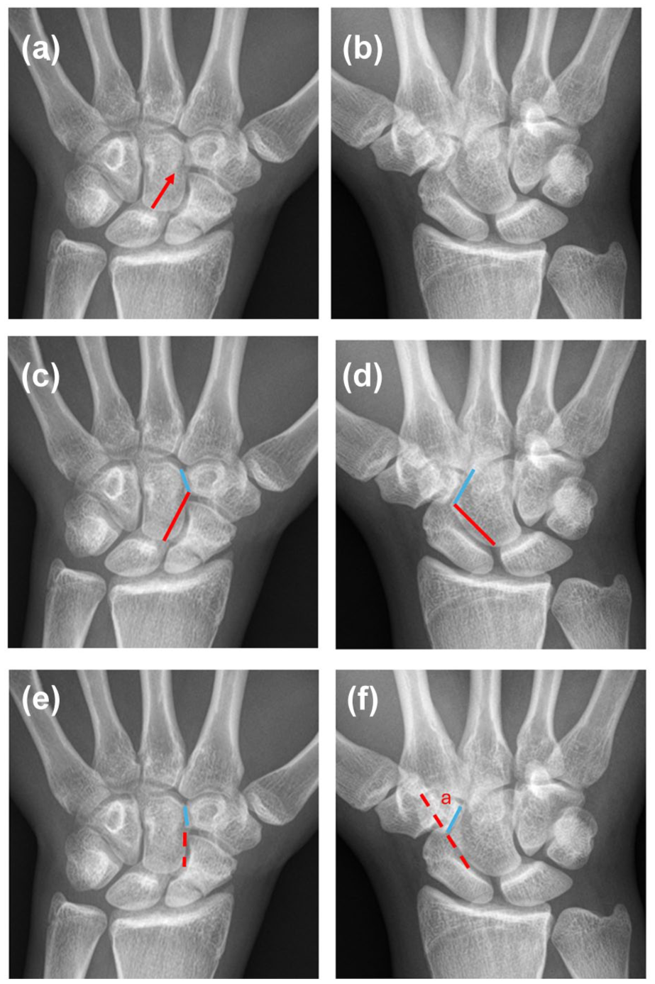
Radiographic characteristics of Type I and Type II capitates: morphological and angular differences. (a) A representative image of a Type I capitate, illustrating its rectangular morphology. A distinct groove (red arrow) is often observed near the scaphotrapeziotrapezoid (STT) joint. (b) Representative image of a Type II capitate, characterized by a pentagonal shape. (c) In Type I capitates, the capitatotrapezoid (CT) to scaphocapitate (SC) joint line ratio (blue line ÷ red line) is typically less than 0.6. (d) In Type II capitates, the CT/SC joint line ratio is generally greater than 0.6. (e) In Type I capitates, the scaphotrapezoid (ST) angle,formed between the CT joint line (blue line) and the extended upper half of the SC joint line (red dashed line) is usually less than 30°. The third Gilula’s arc (red dashed line) typically maintains a smooth contour extending through the CT joint to the base of the second metacarpal. (f) In Type II capitates, the ST angle (angle ‘a’) is generally greater than 30°. The third Gilula’s arc is often disrupted at the trapezoid owing to the enlarged and more oblique CT facet.
Methods
Patient enrolment
This retrospective study received approval from the local institutional review board (no. 24-007716). Informed consent was waived owing to the retrospective nature of the study.
To investigate the role of capitate morphology in surgical outcomes of scaphoid non-union, we conducted a retrograde chart review of patients who underwent either VBG or NVBG for established scaphoid non-union at a single academic medical centre between 2000 and 2019. All procedures were done by fellowship-trained hand surgeons. The decision to carry out a vascularized or non-vascularized bone graft was based on the presence of AVN determined by preoperative radiographic findings and intraoperative punctate bleeding; however our study highlights the limitations of these methods, which is why we propose a new classification.
Exclusion criteria for both groups were: inadequate radiographic images, and less than 3 months’ follow-up after operation. For the VBG group, 172 patients initially underwent surgery; 25 were excluded owing to inadequate images and nine for insufficient follow-up, resulting in 138 included patients. For the NVBG group, from 88 patients, 12 were excluded owing to inadequate images and one for insufficient follow-up, resulting in 75 included patients. Inadequate images were defined as those that did not allow for preoperative identification and measurement of the specific joint lines and angles required to classify capitate and scaphoid morphology. This included cases where patients did not have a preoperative wrist PA view, had undergone previous carpal bone surgery (e.g. trapezial excision and ligament reconstruction) or had radiographs with a cast that obscured the clarity of the joint lines and angles for measurement.
Demographic data, medical history (e.g. diabetes mellitus), a history of previous surgery, tobacco use, fracture location, fixation implant type, bone graft donor site and capitate morphology were collected and analysed as potential risk factors for treatment outcome. The patient enrolment flowchart is illustrated in Figure 2.
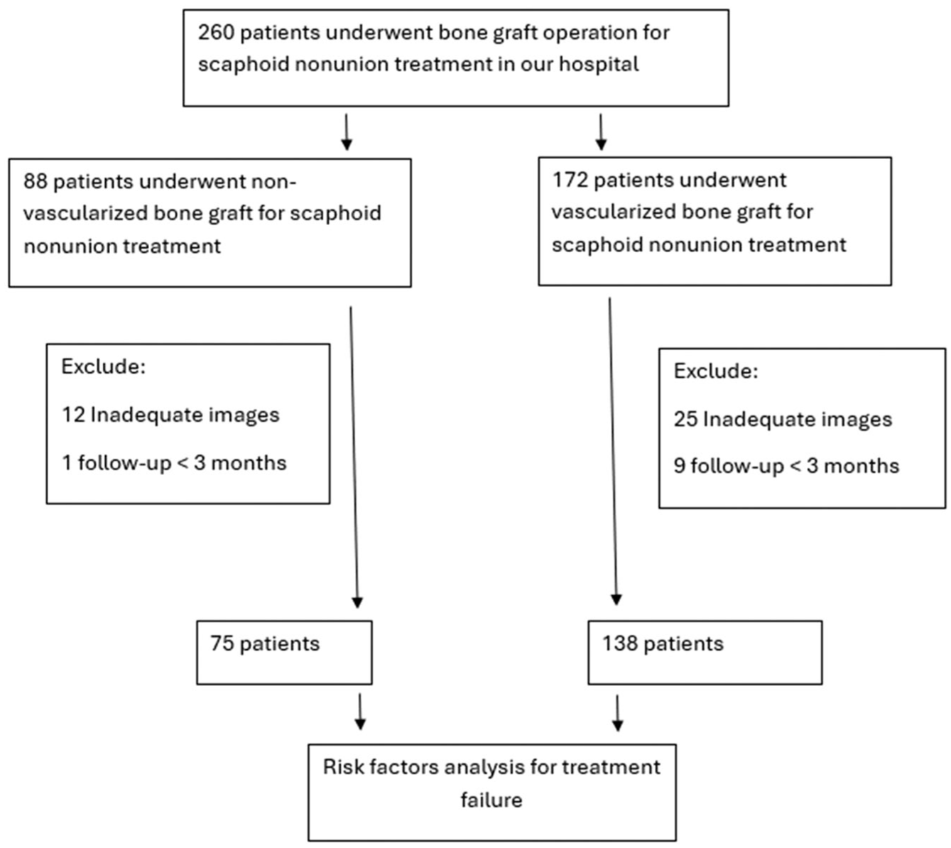
The flowchart of patient enrolment.
Radiographic measurements
All radiographs were obtained with patients positioned for a standard PA wrist view, with neutral forearm rotation, the elbow flexed at 90° and the shoulder abducted at 90°. Scaphoid bone union was defined as more than 50% bridging trabeculae on plain radiographs in more than one view (Zhao et al., 2018). Treatment failure was defined as persistent symptoms, no visibly progressive signs of healing on radiographs for a minimum of 3 months or obvious implant failure recognized by screw loosening or prominence (Ross et al., 2020).
The type of scaphoid in the uninjured wrist was classified into Type I and Type II based on lateral view radiographs, using a modified method from the original article of Morsy et al. (2019). The waist index (WI) was calculated by dividing the narrowest width at the scaphoid waist by the longest length of the scaphoid. A Type II scaphoid has a waist index less than 0.4 and a Type I scaphoid has a WI ⩾ 0.4.
The capitate morphology was classified on PA radiographs into Types I and II, based on specific criteria involving the CT and SC joint lines (Figure 1). A Type I capitate was defined by a CT–SC angle of less than 30° and a CT/SC joint length ratio of less than 0.6. This morphology is characterized by a short CT joint line and a nearly parallel orientation between the CT and SC joint lines. A Type II capitate features a pentagonal shape with a CT–SC angle of 30° or more and a CT/SC joint length ratio of 0.6 or more. Only capitates with both these criteria were classified as Type II. Radiological assessments for capitate classification were made by two hand surgeons (CEH and PV) with over 5 years’ clinical experience. In any discrepancies, the final type was determined through reassessment and discussion.
To investigate the normal relationship between capitate and scaphoid morphology, we analysed radiographic measurements of the contralateral uninjured wrists from a subset of patients with scaphoid non-unions who sustained bilateral wrist trauma and had had radiographs of both wrists but were subsequently found to have negative radiographic findings in one wrist. We compared the scaphoid waist length, total scaphoid length, and scaphoid WI in uninjured wrists with the injured wrists classified with either Type I or Type II capitate morphology. The precision of our measurement method was 0.1 mm. We also analysed the correlation between the type of scaphoid morphology of the uninjured wrist and the type of capitate morphology of the injured wrist.
Statistical analysis
Categorical variables are presented as numbers and percentages and continuous data as median values with the 25th and 75th percentiles (interquartile range, IQR). Baseline characteristics and radiographic classifications were compared between the groups with success or failure of treatment using Fisher’s exact test for categorical variables. Continuous data were compared using the Mann–Whitney U-test. Logistic regression was used to determine the odds ratios with 95% confidence intervals of the risk factors involved. To achieve 80% statistical power with an alpha level of 0.05, sample size calculations were made using reported WIs (Morsy et al., 2019) assuming means of 0.49 (SD 0.036) for Type I capitates and 0.38 (SD 0.023) for Type II capitates. Given a 4:1 group ratio, the minimal required sample size was 24 for the Type I capitate group and six for the Type II capitate group. A p-value <0.05 was considered statistically significant. Statistical analyses were conducted using SPSS v. 28 (IBM, Chicago, IL, USA).
Results
Among the total of 213 patients, the successful treatment rate was 81% (61/75) for patients treated with NVBG and 86% (119/138) for patients treated with VBG. There was no significant difference between them (p = 0.43). Table 1 details the baseline characteristics and proportions of various factors between the groups. There was a significantly higher proportion of tobacco use and screw fixation in the NVBG group. There were significantly more proximal pole fractures, a history of previous surgery and Type II capitate morphology in the VBG group.
Overview of 213 patients treated with non-vascularized and vascularized bone graft for scaphoid non-union.
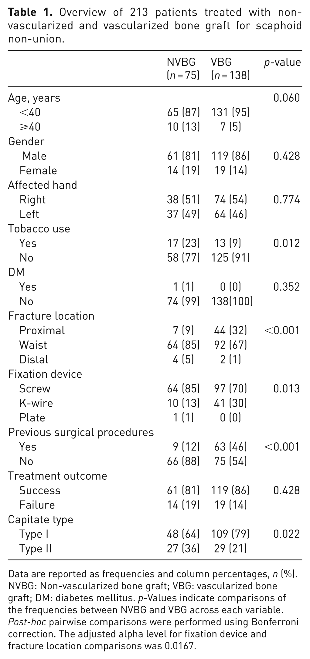
Data are reported as frequencies and column percentages, n (%). NVBG: Non-vascularized bone graft; VBG: vascularized bone graft; DM: diabetes mellitus. p-Values indicate comparisons of the frequencies between NVBG and VBG across each variable. Post-hoc pairwise comparisons were performed using Bonferroni correction. The adjusted alpha level for fixation device and fracture location comparisons was 0.0167.
Non-vascularized bone graft group analysis (n = 75)
The risk factor analysis for the 75 patients treated with NVBG is detailed in Table 2. The mean age was 24 (SD 10.8) years for the 61 patients in the successful union group and 35 (SD 12.5) years for the 14 patients in the failure group. Patients in the success group were significantly younger than those in the failure group (p < 0.001). The mean postoperative follow-up time was 35 (SD 181) months in the success group and 17 (SD 14.6) months in the failure group, with no significant difference between the groups (p = 0.71). A significantly higher proportion of Type II capitate morphology was found in the treatment failure group compared to the treatment success group. Significantly more tobacco use was observed in the treatment failure group than in the successful treatment group. Other variables including gender, affected hand, diabetes mellitus, fracture location, fixation implant and source of bone graft showed no significant difference between the success and failure groups.
Predictive factors for outcome in NVBG-treated scaphoid non-union patients.
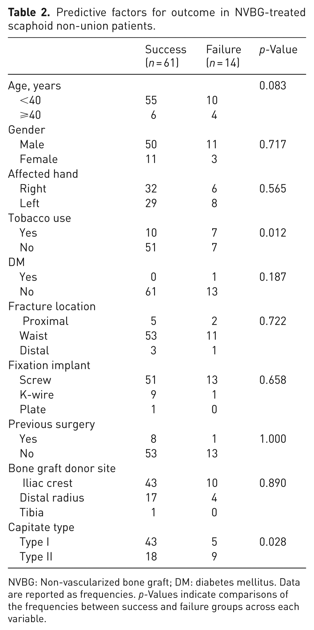
NVBG: Non-vascularized bone graft; DM: diabetes mellitus. Data are reported as frequencies. p-Values indicate comparisons of the frequencies between success and failure groups across each variable.
In the regression analysis (Table 3), tobacco use and a Type II capitate were identified as factors associated with an increased risk of failure of NVBG treatment in scaphoid non-union cases. After multivariate adjustment, age ⩾ 40 years, tobacco use and Type II capitate remained statistically significant.
Multivariate regression analysis of risk factors for non-vascularized bone graft treatment failure.
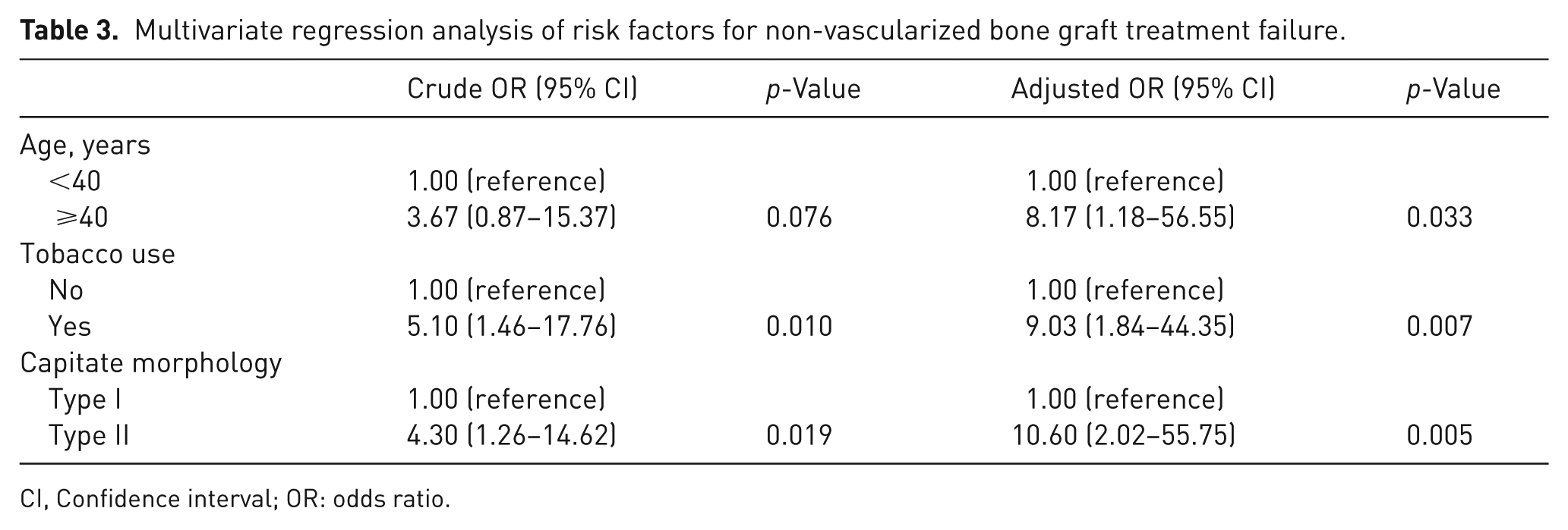
CI, Confidence interval; OR: odds ratio.
Vascularized bone graft group analysis (n = 138)
The failure risk analysis for the 138 patients treated with VBG is presented in Table 4. The mean age was 22 (SD 9) years for the 119 patients in the successful union group and 32 (SD13) years for the 19 patients in the treatment failure group. Patients in the success group were significantly younger than those in the failure group (p < 0.001). The mean postoperative follow-up time was 10 (SD 12.2) months in the success group and 18 (SD 17.4) months in the failure group, with no significant difference (p = 0.96). A significantly higher proportion of patients who had previous surgery was found in the treatment failure group. Other variables, including older age, gender, affected hand, tobacco use, diabetes mellitus, fracture location, fixation implant, bone graft origin and capitate type, showed no significant difference between the treatment success and failure groups.
Overview of 138 vascularized bone graft treated scaphoid fracture non-unions.
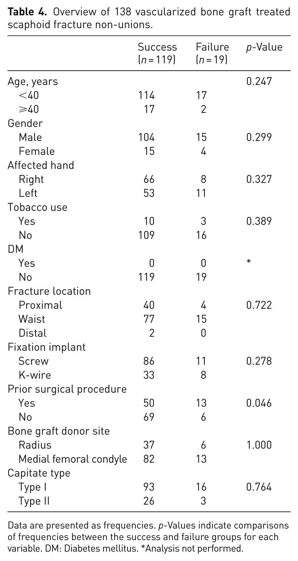
Data are presented as frequencies. p-Values indicate comparisons of frequencies between the success and failure groups for each variable. DM: Diabetes mellitus. *Analysis not performed.
Capitate–scaphoid morphological association
Of the 213 patients, 31 had radiographs available for evaluation of their contralateral uninjured wrists. Among the wrists with scahphoid non-union, 21 patients had a Type I capitate and 10 had a Type II capitate. Table 5 illustrates the relationship between the capitate type in wrists with scaphoid non-union and both the capitate type and scaphoid morphology in the corresponding contralateral uninjured wrists. Contralateral uninjured wrists of individuals with Type II capitate in the affected wrists demonstrated a significantly thinner waist, a lower WI and a significantly higher proportion of slender-type scaphoids than those with a Type I capitate. There was a significantly higher ratio of Type II capitate found in the contralateral uninjured wrists among individuals who had a Type II capitate in the wrists with scaphoid non-union than in those with Type I capitate.
The relationship between the capitate type in wrists with scaphoid non-union and both the capitate type and scaphoid morphology in the corresponding contralateral uninjured wrists.
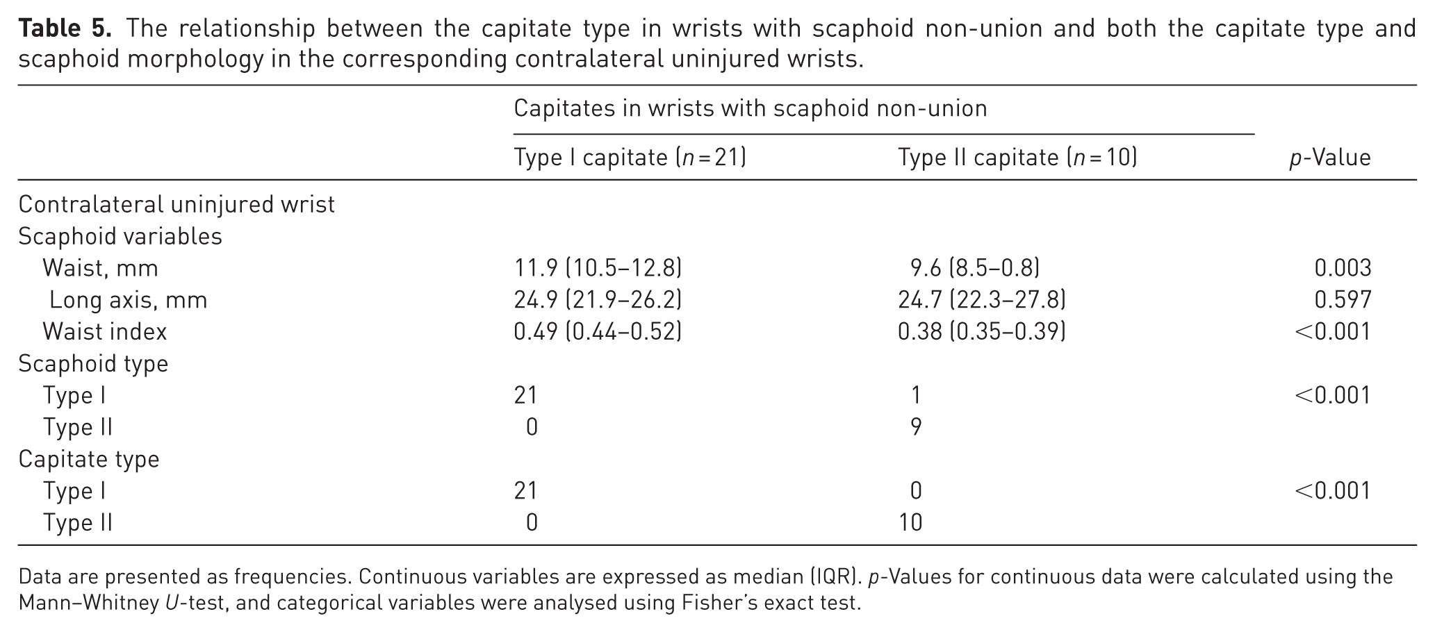
Data are presented as frequencies. Continuous variables are expressed as median (IQR). p-Values for continuous data were calculated using the Mann–Whitney U-test, and categorical variables were analysed using Fisher’s exact test.
Discussion
Our study introduces a new radiographic classification of capitate morphology and establishes its association with scaphoid morphology as a risk factor for treatment failure after NVBG for scaphoid non-union. It provides a clinically applicable method for assessing scaphoid type, which may be difficult to determine on PA radiographs, especially after non-union has occurred. Furthermore, it provides an indication of the vascularity of the non-united scaphoid, based on the type of scaphoid, offering guidance about whether treatment by VBG might be beneficial.
Numerous studies have compared the treatment outcomes of VBG and NVBG for scaphoid non-union; however, the benefits of VBG remain unclear (Braga-Silva et al., 2008; Duncumb et al., 2022; Fujihara et al., 2023; Guria et al., 2025; Ribak et al., 2010). Certain predisposing factors to failure of union, such as poor scaphoid vascularity identified intraoperatively, may indicate potential benefit from VBG. The success rates between VBG and NVBG in our study are similar to those previously reported. However, we studied the effect of various risk factors on treatment success, which offer additional insights for preoperative planning rather than solely relying on intraoperative assessment.
For the NVBG group, age ⩾ 40 years or tobacco use were identified as significant risk factors for treatment failure, consistent with previous research (Dinah and Vickers, 2007; Patterson et al., 2025; Xu et al., 2021). The detrimental effects of tobacco on blood supply and angiogenesis are well-established contributors to non-union (Pathak et al., 2025; Pearson et al., 2016; Waters et al., 2023) and surgical failure (Dinah and Vickers, 2007). Similarly, aging has an adverse effect on bone healing, which is often linked to impaired blood supply, contributing to poorer outcomes (Lu et al., 2008).
However, within the VBG treatment group, age ⩾ 40 years, tobacco use and Type II capitate morphology were not significant risk factors for treatment failure (Table 3). This strongly suggests that the success of NVBG depends heavily on the patient’s intrinsic blood supply, which these factors can compromise. Given that Type II scaphoids have a less robust vascular network (Morsy et al., 2019), the ability of VBG to provide a direct blood supply compensates for these pre-existing vascular deficiencies (Konstantinidis et al., 2022). Our radiographic classification of capitate morphology allows the assessment of scaphoid vascularity before operation. This helps clinicians to decide about the type of surgical intervention required.
The capitate, given its extensive articulation with the scaphoid, serves as an ideal surrogate for predicting scaphoid morphology, as its shape remains largely unaltered during the development of scaphoid non-union (Figure 3). Capitate morphology is much easier to measure and classify on PA radiographs compared with the scaphoid, which is often distorted (Figure 1). In wrists with a Type II capitate, defined by a sharp transection angle between the C) and SC joint lines and a high CT–SC joint line length ratio, the scaphoid is compelled to adopt a more transversely extended configuration owing to the increased capitotrapezoid joint ratio. This configuration, combined with the capitate’s overriding position, contributes to a relatively narrower scaphoid waist (Figure 4). Conversely, wrists with a Type I capitate have nearly parallel CT and SC joint lines and a shorter CT joint line. Here, the trapezoid typically articulates with the distal flattened region of the scaphoid rather than its slope. This arrangement prevents the formation of a narrow waist, as the Type I scaphoid maintains a more upright position, minimizing capitate override. Our findings support this, as most corresponding scaphoids in wrists with a Type I capitate were classified as Type I scaphoid, characterized by a thicker waist and a larger WI.
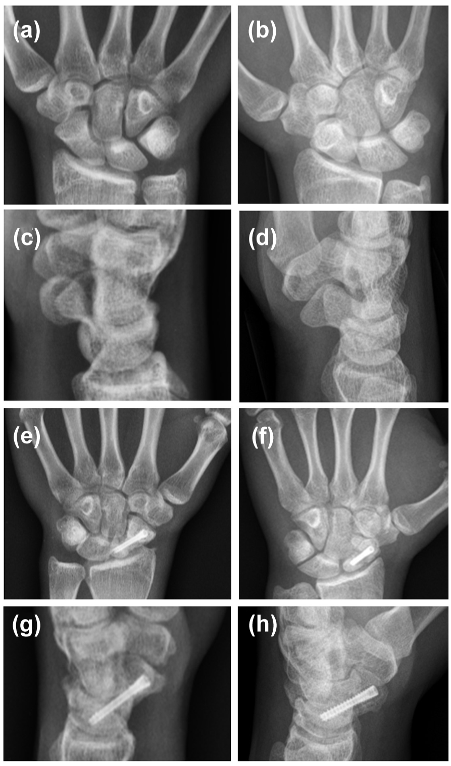
Association between scaphoid and capitate morphologies in uninjured and injured wrists. Representative posteroanterior (PA) radiographs demonstrating capitate morphology in: (a) an uninjured contralateral wrist with a Type I capitate (rectangular morphology); (b) an uninjured contralateral wrist with a Type II capitate (pentagonal morphology). Lateral view radiographs illustrating the corresponding scaphoid morphology: (c) the Type I capitate (from A) accompanying a Type I (full-type) scaphoid, characterized by a thick waist (waist index ⩾ 0.4); (d) the Type II capitate (from B) accompanying a Type II (slender-type) scaphoid, characterized by a narrow waist (waist index < 0.4). Posteroanterior and lateral views of injured wrists, showing capitate morphology stability despite scaphoid changes after the surgery: (e) (PA view) and (g) (lateral view): injured wrist of the same individual as in (a), demonstrating an unaltered Type I capitate morphology despite changes in scaphoid shape from bone graft and internal fixation. (f) (PA view) and (h) (lateral view): injured wrist of the same individual as B, demonstrating an unaltered Type II capitate morphology despite changes in scaphoid morphology from bone graft and internal fixation.
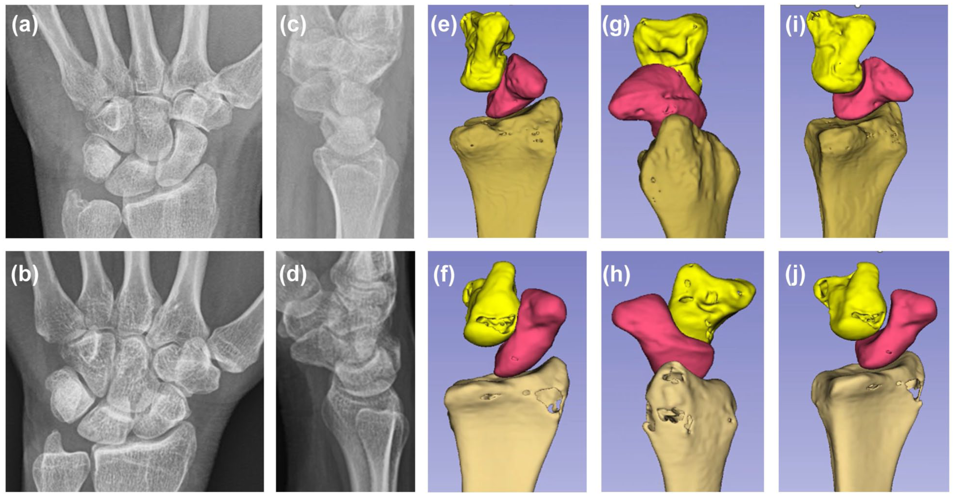
Three-dimensional computed tomography reconstructions of Type I and Type II capitate morphologies in healthy wrists. (a) Posteroanterior (PA) radiograph of a wrist with a Type I capitate. (b) Posteroanterior radiograph of a wrist with a Type II capitate. (c) Lateral radiograph of a wrist with a Type I capitate. This type usually accompanies a thick-waisted scaphoid. (d) Lateral radiograph of a wrist showing a Type II capitate, which commonly appears alongside a scaphoid with a narrow waist. (e) This image presents a posterior three-dimensional (3D) CT reconstruction of a neutrally positioned wrist, highlighting a Type I capitate alongside the radius and scaphoid. A notable feature is the prominent ridge located at the margin of the scaphoid’s capitate facet. This anatomical characteristic indicates a limited extent of capitate overlap on the scaphoid’s waist, contributing to the formation of a thick-waisted scaphoid morphology. (f) 3D CT reconstruction of a wrist with a Type II capitate. The capitate forms a sharp angle at the waist of the scaphoid. The observed overriding motion of the capitate on the scaphoid waist, coupled with the absence of a marginal ridge on the capitate facet, contributes to the narrow scaphoid waist and its association with a slender (Type II) scaphoid morphology. (g) View from the sigmoid notch of a wrist with a Type I capitate (capitate removed for clarity) to visualize the capitate facet on the scaphoid. Note that the capitate facet is primarily limited to the lower half of the scaphoid, featuring a well-defined ridge. The trapezoid is positioned at the distal pole of the scaphoid, beyond the capitate facet. (h) Same view as E, but for a wrist with a Type II capitate. Observe the continuous, undifferentiated scaphocapitate (SC) and scaphotrapezoid (ST) joint surfaces on the scaphoid, which form a narrow waist without a clear septum (i & j). The trapezoid is located closer to the scaphoid waist, in contrast to its more distal position in Type I (e), with its posterior placement also contributing to the narrower waist of a Type II scaphoid (i & j).
Apart from vascularity, biomechanical factors also influence the healing of scaphoid non-unions. Type I scaphoids may provide a larger bone contact area at the fracture site, facilitating bone healing. The wider waist of a Type I scaphoid also allows for optimal placement of a screw along the central one-third region, enhancing strength of fixation (Dodds et al., 2006; McCallister et al., 2003). In contrast, Type II scaphoids typically have a relatively narrow waist and longer axis, which can make the insertion of a screw in the centre of the axis difficult.
There have been previous anatomical classifications of the capitate such as that of Yazaki et al. (2008) based on the relationship between the SC and lunatocapitate joints, delineating flat, spherical and V-shaped capitates (Yazaki et al., 2008). The clinical significance of this earlier classification focused on the V-shaped capitate potentially leading to reduced joint congruency after proximal row carpectomy owing to a smaller articular surface with the lunate. Our study, however, used both the CT–SC joint line transection angle and the CT/SC joint line ratio to classify the capitate. It places greater emphasis on the specific anatomical relationship between the capitate, trapezoid and scaphoid. Our method is based on measurable ratios and angles and offers a more objective and quantitative approach than previous classifications that relied on more abstract shape descriptions, thereby enhancing its applicability and reliability in clinical settings.
This study has several limitations. First, a significant selection bias was present in the VBG treatment group, characterized by a higher proportion of proximal pole fractures, a history of previous surgical procedures and Type II capitates. This bias may have influenced the reported success rates and the risk factor analysis. Second, as a retrospective, single-institution study, only 31 out of 213 patients had bilateral wrist radiographs available to analyse the relationship between the morphology of the normal capitate and scaphoid. Although a statistically significant association was observed, larger case studies are needed to confirm this association. Third, the 3 month minimum follow-up period may not be sufficient to identify all delayed treatment failures or delayed unions and future studies should include longer follow-up. Fourth, although vascular insufficiency is strongly suspected as a cause of treatment failure, direct evidence from advanced imaging (e.g. MRI or contrast micro-CT) was not available to confirm poor blood supply in failed cases. Future studies incorporating such imaging would provide a better understanding.
Footnotes
Acknowledgements
We are deeply grateful to Taichung Veterans General Hospital for their unwavering financial and spiritual support.
Declaration of conflicting interests
The authors declared no potential conflicts of interest with respect to the research, authorship, and/or publication of this article.
Funding
The authors received no financial support for the research, authorship, and/or publication of this article.
IRB approval and waiver of consent
The Institutional Review Board at Mayo Clinic, Rochester, MN (IRB no. 24-007716), approved this retrospective study. Informed consent was not obtained as a result of the study’s retrospective design.
Ethical statement
Approval from our institutional review board was obtained for this retrospective study. All procedures performed in studies involving human participants were in accordance with the ethical standards of the institutional and/or national research committee and with the 1964 Declaration of Helsinki and its later amendments or comparable ethical standards.