
Abstract
Introduction:
This systematic review aimed to investigate the use of single-photon emission computed tomography/computed tomography (SPECT/CT) in diagnosing and managing hand and wrist disorders, especially hand and wrist pain of unknown aetiology.
Methods:
A systematic review of the Embase, MEDLINE and Cochrane databases was performed in accordance with Preferred Reporting Items for Systematic Review and Meta-Analyses guidelines. Author experiences with SPECT/CT and, where available, sensitivity and specificity data were analysed.
Results:
The search yielded 3546 records, of which 3477 were excluded, leaving 69 for full-text analysis. Fourteen studies were eligible for inclusion. Overall, SPECT/CT demonstrated sensitivities and specificities in the ranges 0.6–0.83 and 0.9–0.1 for non-specific hand and wrist pain (n = 6), 0.75–1.0 and 0.82–0.93 for osseous disorders (n = 5) and 0.72–1.0 and 0.65–0.82 for rheumatic disorders (n = 3). For hand and wrist disorders, SPECT/CT demonstrated comparable sensitivity and specificity to magnetic resonance imaging (MRI), but greater diagnostic accuracy than routine imaging modalities. In cases where the pathology was subtle or ambiguous, SPECT/CT could help confirm the diagnosis, estimate the severity of the condition and identify clinically relevant anatomy.
Conclusion:
Although SPECT/CT has comparable diagnostic accuracy to other cross-sectional imaging modalities, such as MRI, the additional information it provides influences surgical management in only a minority of cases. Its substantial radiation exposure may prevent SPECT/CT from being used routinely, but it can be an effective problem-solving tool in cases where the diagnosis remains unclear despite prior imaging.
Introduction
Hand and wrist pain of unknown aetiology is a challenging, yet common, presentation for hand surgeons (Bay et al., 2020; Walker-Bone et al., 2004). Clinical assessment may be inconclusive, given the overlap in symptoms and pain location amongst differential diagnoses (Krastman et al., 2020), and many patients are unable to localize their pain. Although imaging plays a critical role in diagnosis, there is a group of patients for whom standard imaging does not correlate with clinical findings, meaning the underlying pathology remains unclear.
Diagnostic imaging for hand and wrist conditions typically follows a stepwise approach. Conventional radiography is the first-line investigation owing to its accessibility and ability to detect gross osseous abnormalities. (Forman et al., 2005; Van Vugt et al., 1999). However, its low sensitivity and specificity in detecting early rheumatological and soft tissue abnormalities are well recognized (Copeland and Byerly, 2025). Consequently, ultrasound and cross-sectional imaging modalities such as computed tomography (CT) and magnetic resonance imaging (MRI) are often used as second-line diagnostic tools. Ultrasound provides the ability to perform a dynamic assessment, while CT offers high-resolution visualization of bony structures. Magnetic resonance imaging is optimal for assessing soft tissue, cartilage and ligamentous pathology. However, both modalities are limited in their ability to provide dynamic insights into bone metabolism and physiological activity (Deveza et al., 2017; Fogelman and Carr, 1980).
Single-photon emission computed tomography combined with computed tomography (SPECT/CT) is a hybrid imaging technique that combines functional nuclear imaging with high-resolution cross-sectional imaging (Buck et al., 2008). By combining the metabolic insights of SPECT with the anatomical resolution of CT, SPECT/CT may overcome the key limitations of conventional imaging modalities. Despite this potential, SPECT/CT is not routinely used in the diagnostic work-up of hand and wrist disorders as its clinical utility in this context remains unknown.
This systematic review evaluated the diagnostic usefulness of SPECT/CT for disorders of the hand and wrist. Furthermore, we aimed to establish its potential as an adjunctive imaging modality for patients with persistent or unexplained symptoms where the primary diagnosis remains unclear despite standard assessments and imaging.
Methods
This systematic review followed Preferred Reporting Items for Systematic Review and Meta-Analyses (PRISMA) reporting guidelines (Page et al., 2021) and was registered with the international prospective systematic review registry, PROSPERO (ID: CRD42024621940).
Eligibility criteria
Studies were included if they reported on the use of SPECT/CT, with or without a comparator, for the diagnosis or prognosis of any disorder affecting the hand and/or wrist. A hand and/or wrist disorder was defined as any primary or secondary pathology that occurred in the area from the radiocarpal joint distally.
Search strategy and study selection
A systematic search of the Embase, MEDLINE and Cochrane databases was performed from inception to 1 November 2024. Studies were limited to those published in English. The references of the included studies were manually checked to identify any further eligible articles. A comprehensive search strategy was designed in collaboration with a clinical librarian, consisting of index, free-text and Medical Subject Headings terms relevant to the following key concepts: hand, wrist, SPECT, CT and relevant pathologies, see Online Table S1.
All duplicates were removed, after which the remaining records underwent title and abstract screening using the Rayyan systematic review management platform (Ouzzani et al., 2016), with two independent, blinded reviewers (S.S.S., S.Y.). Any conflicts were resolved by a third reviewer (O.M.). A list of potentially eligible articles was then reviewed in full.
Data collection
Two independent reviewers (O.M., S.Y.) collected the data from the included studies using a pre-designed proforma. The following data variables were extracted: first author, year of publication, country, study design, sample size, comparator modality (if applicable), pathology of interest and sensitivity/specificity (if applicable). Further technical details were collected from included articles where applicable, including the type and dose of radioisotope, the timing of scans, and the standardized uptake value (SUV)/details of qualitative assessment of scans.
Risk of bias assessment and statistical analysis
The revised Quality Assessment of Diagnostic Accuracy Studies (QUADAS-2) tool was used to evaluate the risk of bias and the applicability of the included studies. Owing to the small number of studies and clinical heterogeneity between the included studies, data pooling was not performed. This differed from our PROSPERO protocol owing to the type and number of studies included in our review.
The search yielded 3546 records, of which 3477 were excluded. A full-text review was conducted on the remaining 69 articles. Of these, 55 were excluded. See the PRISMA flow chart for further details (Figure 1). No further eligible studies were identified through hand searching the reference lists of the included articles. Fourteen articles were included. (Abdelhafez et al., 2021; Bhure et al., 2015, 2020; Evbuomwan et al., 2023; Huellner et al., 2012, 2013; Kim et al., 2021; Le Corre et al., 2016; Lee et al., 2020; Lim et al., 2022; Querellou et al., 2014; Schleich et al., 2012; Shirley et al., 2016; Strobel et al., 2014).
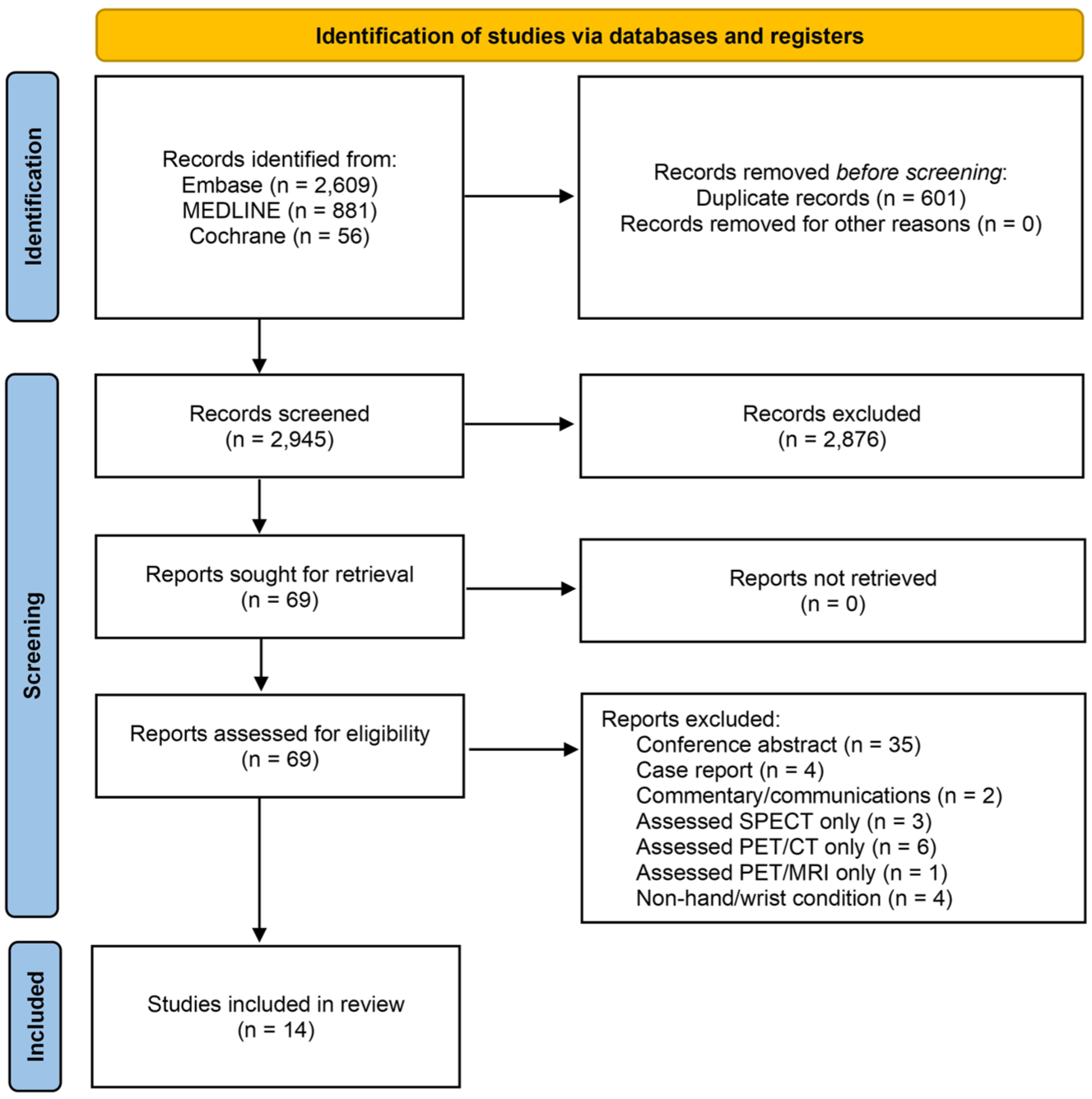
The Preferred Reporting Items for Systematic Reviews and Meta-Analyses (PRISMA) flow chart depicting identification, screening, and inclusion of eligible studies. SPECT: single-photon emission computed tomography; PET/CT: positron emission tomography/computed tomography; PET/MRI: positron emission tomography/magnetic resonance imaging.
Study characteristics
The 14 included studies reported data on 464 patients with hand and/or wrist conditions undergoing SPECT/CT in Switzerland (n = 6), South Korea (n = 3), the UK, the USA, Australia, France and South Africa (n = 1, respectively) from 2012 to 2023. The detailed characteristics of the studies are reported in Table 1, and the imaging procedures in Table 2.
Demographic breakdown of included studies.
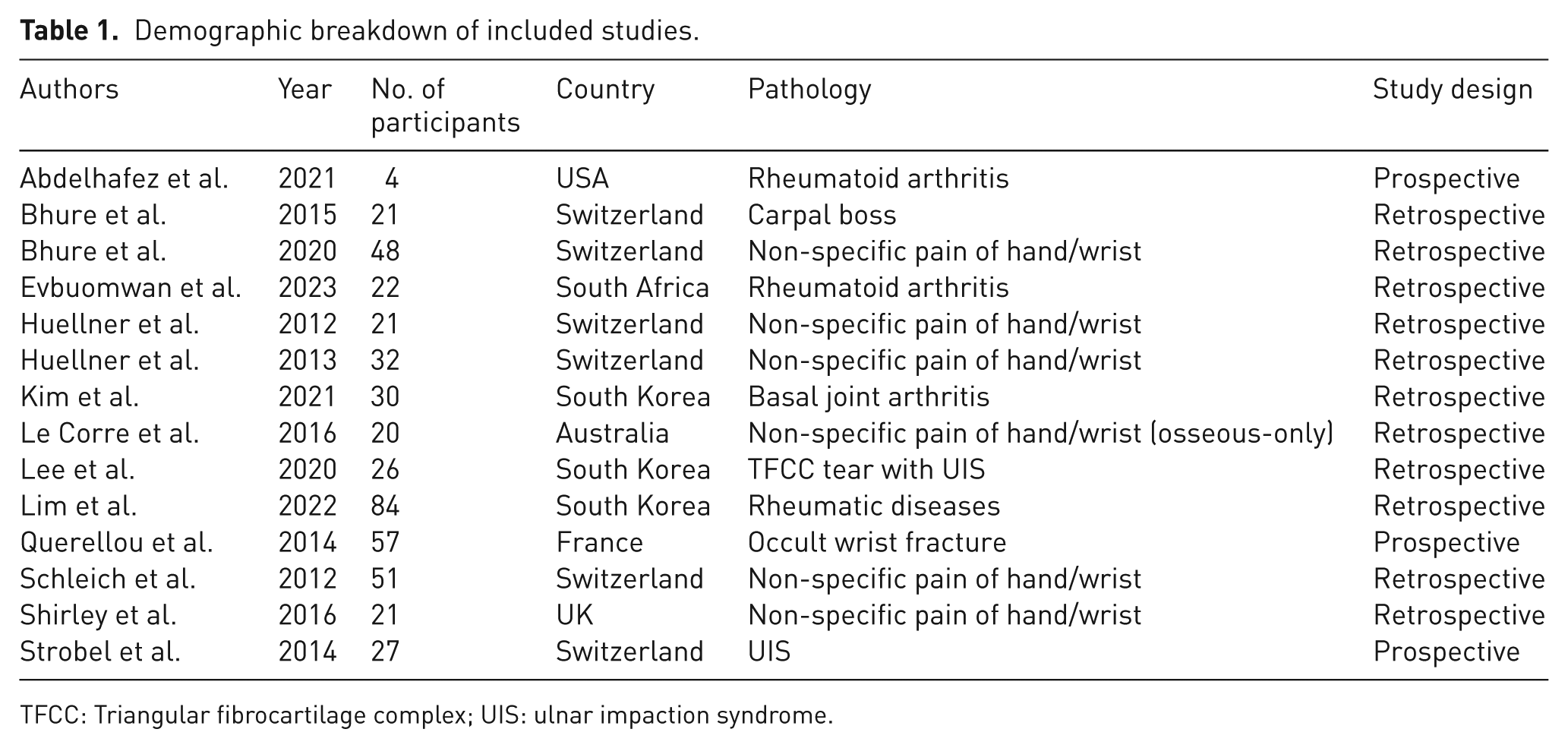
TFCC: Triangular fibrocartilage complex; UIS: ulnar impaction syndrome.
SPECT/CT imaging procedures and interpretation.
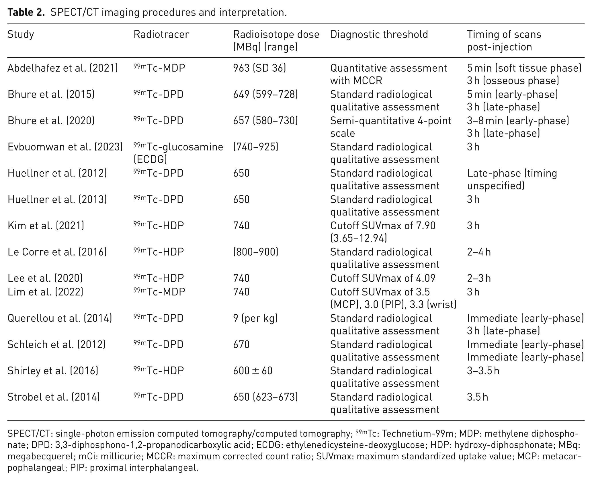
SPECT/CT: single-photon emission computed tomography/computed tomography; 99mTc: Technetium-99m; MDP: methylene diphosphonate; DPD: 3,3-diphosphono-1,2-propanodicarboxylic acid; ECDG: ethylenedicysteine-deoxyglucose; HDP: hydroxy-diphosphonate; MBq: megabecquerel; mCi: millicurie; MCCR: maximum corrected count ratio; SUVmax: maximum standardized uptake value; MCP: metacarpophalangeal; PIP: proximal interphalangeal.
Risk of bias
The studies showed a moderate risk of bias (domains 1–4), however with overall strong applicability (domains 5–7). A full breakdown of our risk of bias assessment is shown in Figure 2.
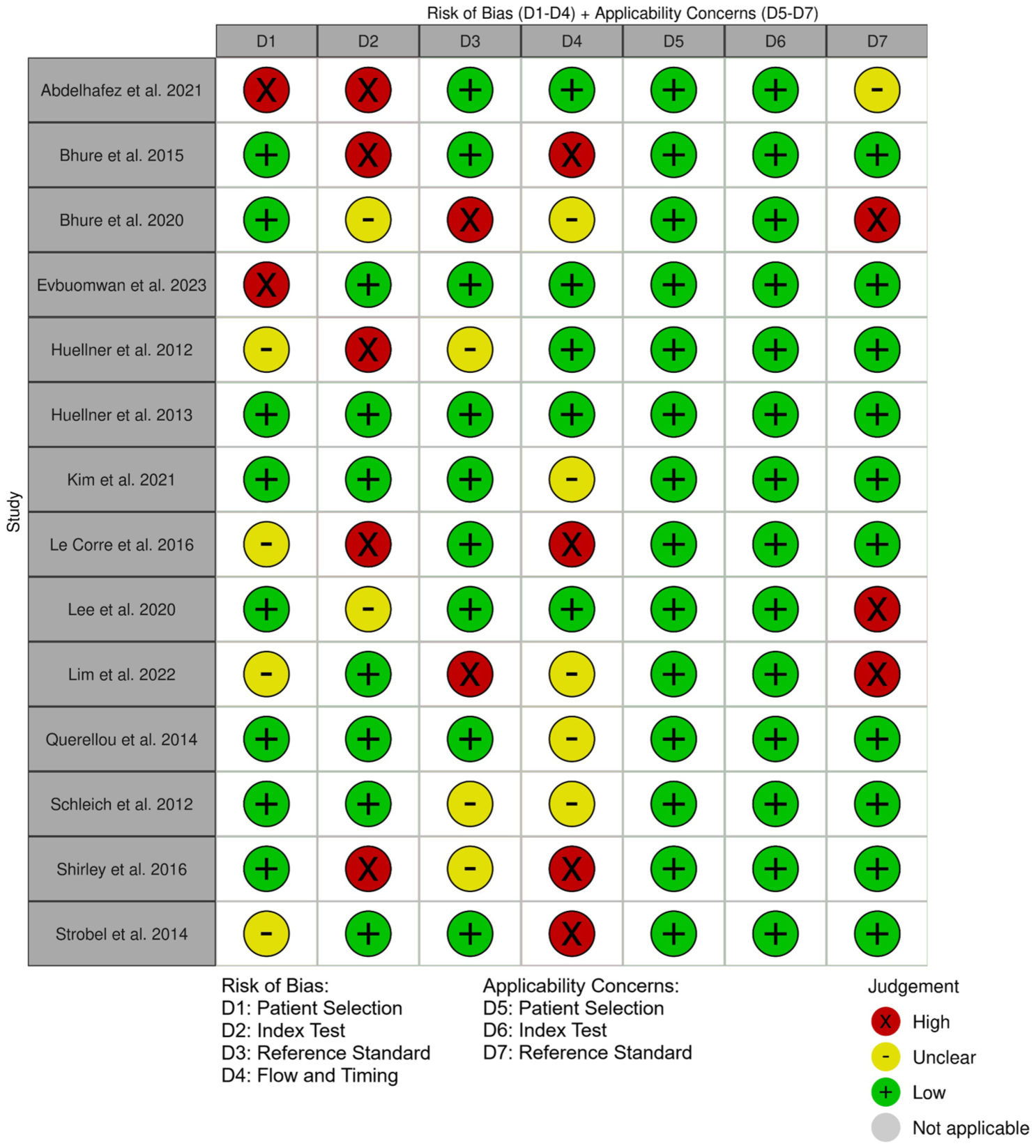
The revised Quality Assessment of Diagnostic Accuracy Studies (QUADAS-2) assessment of included studies. D(n): Domain number.
Results
Non-specific hand and wrist pain
Six of the 14 studies examined the diagnostic value of SPECT/CT for new presentations of non-specific hand and wrist pain (Table 1). All studies found that SPECT/CT had a comparable diagnostic accuracy to MRI, with a sensitivities and specificities ranging from 0.6 to 0.83 and from 0.9 to 0.1 respectively. In general, SPECT/CT had a lower sensitivity but greater specificity than MRI. All studies that measured interobserver agreement between radiologists also found that SPECT/CT demonstrated greater agreement than MRI. Several studies found that SPECT/CT changed the diagnosis or management despite prior use of conventional imaging (Bhure et al., 2015; Le Corre et al., 2016; Schleich et al., 2012; Shirley et al., 2016) – see Online Table S2. Two studies used SPECT/CT arthrography with intraarticular contrast to enhance ligament and cartilage visualisation (Bhure et al., 2020; Strobel et al., 2014), finding high concordance between the two modalities but greater overall sensitivity with SPECT/CT (Online Table S2).
Osseous disorders
Five studies examined ulnar impaction syndrome (n = 2), occult wrist fractures (n = 1), basal joint arthritis (n = 1) and carpal boss (n = 1). The sensitivity of SPECT/CT ranged from 0.75 to 1.0, with a specificity ranging from 0.82 to 0.93 (Online Table S2). Three studies demonstrated that SPECT/CT showed uptake in areas that were previously negative on CT/MRI. Overall, SPECT/CT findings influenced surgical management in four out of 11 eligible studies (Bhure et al., 2015; Le Corre et al., 2016; Schleich et al., 2012; Shirley et al., 2016), demonstrating an overall positive concordance between wrists showing additional uptake on SPECT/CT and those responding to surgery (Online Table S2). However, a change in the management plan occurred for only a minority of patients scanned.
Rheumatic disorders
Two studies evaluated the prognostic value of SPECT/CT in rheumatoid arthritis (RA), while a third study assessed its use in diagnosing rheumatic diseases in general. In all studies, SPECT/CT uptake reliably correlated with active synovitis and the clinical severity of RA. The sensitivity for active synovitis ranged from 0.72 to 1.0, and the specificity ranged from 0.65 to 0.82. Ultrasound was more specific than SPECT/CT; however, SPECT/CT could differentiate between rheumatic conditions by utilizing SUVs to determine cutoff points for each condition (Lim et al., 2022).
Discussion
This systematic review found that SPECT/CT has comparable sensitivity and specificity to established cross-sectional imaging modalities and can outperform these modalities in equivocal or borderline cases, providing information to inform management decisions. However, there is little evidence to support its routine use in diagnosing hand and wrist conditions. SPECT/CT only changed surgical management in a minority of cases and thus may not be cost-effective or justify the risks associated with additional radiation exposure or adverse reactions to radiopharmaceuticals. Further evidence, however, is needed to determine this.
This study had several limitations. Firstly, high clinical heterogeneity made statistical pooling of sensitivities and specificities unreliable and prevented its implementation. Most studies did not report their true positive, true negative, false positive, and false negative rates, making it impossible to verify their calculations with a contingency table. Differences in the pathology being assessed, the study protocol, the timing of scans after injury, and the time between the reference test and the SPECT/CT scan could also limit the reliability of the conclusions (Table 2). However, considering the chronic nature of the conditions, it is unlikely that they would have resolved between reference and index testing. Since no treatment was given, differences in protocol would have less impact. Secondly, the studies included in this review had small sample sizes. It is known that small studies have lower statistical power and tend to overestimate effect sizes in diagnostic studies (Song et al., 2002). We may have limited our evidence pool by only including studies written in English. Thirdly, several retrospective studies interpreted the index test (SPECT/CT) while knowing the results of the reference test (Abdelhafez et al., 2021; Bhure et al., 2015; Le Corre et al., 2016; Huellner et al., 2012; Shirley et al., 2016) (Figure 2). This could bias the interpreter; however, it is difficult to determine whether this has a noteworthy impact on the diagnostic accuracy of the index test. Well-designed retrospective studies can allow for reliable conclusions, and most of our clinical knowledge comes from retrospective reviews (Brand, 2008). Future studies should aim to use robust blinding measures as some of the higher-quality studies included in this review have done. Finally, evidence of SPECT/CT’s performance in non-autoimmune cases of soft tissue pathology was sparse. Ligament and cartilage injuries were underrepresented in the pathologies across all studies, probably owing to the presumed superior performance of MR in soft tissue pathology compared with SPECT/CT.
In cases of non-specific pain, SPECT/CT scans were found to be more accurate than radiographs, CT scans and bone scintigraphy. This is to be expected given the hybrid anatomical-functional nature of the scan, which provides information that other modalities cannot (Buck et al., 2008). Although SPECT/CT was generally less sensitive than MRI, it was more specific (Huellner et al., 2013), yielding comparable diagnostic accuracy. The diagnostic performance of SPECT/CT vs. MRI is consistent across other non-hand musculoskeletal conditions, including ankle, foot and complex spine conditions (Ha et al., 2015; Thurston et al., 2024). In several studies, SPECT/CT diagnosis directly changed management plans for non-specific pain (Online Table S2), and one study reported positive outcomes as a result (Shirley et al., 2016). Our findings suggest that SPECT/CT may be valuable in confirming diagnoses and aiding surgical planning. However, concordance across modalities was generally high, and surgical cases in which SPECT/CT findings directly altered management plans were in the minority. This suggests that SPECT/CT may not provide a major benefit if used routinely in diagnostic workups.
The clinical use of SPECT/CT has predominantly focused on osseous and non-osseous malignant lesions (Alqahtani et al., 2020). Where a diagnosis had already been made using prior imaging, the main benefit of SPECT/CT was to differentiate clinically noteworthy areas and detect concomitant pathology that had not been seen in the initial imaging. For instance, this has been used to detect concomitant ulnar impaction syndrome and triangular fibrocartilage tears (Lee et al., 2020; Strobel et al., 2014) and to differentiate rheumatic joint diseases (Lim et al., 2022). This is possible due to the quantitative measurements that scintigraphy can provide, specifically localising areas of high SUV, which allows for better radiological correlation with clinical symptoms. This allows for the specific targeting of the most clinically relevant anatomy and can theoretically create diagnostic cutoffs for certain conditions (e.g. RA vs. fibromyalgia; Lim et al., 2022). SPECT/CT showed higher interobserver agreement among both experienced and inexperienced readers than MRI or radiography (Huellner et al., 2013). In a diagnostic workup, this may be beneficial if rapid interpretation is required; however, this scenario is unlikely to arise in the context of hand and wrist conditions.
Prognostic studies have demonstrated that SPECT/CT uptake, as measured by SUVmax or qualitative interpretation, reliably correlates with disease activity. The SUVmax has also been found to stratify risk in amyloid transthyretin-related cardiomyopathy (Caobelli et al., 2024) and to monitor treatment response and predict survival in prostate cancer (Dittmann et al., 2021; Neubauer et al., 2024). However, multiple studies in our review noted that SPECT/CT can overestimate the clinical importance of some areas of uptake. This can make correlating these to clinical symptoms more challenging and may lead to overtreatment. The same has been found to be the case in sentinel lymph node biopsies of head and neck malignancies (Remenschneider et al., 2015). In the case of hand and wrist disorders, this phenomenon could cause more harm than good. Given the widespread availability and low cost and absence of radiation exposure of ultrasound, SPECT/CT may have no role in the routine determination of prognosis of autoimmune hand and wrist conditions.
The included studies provided no details or discussion surrounding the economic burden or cost-effectiveness of SPECT/CT. In non-hand literature, however, there is some evidence that SPECT/CT can save costs as a strategy to avoid surgery in cases of persistent knee pain after total knee arthroplasty and in small renal masses (Su et al., 2021; Van den Wyngaert et al., 2018). However, SPECT/CT was applied in specific and uncommon situations in these studies, so there is little evidence of its broad cost-effectiveness as a routine modality.
While the included studies reported no adverse reactions, technetium-99m (99mTc)-based radiopharmaceuticals commonly cause nausea, vomiting and erythematous skin reactions (Meher et al., 2021). In addition, an intravenous dose carries substantial radiation exposure. The most commonly used radiopharmaceutical was 99mTc-3,3-diphosphono-1,2-propanodicarboxylic acid (99mTc-DPD), which was used in seven of the 14 included studies (Table 2). A SPECT/CT scan using 700 MBq of 99mTc-DPD results in radiation exposure equivalent to 6.7 mSv (Quarta et al., 2021), which translates to an additional fatal cancer case for every 3000 patients scanned (Lin, 2010). Considering that the radiation exposure for a standard CT wrist scan is 0.15 mSv (Iordache et al., 2017) and is negligible for an MRI scan, both surgeons and patients need to give serious consideration to these risks for informed decision-making.
Overall, despite having comparable sensitivity and specificity to other cross-sectional imaging modalities, such as MRI, the evidence suggests that it cannot replace them. This is demonstrated by the fact that it only influences surgical management in a minority of cases. While its dual anatomical and functional nature is advantageous in specific scenarios, it can also lead to the overestimation of the clinical relevance of areas of uptake in others. Furthermore, SPECT/CT may carry a financial and radiation burden that is not justified by its demonstrated benefits. This is particularly the case when it comes to predicting autoimmune conditions, where ultrasound carries zero radiation and radiopharmaceutical risks and costs a fraction of the price. Based on the current evidence, SPECT/CT should not be used routinely in diagnostic procedures and offers little benefit in cases of obvious pathology. However, in select circumstances where the pathology is subtle or the prior imaging is inconclusive, SPECT/CT can be an effective problem-solving tool.
Supplemental Material
sj-docx-1-jhs-10.1177_17531934251404743 – Supplemental material for The diagnostic utility of single-photon emission computed tomography combined with computed tomography (SPECT/CT) in hand and wrist disorders: a systematic review
Supplemental material, sj-docx-1-jhs-10.1177_17531934251404743 for The diagnostic utility of single-photon emission computed tomography combined with computed tomography (SPECT/CT) in hand and wrist disorders: a systematic review by Onyedi Moses, Sharon Yohannes, Shaikh S Seraj, Luke Geoghegan and Maxim D Horwitz in Journal of Hand Surgery (European Volume)
Supplemental Material
sj-docx-2-jhs-10.1177_17531934251404743 – Supplemental material for The diagnostic utility of single-photon emission computed tomography combined with computed tomography (SPECT/CT) in hand and wrist disorders: a systematic review
Supplemental material, sj-docx-2-jhs-10.1177_17531934251404743 for The diagnostic utility of single-photon emission computed tomography combined with computed tomography (SPECT/CT) in hand and wrist disorders: a systematic review by Onyedi Moses, Sharon Yohannes, Shaikh S Seraj, Luke Geoghegan and Maxim D Horwitz in Journal of Hand Surgery (European Volume)
Footnotes
Acknowledgements
Evidence search strategy: Ms Assad Lahlou, Barts Health Knowledge and Library Services.
Declaration of conflicting interests
The authors declared no potential conflicts of interest with respect to the research, authorship, and/or publication of this article.
Funding
The authors received no financial support for the research, authorship, and/or publication of this article.
Ethical approval
Not applicable.
Informed consent
Not applicable.
Supplementary material
Supplemental material for this article is available online.
References
Supplementary Material
Please find the following supplemental material available below.
For Open Access articles published under a Creative Commons License, all supplemental material carries the same license as the article it is associated with.
For non-Open Access articles published, all supplemental material carries a non-exclusive license, and permission requests for re-use of supplemental material or any part of supplemental material shall be sent directly to the copyright owner as specified in the copyright notice associated with the article.