






Abstract
Background
The incidence of headaches during pregnancy is 35%. Although ruling out pre-eclampsia as a possible cause for headache is important in the pregnant population, acute sinusitis should remain on the differential as it occurs six times more frequently in pregnant women. Untreated disease can lead to rare intracranial complications such as a subdural empyema.
Conclusion
Maintaining a clinical suspicion for acute sinusitis as a cause of headache in a pregnant woman is important for prompt diagnosis and treatment before it develops into a rare intracranial complication.
Introduction
Headaches are classified as either primary or secondary. Primary headaches, such as tension headaches and migraines, are common in women of childbearing age. 1 Secondary headaches indicate an underlying pathology such as acute sinusitis, arterial dissection, or meningitis. 2 The incidence of primary and secondary headaches during pregnancy is around 35% and careful consideration of the underlying cause is required due to the differential diagnosis that includes both pregnancy-related and nonpregnancy-related pathologies (for example, pre-eclampsia and intracranial pathology, respectively). 3
Infectious sinusitis may occur in 1.5% of the pregnant population. 4 If untreated, sinusitis may lead to intracranial complications and spread to the extradural, subdural, or intracerebral spaces due to the close proximity of the brain to the sinuses. 5 Here described is a pregnant woman who developed a subdural empyema secondary to persistent sinusitis of odontogenic source.
Case presentation
A 21-year-old woman in her second pregnancy presented at 33 weeks’ gestation to an acute obstetric triage with a 8/10 pulsatile headache localized to the left forehead associated with sinus pain, fever of 39.4°C, photophobia, vomiting, and diarrhea. She was pregnant with dichorionic, diamniotic twins and had previously been diagnosed with gestational hypertension. She had never had similar symptoms before and there was no history of migraine. Routine hematology and biochemistry were all normal. She was diagnosed with sinusitis and gastroenteritis and treated with a seven-day course of amoxicillin–clavulanic acid and overnight observation.
The woman subsequently called the acute obstetric triage a number of times after her initial evaluation, as she had an ongoing dull headache associated with nasal congestion and sinus pressure that had initially resolved but returned after antibiotic completion. Assessment showed no evidence of pre-eclampsia and she was eventually given the diagnosis of migraine.
Twenty-one days after initial evaluation, her headache was re-evaluated due to worsening pain associated with intractable nausea and vomiting. Blood pressure was 139/78 mmHg. Blood tests showed that her white blood cell count was elevated at 16.2 ×10
3
/
Following delivery, her mental status worsened and was associated with right sided anisocoria. Blood pressure was 174/87 mmHg. Computerized tomography (CT) head was obtained due to concern for an intracranial hemorrhage and demonstrated a left-sided subdural collection with a left to right midline shift (Figure 1). She subsequently underwent emergency craniectomy under the care of the neurosurgical team and was diagnosed with a subdural empyema. The cranium was not replaced at the end of the procedure due to the presence of cerebral edema.
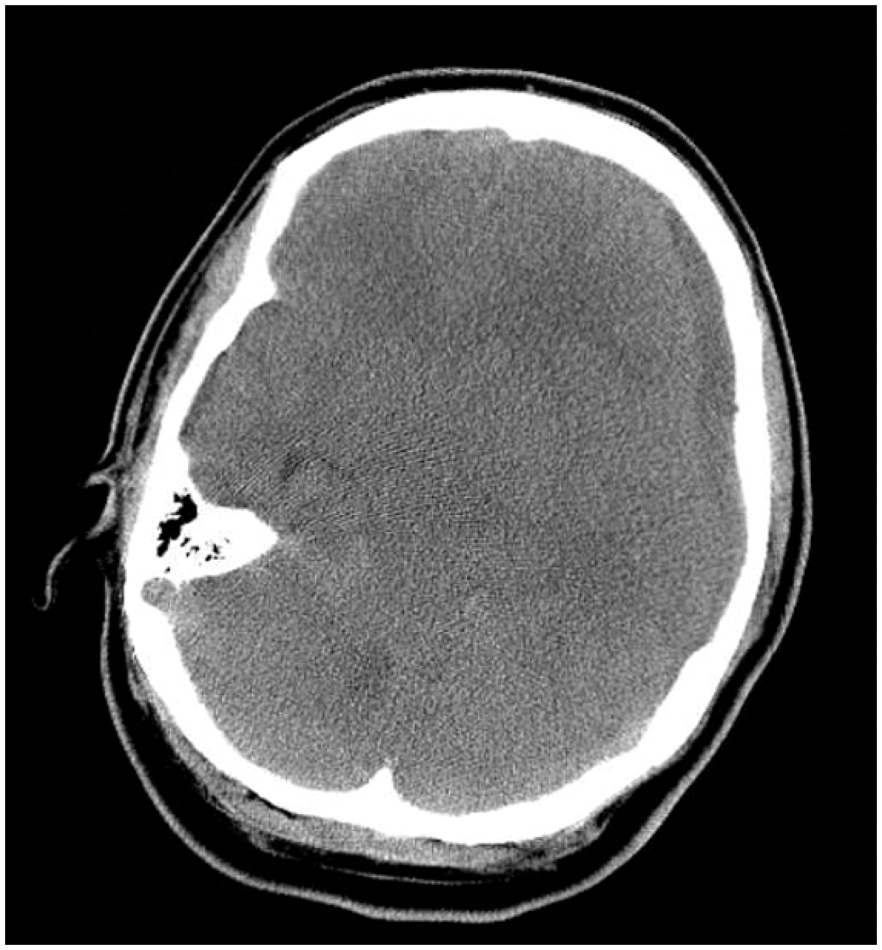
Axial section of CT head demonstrating a left-sided subdural collection with mass effect, brain edema, and effacement of the ventricles and basilar cisterns.
Complete opacification of the left maxillary and anterior ethmoid sinuses was also noted on CT, so otolaryngology performed bedside nasal endoscopy, which revealed purulent drainage from the left maxillary sinus. Endoscopic sinus surgery was performed the same day. Cultures grew Streptococcus intermedius. There was also concern on the CT about a periapical abscess concerning for an odontogenic source (Figure 2) and she went on to have removal of two teeth with the oral surgery service two days later. Six weeks of intravenous ampicillin–sulbactam was recommended. The woman required rehabilitation upon discharge due to persistent expressive aphasia and residual right-sided weakness.
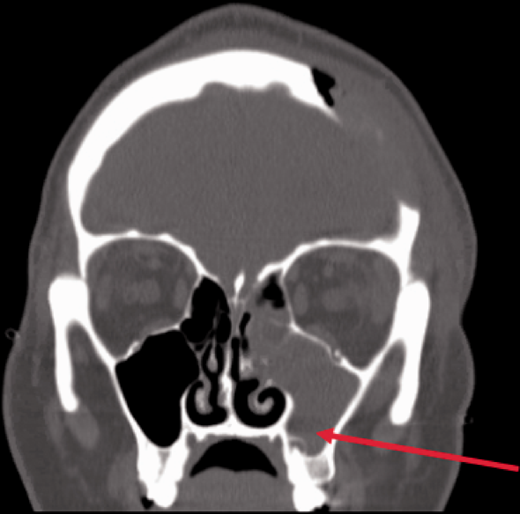
Coronal section of CT sinus demonstrating complete opacification of the left maxillary sinus and a peri-apical hyperlucency depicted by the arrow.
When seen for outpatient evaluation two months later, endoscopic examination revealed patent sinuses without infection. She underwent a cranioplasty 10 months later and was felt to be neurologically intact with equal pupils and normal motor and sensory examination.
Discussion
Pre-eclampsia is an important cause for headache in pregnancy as it accounts for the world’s largest maternal death rates, estimated at around 60,000 per year. 6 Often, acute cerebral complications such as eclampsia, hemorrhagic stroke, and seizures account for the fatalities. 7 The woman described here had a higher risk of developing pre-eclampsia due to prior gestational hypertension, obesity (body mass index of 43.8 kg/m2), and twin pregnancy. She had been given aspirin during this pregnancy in recognition of this.
Acute sinusitis should remain on the differential as it can occur six times more frequently in the pregnant population, thought to be due to increased mucosal thickening secondary to hormonal effects.8,9 Symptoms of acute sinusitis include nasal congestion, purulent discharge, facial pain/pressure, and reduction of smell lasting less than four weeks. 10 Classic symptoms may be absent in nearly half of women with documented purulent sinusitis during pregnancy, making diagnosis difficult. 4
A handful of case reports have described rare intracranial complications from untreated acute sinusitis in pregnant women. A woman developed a pituitary abscess due to sphenoid sinusitis at 34 weeks of gestation. 10 A temporal lobe abscess developed in a woman at 36 weeks of gestation due to maxillary and ethmoid sinusitis. 11 A woman at 35 weeks of gestation developed a Pott’s puffy tumor (subperiosteal abscess and osteomyelitis of the frontal bone) secondary to frontal sinusitis. 12
The woman described here was treated with a course of antibiotics for acute sinusitis on initial presentation. It is possible that sinusitis was no longer considered on the differential since she had received treatment with initial symptom improvement. During the subsequent evaluations, she was evaluated for pre-eclampsia. Headaches associated with pre-eclampsia often present as a bilateral pulsatile headache associated with visual disturbances and may be exacerbated by physical activity. 13 A unilateral, focal headache along with nasal congestion and sinus pressure, especially following the cessation of antibiotics is suggestive of persistent sinusitis. Otolaryngology input is beneficial in this setting as a bedside nasal endoscopy can be performed to assess for purulent drainage from the sinus outflow tracts. Treatment options may include switching antibiotic therapy, sinus irrigations, and topical intranasal steroids to completely address the sinus infection. If complications are suspected, then urgent cranial imaging should be performed, and whilst MRI is generally preferred in pregnancy, the use of CT is not contra-indicated and the choice of imaging modality should not lead to delay in diagnosis and intervention.
Unilateral sinusitis is due to odontogenic origin in up to 72.6% of cases. 14 A peri-apical abscess of an upper maxillary tooth was the likely source of infection in the case described here, but was not recognized on the official review of imaging. Therefore, careful radiologic review is important to rule out associated odontogenic disease in unilateral sinusitis.
Pregnant women commonly present with headache, which may be difficult to diagnose given their variable presentations and etiologies. This case illustrates the importance of a thorough history and examination. Although this woman was diagnosed with pre-eclampsia, a clinical suspicion of sinusitis should be investigated. The periodontal infection led to unilateral sinusitis and ultimately a subdural empyema. Otolaryngology input may be beneficial to help evaluate for sinusitis as untreated or refractory disease can lead to rare intracranial complications.
Footnotes
Declaration of conflicting interests
The author(s) declared no potential conflicts of interest with respect to the research, authorship, and/or publication of this article.
Funding
The author(s) received no financial support for the research, authorship, and/or publication of this article.
Ethical approval
This case report received approval from Loyola University Medical Center’s IRB, entitled “The Insidious Headache in a Pregnant Woman”.
Informed consent
Written consent for publication was obtained from the participant of the case report, which is filed with the corresponding author’s records and available upon request.
Guarantor
MP.
Contributorship
MP and SY researched literature and conceived the case report. SY was involved in protocol development, gaining ethical approval, and consenting patient for the written report. SY and JH wrote the first draft of the article. All authors reviewed and edited the article and approved the final version of the article.
Acknowledgements
We would like to thank Ioana Danci, MD for her guidance in the writing of this case report.
