






Abstract
Proteus syndrome is a rare, multi-system, genetic syndrome characterised by atypical and excessive growth of skeletal tissue. Clinical presentations include abnormal musculoskeletal growth and cutaneous lesions. Due to its rarity, there have been a limited number of published case reports of Proteus syndrome. This is the first case report on the management of Proteus syndrome in pregnancy. We present the case of a pregnant woman with Proteus syndrome in her first pregnancy in a large teaching hospital and discuss the considerations and challenges faced in her antenatal, intrapartum and postnatal care.
Keywords
Background
Proteus syndrome is a very rare genetic disorder. The hallmark of the disorder is atypical, asymmetrical and aberrant growth of musculoskeletal tissue affecting multiple body parts, 1 as the mutation affects tissues from all three embryological germ layers. Other manifestations may involve vascular malformations (such as arterio-venous malformations), neurological disability (e.g. seizures and intellectual impairment) 2 and cutaneous lesions (such as epidermal naevi and lipomas). 1
Proteus syndrome is caused by a gain-of-function, somatic mutation of the oncogene AKT1 (AKT serine/threonine protein kinase 1) gene (specifically c.49G → A, p.Glu17Lys) and displays mosaicism, as only some cells will have the mutation. 3 As this is a mosaic mutation, there is no increased risk of a sibling inheriting the condition. At present, there has been no proven transmission of Proteus syndrome from parent to child. It has been proposed that should this mutation be present as a germ-line mutation and passed on to an embryo, this may be lethal in early pregnancy. 4
The AKT1 gene is responsible for producing AKT1 kinase, an enzyme involved in many cell proliferation and differentiation signalling pathways, and therefore mutations in the AKT1 gene can cause aberrant and uncontrolled cell division. 1 AKT1 gene mutations have been implicated in several other disease processes and plays a central role in many types of cancers such as breast cancer 5 and neuroblastoma. 6
The incidence is believed to be less than 1 in 1 million live births; however, due to the previous diagnostic challenges with Proteus syndrome, this quoted incidence is most likely not reflective of the true prevalence of the disorder. 7 The significant variability in severity and phenotype of the disease can make clinical diagnosis challenging; however, in 2004, Turner et al. 8 produced a set of diagnostic criteria for Proteus syndrome (which was later updated by Cohen et al. 1 ). These criteria, the increasing accessibility and reducing costs of genetic analysis and the use of a clinical geneticist, should allow for easier diagnosis and treatment for those with the disorder. Although diagnosis may now be relatively straightforward, this may still be challenging in resource-limited settings.
There is also overlap of clinical presentation between several different rare overgrowth syndromes such as Klippel-Trenaunay syndrome 9 and Parkes-Weber syndrome. 10
The clinical manifestations and severity of the syndrome are highly variable. There are very few case reports of women with Proteus syndrome, and we have not identified any involving pregnant women in our literature search. We believe our case is the first case report and discussion of a pregnant woman with Proteus syndrome.
Case
A 19-year-old woman in her first pregnancy was referred to our tertiary hospital for specialist antenatal care in our maternal medicine antenatal clinic at 19 weeks’ gestation.
She was born in Russia where she was noted to have macrodactyly of the second and third right finger at birth. In childhood, she underwent an amputation of her second right finger and transplantation of her second toe. The woman later underwent a total left pneumonectomy aged 14 due to complete emphysematosis and multiple benign cysts. She had no cutaneous lesions, no organomegaly, no distinctive pathognomonic facial features and no known large vascular malformations.
Initially, she had serum genetic testing for protein kinase B (AKT1), phosphatase and tensin homolog and phosphatidylinositol-4,5-bisphosphate 3-kinase, catalytic subunit alpha (PIK3CA) mutations, all of which were negative. She subsequently had AKT1 genetic testing of the lung tissue from her pneumonectomy, which revealed a heterozygous mutation in the second exon in 50% of the tissue, indicating somatic mosaicism with the specific c.49G → A, p.Glu17Lys mutation, confirming a diagnosis of Proteus syndrome.
This woman had a mild phenotype and had always been completely independent in daily activities. She did not take any medications and did not suffer from any significant symptoms as a result of her Proteus syndrome. However, she did report long-standing dizziness and palpitations, and prior to conception, she underwent several cardiac investigations. An echocardiogram was performed which was normal, and a 48-h Holter monitor was also normal (Figure 1). She also underwent a high-resolution CT scan which showed her left pneumonectomy, scattered emphysematous changed in the right lung, no mediastinal or hilar lymphadenopathy, no airway or bony abnormalities.
The woman was reviewed in our specialist maternal medicine clinic. She underwent lung function testing, which was acceptable with a forced expiratory volume in 1 s of 70% of predicted and forced vital capacity of 78% of predicted; therefore, there was no evidence of overt obstructive lung disease in her remaining lung.
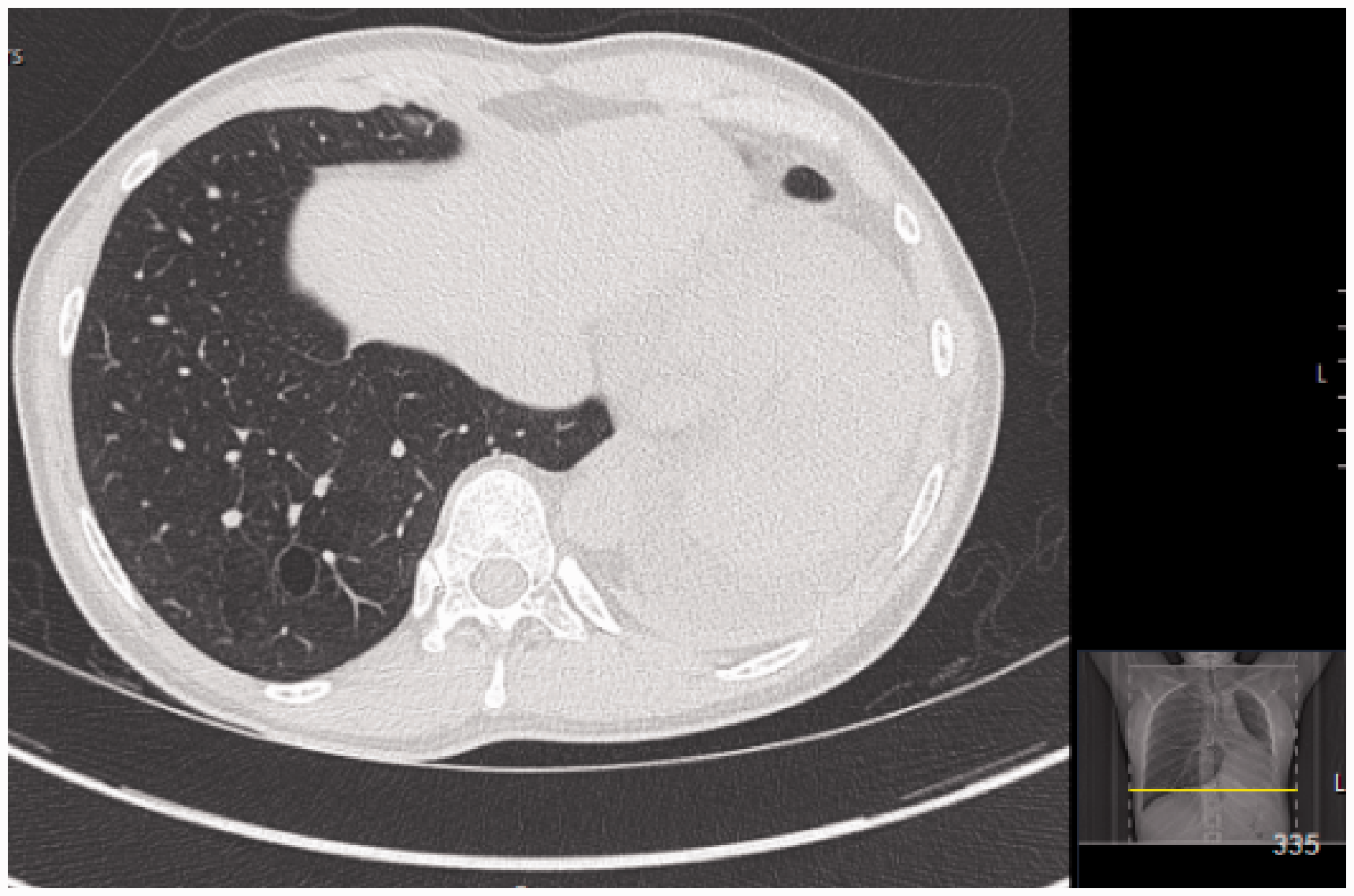
CT chest (high resolution) – left pneumonectomy and emphysematous changes in the right lung.
Proteus syndrome is also known to increase the risk of developing venous thromboemboli (VTE), 11 and pulmonary embolism is one of the most common cause of premature death in these women. 12 Therefore, at 28 weeks’ gestation, she was commenced on thromboprophylaxis with low-molecular weight heparin and advised to continue this for a further six weeks postnatally. There have been case reports of death from venous thromboembolism in Proteus syndrome despite therapeutic anticoagulation, 13 and so during her antenatal care, she was advised to be vigilant for any symptoms of VTE including unilateral calf pain and swelling, pleuritic chest pain, persistent abdominal pain and difficulty in breathing.
She was reviewed every four weeks in our maternal medicine clinic and remained well throughout the pregnancy, only complaining of mild shortness of breath. Fetal growth scans were normal. As Proteus syndrome is not an inherited condition (inheritance of the mutation would likely cause pregnancy loss), no fetal genetic screening was performed.
The woman was also seen in a consultant-led high-risk anaesthetic clinic, where it was concluded that it would be safe to have regional anaesthesia, with a view to avoiding a general anaesthetic. The only constraint for her birth plan was that a consultant-led labour ward would be the most suitable setting for labour and that she should stop administration of low-molecular weight heparin if she should have any signs of labour.
The woman presented to our obstetric triage in labour at 39 weeks’ gestation and had a spontaneous vaginal delivery without regional analgesia with an estimated blood loss of 500 ml. The baby was born in good condition with a birth weight 3260 g. She recovered well and had no postnatal complications. Both she and her baby were discharged the following day with a six-week course of prophylactic low-molecular weight heparin.
Discussion
Due to the rare incidence of Proteus syndrome and the lack of case reports of pregnant women with Proteus syndrome, there was no set standard of care to follow.
Antenatal care
We predicted that Proteus syndrome would increase obstetric risk due to the increased pre-existing risk of developing thromboembolism in this disease (compounded by the hormonal effects of pregnancy on thromboembolic risk) and haemorrhage due to exacerbation of pre-existing arterio-venous malformations. 14 Therefore, individual bleeding risk assessments need to be made for these women when commencing anticoagulation. Women should be counselled regarding symptoms of VTE and advised to promptly report these to a healthcare professional. Women should also be seen in a specialist high-risk obstetric anaesthetic clinic for a detailed risk assessment and appropriate anaesthetic management plan.
Tissue atrophy is also a feature of Proteus syndrome, and therefore nutritional assessment in the antenatal and postnatal period should be considered. 15
Proteus syndrome is caused by a somatic mutation; therefore, routine fetal ultrasound scans will not identify women with the disorder, as clinical features manifest after birth. Routine midwifery care should occur alongside the specialist obstetric medicine reviews.
Anaesthetic considerations
The use of anticoagulation as mentioned above can complicate obstetric care, should the woman present with an obstetric emergency needing immediate intervention. Ideally, women requiring regional anaesthesia would not have had prophylactic low-molecular weight heparin within 12 h or 24 h if taking a treatment dose to reduce the risk of developing a spinal haematoma. 16 The use of anticoagulants in pregnancy inherently subjects women to a higher risk of needing a general anaesthetic in the event of an obstetric emergency. General anaesthesia in itself carries a higher risk to obstetric women compared with the general population, such as failed intubation, airway oedema and ventilation difficulties. 17 Additionally, women with Proteus syndrome may have obstructive or restrictive lung disease which further increases their risk during general anaesthesia. 18 Our patient did not have any obstruction or restrictive lung disease.
Scoliosis is also a feature of the syndrome and may present further challenges for administration of regional anaesthesia as well as increasing patient discomfort as the pregnancy progresses.
Vertebral distortion including fixed flexion, limited range of mobility and torticollis may also be present, and therefore difficulties in intubation during general anaesthesia should be anticipated. 18
Meningiomas and hyperostosis of cranial and facial bones are commonly found in Proteus syndrome. One retrospective cohort study found these to be present in 6.5% and 48%, respectively, in a group of 61 women with Proteus syndrome. 19 Pregnancy is associated with an increase in meningioma growth, theories for which include hormonal changes (such as placental hPL secretion 20 and increased levels of progesterone 21 ) and physiological vascular adaptations resulting in increased plasma volume, blood flow and cerebral oedema. 22
Thus far, there have been no studies investigating meningiomas in pregnant women with Proteus syndrome; however, consideration of current evidence, it is reasonable to presume that pregnant women with Proteus syndrome are at an increased risk of meningioma growth. This would warrant advice for women to report symptoms such as visual disturbances, headaches and focal neurology.
Revised Proteus syndrome diagnostic criteria.
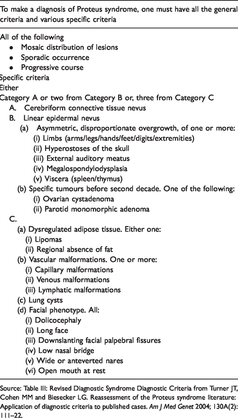
Source: Table III: Revised Diagnostic Syndrome Diagnostic Criteria from Turner JT, Cohen MM and Biesecker LG. Reassessment of the Proteus syndrome literature: Application of diagnostic criteria to published cases. Am J Med Genet 2004; 130A(2): 111–22.
Surgical considerations
Surgical complications should also be anticipated in women with Proteus syndrome. One of the hallmarks of the syndrome are unencapsulated lipomas, which manifest as localized overgrowth of adipose tissue. 15 These lipomas may be highly vascular and may cause additional intra-operative blood loss. Furthermore, these women may have previously unidentified vascular abnormalities, organomegaly and soft tissue masses.
Our patient had a mild phenotype and therefore did not require many specialist services during her antenatal care; however, this case highlights the significant variability in clinical manifestations of Proteus syndrome and that antenatal care needs to be individualised on a case-by-case basis.
Management of rare conditions in pregnancy should always be managed with a multi-disciplinary approach. This woman was reviewed by a maternal medicine specialist, obstetric anaesthetist and respiratory physician which led to the development of an individualised management plan. This approach should be employed for all pregnant women with rare and complex medical conditions.
Footnotes
Acknowledgements
We would like to thank the patient for allowing us to discuss her case.
Declaration of conflicting interests
The author(s) declared no potential conflicts of interest with respect to the research, authorship, and/or publication of this article.
Funding
The author(s) received no financial support for the research, authorship, and/or publication of this article.
Ethical approval
The Royal London Hospital (Barts Health) does not require ethical approval for reporting individual cases.
Informed consent
Written consent was obtained from the patient for their anonymized information to be published in this article.
Guarantor
Dr Niccole Ranaei-Zamani is the guarantor of the present work.
Contributorship
NR and MK wrote the article, and was approved by RK. The article was reviewed and edited by all authors.
