






Abstract
Background:
Nongranulomatous acute anterior uveitis (NG-AAU) is the most common extra-articular manifestation of spondyloarthritis (SpA) and may be a warning sign of previously undiagnosed axial SpA (ax-SpA).
Objective:
Given the known diagnostic delay of ax-SpA, this study aimed to determine the prevalence of previously undiagnosed ax-SpA among NG-AAU patients and to characterize the clinical features of ax-SpA identified through uveitis.
Design:
Cross-sectional study.
Methods:
This monocentric study included consecutive NG-AAU patients referred to a tertiary ophthalmologic center between August 2024 and August 2025. All patients underwent a comprehensive rheumatologic assessment, including human leukocyte antigen B27 (HLA-B27) testing and sacro-iliac joint magnetic resonance imaging (MRI). Ultrasound evaluation of entheses and symptomatic joints was also performed. SpA diagnosis was established by expert opinion based on clinical, laboratory, and imaging findings.
Results:
Among 68 NG-AAU patients, 38 without a previous rheumatic diagnosis were included. Sixteen (43.2%) received a new diagnosis of ax-SpA, and none with peripheral SpA. HLA-B27 positivity was more frequent in ax-SpA than in non-SpA patients (56% vs 33%). All ax-SpA patients reported prior episodes of back pain, although inflammatory back pain was present in only 56.3% at the time of evaluation. Mean Bath Ankylosing Spondylitis Disease Activity Index (BASDAI) scores and symptom duration were significantly higher in ax-SpA patients (p = 0.005 and p = 0.041, respectively). MRI revealed active sacroiliitis in 62.5% and erosions in 75% of newly diagnosed ax-SpA patients. Only 68.7% of new diagnoses fulfilled the 2009 Assessment of Spondyloarthritis International Society (ASAS) classification criteria for ax-SpA.
Conclusion:
Nearly half of NG-AAU patients had previously unrecognized ax-SpA, often presenting with mild or atypical axial symptoms. These findings highlight the importance of integrated ophthalmologic and rheumatologic evaluation to facilitate earlier ax-SpA diagnosis.
Introduction
Axial spondyloarthritis (ax-SpA) is an inflammatory rheumatic disease within the spondyloarthritis (SpA) spectrum. It primarily affects the axial skeleton and presents with chronic back pain (CBP) that typically has inflammatory characteristics such as insidious onset, morning stiffness, nocturnal pain, and improvement with exercise rather than rest. 1 Peripheral articular manifestations and extra-articular domains, such as the eyes, skin, and bowel, may also be involved.
Non-infectious acute anterior uveitis (AAU), particularly nongranulomatous AAU (NG-AAU), represents the most frequent extra-articular SpA manifestation and is associated with higher disease activity and greater physical impairment. 2 In SpA, uveitis typically presents as recurrent or alternating unilateral NG-AAU with conjunctival and ciliary injection, causing noticeable eye redness.3,4 Recurrent episodes may lead to serious complications such as glaucoma, cataract, and even vision loss. 5
However, in some cases, NG-AAU may be a warning sign of previously undiagnosed SpA. Studies suggest that about 40% of patients presenting with AAU may have underlying SpA that remains unrecognized at the time of ophthalmological evaluation.6 –8 This proportion is even higher among human leukocyte antigen B27 (HLA-B27)-positive AAU cases, with estimates ranging from 58% to 78%. 9 Although structured algorithms have been proposed to support early detection of SpA in AAU patients, there are still no formal recommendations on when these patients should be referred to rheumatologists.
The diagnostic delay for ax-SpA remains substantial, estimated at 2–8 years, 10 and CBP is often underestimated as a non-specific symptom, particularly when inflammatory features are not clearly evident. In this study, we aimed to identify the clinical features of patients in whom uveitis served as the red flag leading to a diagnosis of ax-SpA.
Methods
Study design and objectives
We conducted a cross-sectional study on consecutive patients with NG-AAU referred to our Rheumatology Unit by the Ocular Immunology Unit of AUSL-IRCCS di Reggio Emilia, Italy, between August 2024 and August 2025. The primary aim was to determine the prevalence of newly diagnosed ax-SpA in this monocentric series of NG-AAU patients. The secondary aim was to characterize the clinical features of patients whose ax-SpA diagnosis was prompted by the onset of uveitis.
The inclusion criterion was an ophthalmologic diagnosis of NG-AAU, defined according to the Standardization of Uveitis Nomenclature (SUN) Working Group guidelines as acute onset anterior uveitis with an anterior chamber reaction but without granulomatous keratic precipitates. 11 Exclusion criteria included other causes of uveitis (e.g., infectious uveitis), a prior diagnosis of rheumatic disease, age under 18 years, or the absence of HLA test results.
Data were collected as part of routine clinical practice. The study protocol was approved by the Local Ethics Committee and conducted in accordance with the Declaration of Helsinki and the Principles of Good Clinical Practice. The reporting of this study conforms to the Strengthening the Reporting of Observational Studies in Epidemiology (STROBE; Supplemental Material) statement 12 .
Ophthalmic evaluation
All patients underwent a comprehensive ophthalmic evaluation, including best-corrected visual acuity, intraocular pressure, slit-lamp examination of the anterior segment with clinical evaluation of anterior chamber inflammation, and fundus examination after pupillary dilatation. Uveitis laterality (unilateral, bilateral, or alternating) and the presence of complications, such as posterior synechiae, were recorded. Patients were subsequently referred to a rheumatologist to assess for previously undiagnosed ax-SpA.
Rheumatologic evaluation and imaging
Patients underwent a structured evaluation by a rheumatologist expert in SpA at AUSL-IRCCS di Reggio Emilia, which included the collection of demographic data and disease history. Family history of immune-mediated diseases, including SpA, psoriasis (PsO), and inflammatory bowel diseases (IBD), was documented. Additional parameters recorded included body mass index (BMI), smoking status, comorbidities, and uveitis characteristics (age at first episode, recurrence, laterality, presence of posterior synechiae, and prior local or systemic treatments). The presence and age at onset of peripheral or axial symptoms were also assessed. Peripheral symptoms included current or previous episodes of arthritis and dactylitis. Axial symptoms included current or past episodes of CBP lasting >3 months, classified as inflammatory or non-inflammatory according to the Assessment of Spondyloarthritis International Society (ASAS) criteria for inflammatory back pain (IBP): CBP lasting >3 months, with at least four of the following features—age at onset <40 years, insidious onset, improvement with exercise, no improvement with rest, and nocturnal pain improving upon rising. 13
Laboratory assessments included HLA-B27, C-reactive protein, and erythrocyte sedimentation rate.
Clinical examination evaluated spinal mobility (Schober’s test), peripheral count of tender and/or swollen joints (68/66), and evaluation of the following enthesis sites: plantar fascia, Achilles tendon, proximal and distal patellar tendon insertions, quadriceps tendon insertion, common extensor, and triceps tendon insertions at the elbow. Enthesis pain was recorded as present or absent without grading severity.
Disease activity was assessed using the Bath Ankylosing Spondylitis Disease Activity Index (BASDAI), the Axial Spondyloarthritis Disease Activity Score (ASDAS), and the Leeds Enthesitis Index (LEI).14,15
At the end of the clinical evaluation, patients underwent ultrasound (US) examination of the same entheses to detect inflammatory and structural lesions, defined according to current Outcome Measures in Rheumatology (OMERACT) definition. 16
Enthesitis was defined as a hypoechoic and/or thickened tendon insertion within 2 mm of the bony cortex, with a Doppler signal indicating active inflammation. Chronic enthesopathy features, including calcifications, erosions, and enthesophytes, were also recorded. 17
In a small number of patients with articular pain, the US examination was extended to the symptomatic joints. The US examination of the entheses was performed by a single examiner (A.R.) using the same device, an Esaote Mylab Xpro80 equipped with a Linear Array probe (frequency 4–15 and 8–24 MHz), optimizing the machine settings to improve detection of the entheseal structure. The focus was set on the cortical bone of the explored entheses. Both B-mode analysis and power Doppler (PD) imaging were performed. The pulse repetition frequency was set at 690 Hz to optimize the detection of slow blood flow.
All patients without contraindications, regardless of backpain, underwent magnetic resonance imaging (MRI) of the sacro-iliac joints (SIJ) with short tau inversion recovery (STIR) and T1-weighted sequences in semicoronal and axial planes. Some MRIs were performed outside our center, depending on patients’ area of residence, in line with usual clinical practice. However, all MRI scans were reviewed by a local rheumatologist with long-standing experience in SpA and SpA imaging (N.P.).
Active inflammatory and structural lesions were scored according to the updated ASAS MRI working group definitions of SIJ lesions in SpA. 16 MRI was considered positive for active sacroiliitis when bone marrow edema (BMO) was detected on fluid-sensitive sequences (STIR or T2FS, or post-contrast T1) in typical subchondral areas, indicating a high likelihood of SpA. The presence of BMO in the posterior lower ilium or upper anterior sacrum, especially small lesions, was not considered specific for sacroiliitis, whereas extensive BMO or small lesions near a sacroiliac erosion were deemed indicative of active sacroiliitis.
In addition, in cases not fulfilling the ASAS classification criteria for ax-SpA but showing SpA consistent changes (e.g., SIJ erosions), MRI was still considered suggestive of the diagnosis based on clinical judgment and expert opinion. In one patient, SIJ erosions were confirmed by subsequent CT evaluation.
In all the cases, the gold standard for diagnosis was the expert opinion, in line with routine clinical practice.
Statistical analysis
Statistical analyses were performed with IBM SPSS Statistics version 29.0 (IBM Corp., Armonk, NY, USA). Continuous variables were expressed as mean ± standard deviation (SD) or median with interquartile range, as appropriate. Categorical variables were reported as frequencies and percentages. Comparisons between groups (ax-SpA vs non-SpA) were conducted using Student’s t test or Mann–Whitney U test for continuous variables, and Chi-square test or Fisher’s exact test for categorical variables. A two-sided p-value <0.05 was considered statistically significant.
No formal sample size calculation was performed, as this was an exploratory, monocentric, cross-sectional study including all consecutive patients referred during the study period. Due to the relatively small sample size and because identifying independent predictive factors for SpA was not a study objective, no multivariate analyses were performed.
Results
Sixty-eight consecutive patients with NG-AAU were evaluated at our uveitis clinic between August 2024 and August 2025: 31 males (45.5%) and 37 females (54.4%), with a mean age of 43.1 ± 13.4 years (range 18–74). Among them, 30 patients (44.1%) had a previously diagnosed rheumatic disease (17 (56.7%) SpA, 9 (30%) Juvenile Idiopathic Arthritis, 3 (10%) psoriatic arthritis, and 1 (0.3%) Behçet disease, respectively) and were therefore excluded from the present analysis.
The remaining 38 patients (55.8%) underwent SIJ MRI and US evaluation and were subsequently classified into two groups (ax-SpA vs non-SpA) based on expert opinion. MRI was unavailable for one patient. Table 1 reports findings stratified by newly diagnosed ax-SpA.
Demographic, clinical, radiologic, and serological features of 37 patients with NG-AAU enrolled in the study.
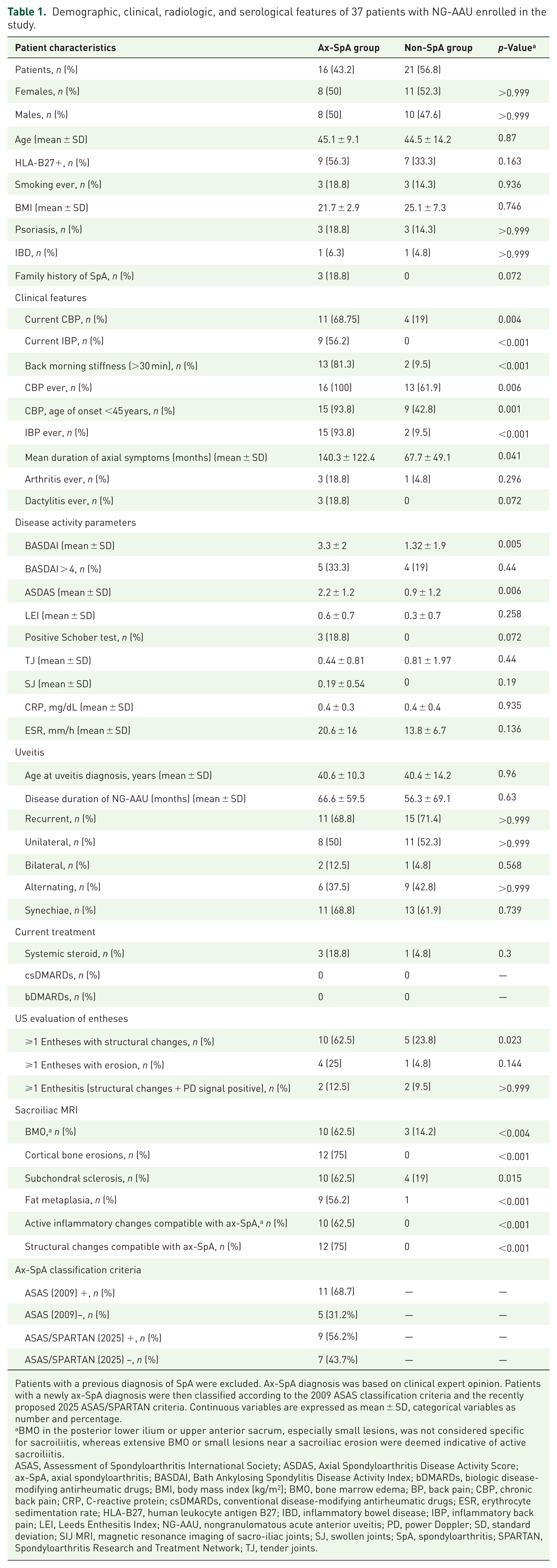
Patients with a previous diagnosis of SpA were excluded. Ax-SpA diagnosis was based on clinical expert opinion. Patients with a newly ax-SpA diagnosis were then classified according to the 2009 ASAS classification criteria and the recently proposed 2025 ASAS/SPARTAN criteria. Continuous variables are expressed as mean ± SD, categorical variables as number and percentage.
BMO in the posterior lower ilium or upper anterior sacrum, especially small lesions, was not considered specific for sacroiliitis, whereas extensive BMO or small lesions near a sacroiliac erosion were deemed indicative of active sacroiliitis.
ASAS, Assessment of Spondyloarthritis International Society; ASDAS, Axial Spondyloarthritis Disease Activity Score; ax-SpA, axial spondyloarthritis; BASDAI, Bath Ankylosing Spondylitis Disease Activity Index; bDMARDs, biologic disease-modifying antirheumatic drugs; BMI, body mass index (kg/m2); BMO, bone marrow edema; BP, back pain; CBP, chronic back pain; CRP, C-reactive protein; csDMARDs, conventional disease-modifying antirheumatic drugs; ESR, erythrocyte sedimentation rate; HLA-B27, human leukocyte antigen B27; IBD, inflammatory bowel disease; IBP, inflammatory back pain; LEI, Leeds Enthesitis Index; NG-AAU, nongranulomatous acute anterior uveitis; PD, power Doppler; SD, standard deviation; SIJ MRI, magnetic resonance imaging of sacro-iliac joints; SJ, swollen joints; SpA, spondyloarthritis; SPARTAN, Spondyloarthritis Research and Treatment Network; TJ, tender joints.
Among these patients, 16 (43.2%) were newly diagnosed with ax-SpA, while the remaining 21 showed no evidence of articular involvement consistent with SpA (Figure 1). No diagnoses of peripheral SpA (p-SpA) were made.
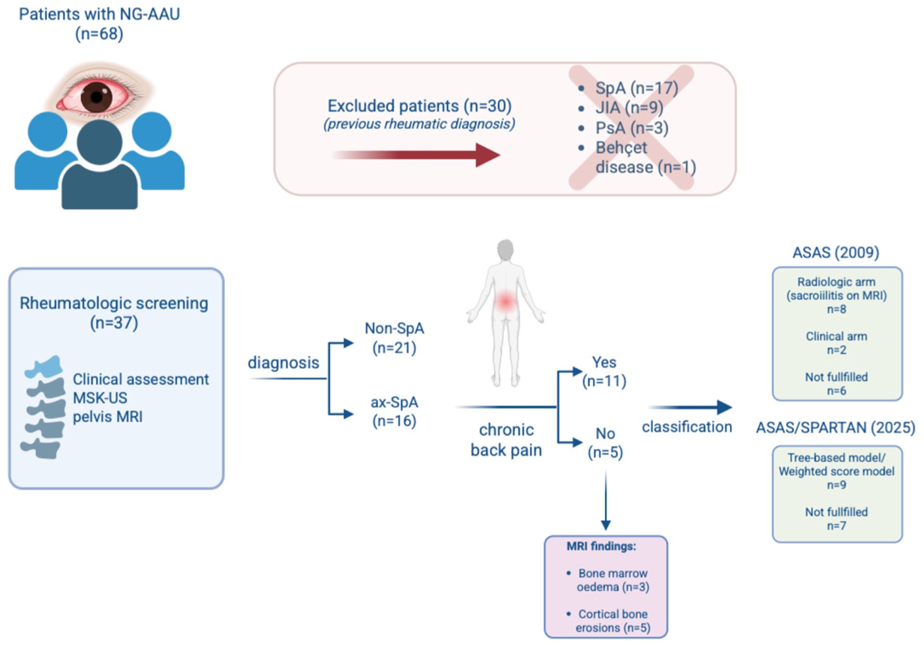
Flow diagram of patient selection, diagnostic assessment, and classification outcomes. Diagnosis was established based on expert clinical judgment. Patients with a new diagnosis of ax-SpA were subsequently classified according to the 2009 ASAS classification criteria and the recently proposed 2025 ASAS/SPARTAN criteria.
No significant differences in age, BMI, sex, or smoking status were observed between the ax-SpA and non-SpA groups. Comorbidities such as PsO and IBD were similarly distributed, while a positive family history of SpA was reported exclusively in the ax-SpA group (18.8%). HLA-B27 positivity was more frequent in ax-SpA patients than in non-SpA patients group (56% vs 33.3%). None of the patients were receiving conventional or biologic disease-modifying antirheumatic drugs, and only 4 patients (10.8%) were on systemic glucocorticoids prescribed by an ophthalmologist, with a mean prednisone dose of 7.5 mg/day.
The mean age at uveitis onset was similar between the two groups (40.6 vs 40.4 years). No significant differences were observed in ophthalmologic features, including disease duration, recurrence, laterality, or posterior synechiae. Uveitis was most often recurrent and unilateral or alternating in both groups. Only 11 patients (28.9%) presented with a first episode of uveitis, of whom 45.4% were diagnosed with ax-SpA. Regarding articular symptoms, all patients diagnosed with ax-SpA reported a history of CBP, showing inflammatory characteristics with an onset before 45 years in 93.8% of cases (p < 0.001). However, at the time of rheumatologic evaluation, only 68.7% of ax-Spa patients reported CBP, IBP was present in 56.2%, and a BASDAI score > 4 was observed in 33.3%. Despite this, mean BASDAI values were significantly higher in ax-SpA patients compared to non-SpA patients (3.3 ± 2.0 vs 1.3 ± 1.9, p = 0.005), and the mean duration of axial symptoms was longer in ax-SpA patients (140.3 ± 122.4 vs 67.7 ± 49.1 months, p = 0.041). Previous episodes of peripheral arthritis or dactylitis were reported in three ax-SpA patients (18.8%), and only one patient had active arthritis at the time of evaluation, confirmed by US.
On SIJ MRI evaluation, active sacroiliitis was detected in 10 newly diagnosed ax-SpA patients (62.5%, p < 0.004). Erosions, one of the most specific structural lesions, were observed in 12 patients (75%, p < 0.001) with ax-SpA. Other structural changes, including subchondral sclerosis and fat metaplasia, were detected in both groups but were significantly more prevalent in ax-SpA patients (62.5% vs 19.0%, p = 0.015; and 56.2% vs 4.8%, p < 0.001, respectively), while no evidence of backfill, ankylosis, or bone buds was observed. Among non-SpA patients, BMO was detected in three patients but without the typical localization or extent indicative of sacroiliitis. 17 Notably, five ax-SpA patients showed only erosions without inflammatory edema on MRI; these were the same individuals who did not report current IBP at the time of rheumatologic evaluation. Further details on MRI findings are provided in Table 1.
US examination did not reveal significant differences between the two groups in detecting active enthesitis. Chronic structural changes at the entheseal level (e.g., enthesophytes, calcifications, and thickening of the tendon insertion without PD signal) were more prevalent in ax-SpA patients (p = 0.023). Bony cortex erosions at the entheses insertion were observed in four (25%) Spa patients versus one (4.8%) non-SpA patients, suggesting higher specificity of this finding.
Only patients with current CBP at the rheumatologic evaluation (n = 11, 68.7%) fulfilled the entry criterion of the 2009 ASAS classification criteria for ax-SpA (Figure 1). 13 Among these, eight patients showed sacroiliitis on MRI. Two patients, despite the absence of active sacroiliitis on MRI, were classified via the clinical arm due to HLA-B27 positivity and other SpA features, while one patient could not be classified (absence of active sacroiliitis on MRI in the presence of SIJ erosions, and HLA-B27 negativity). The remaining five patients without CBP were also unclassifiable according to ASAS criteria. No patients were classified as p-SpA. 18 When applying the recently proposed ASAS/SPARTAN (Spondyloarthritis Research and Treatment Network) 2025 revised classification criteria for ax-SpA, 19 all newly diagnosed ax-SpA patients (n = 16) fulfilled the entry criterion. Among these, 9 patients (56.2%) were classifiable according to either the tree-based model or the weighted score model, whereas 7 patients did not reach the minimum score required for classification.
Discussion
To our knowledge, this monocentric study is the first to assess a consecutive series of NG-AAU patients using both MRI and US. Notably, another distinctive feature of our cohort is the high prevalence of recurrent uveitis (68%), a subgroup more strongly associated with underlying ax-SpA.3,4
In line with this, more than 40% of patients with NG-AAU in our study had previously undiagnosed SpA (43.2%). Considering all NG-AAU patients assessed in our uveitis clinic, nearly half of the cohort had SpA (49.2%). This aligns with estimates from larger studies reporting a prevalence of approximately 50%, supporting the robustness of our findings.3,7,8,20 In the SENTINEL study, a large multicenter cohort of AAU patients, 50.2% met the ASAS criteria for ax-SpA. HLA-B27-positive AAU patients were more frequently diagnosed with SpA and more often reported IBP (p < 0.0001). 8 Similarly, in a recent cohort, 56% of AAU patients undergoing systematic SIJ MRI had concomitant ax-SpA (70% newly diagnosed), with 93% HLA-B27 positivity. 7 In the study by Haroon and colleagues, after excluding patients with a prior SpA diagnosis, the proportion of undiagnosed SpA was comparable to our cohort (41.6%), with 90% HLA-B27 positivity. 20 By contrast, in our study, HLA-B27 prevalence among ax-SpA patients was lower (56.3%) and showed no clear correlation with IBP. The prevalence of HLA-B27 in the Italian general population is relatively low (approximately 2%). 21 More importantly, the strength of the association between HLA-B27 and ax-SpA appears to be weaker in Italy compared with Northern European countries. In support of this, a large Italian series of patients with ax-SpA fulfilling the ASAS 2009 classification criteria reported HLA-B27 positivity in 40.9% of cases. 22 Taken together, these data may help explain the relatively low prevalence of HLA-B27 positivity observed in our cohort of patients with newly diagnosed ax-SpA and uveitis.
Another relevant observation concerns the clinical phenotype of uveitis patients with undiagnosed ax-SpA. In our series, not all newly diagnosed patients exhibited the classic chronic IBP phenotype, observed in only 56.3% of cases. This may be related to the lower prevalence of HLA-B27 positivity compared with the cited cohorts, where ASAS classification criteria have been used for diagnosis.8,23 In the remaining patients (43.7%), back pain, if present, was intermittent, short-lasting, and mild, which may explain the relatively low mean value of BASDAI score (3.3 ± 2.0 SD) and, importantly, the considerable diagnostic delay. Nevertheless, in nearly all undiagnosed ax-SpA patients (93.8%), back pain onset occurred before age 45, with a significantly longer mean duration of symptoms compared to non-SpA patients with back pain (11.7 ± 10.2 vs 5.6 ± 4.1 years, p = 0.041). This delay exceeds the average diagnostic delay reported in the literature, which ranges from 2 to 8 years. 10
These findings suggest that most patients, particularly those with atypical IBP, might not have received a SpA diagnosis if not for the occurrence of uveitis. Since delayed diagnosis is associated with poorer outcomes, greater efforts are needed to achieve an early diagnosis of ax-SpA. 24
Furthermore, when applying the recently proposed ASAS/SPARTAN 2025 revised classification criteria for ax-SpA, 19 a proportion of patients remained unclassifiable compared with the clinical expert diagnosis. This highlights the persistence of a subset of patients with clinically diagnosed ax-SpA who fall outside current classification criteria sets.
This study highlights the importance of close collaboration between rheumatologists and ophthalmologists for careful screening of patients with NG-AAU, especially those who are HLA-B27 positive.
Among the strengths of this study, the monocentric design ensured homogeneous rheumatologic and ophthalmologic evaluation. Another relevant aspect is the referral from a tertiary ophthalmologic center with expertise in ocular immunology. Importantly, only non-granulomatous forms of AAU, which are more strongly associated with SpA than the granulomatous form, were included, a distinction rarely made in previous studies.
Finally, the combined use of MRI and US enabled a comprehensive assessment of both axial and peripheral articular manifestations. In particular, US as a non-invasive and easily accessible tool in routine outpatient settings, may help to detect enthesitis or arthritis and support the diagnostic process, especially when axial symptoms and MRI findings are subtle or equivocal. In our cohort, no significant differences were observed between groups in the prevalence of active enthesitis; however structural entheseal changes, such as erosions, were significantly more frequent in patients with ax-SpA. Larger studies are needed to validate these findings and further define the role of US in this clinical context.
Limitations
Several limitations should be acknowledged. The absence of a priori sample size calculation and the relatively small sample size may reduce the statistical power of the analysis, although it remains considerable for a monocentric study of uveitis patients. The cross-sectional design precluded longitudinal follow-up, thereby limiting the evaluation of long-term progression or the cumulative incidence of SpA. Another limitation of this study is that all MRI scans were reviewed by a single expert rheumatologist without blinded radiologist assessment, which may have limited the diagnostic accuracy of imaging interpretation. However, this approach reflects routine clinical practice in many rheumatology settings, where MRI evaluation is often performed directly by rheumatologists without real-time multidisciplinary review. A further potential limitation of this study is that US findings may have influenced the attribution of a diagnosis of ax-SpA in some cases. This could have contributed to the relatively low rate of HLA-B27 positivity observed in our cohort of patients with newly diagnosed ax-SpA and uveitis.
In addition, newly diagnosed SpA included some patients not fulfilling ASAS classification criteria, a decision that may reduce cohort homogeneity but was made to prioritize sensitivity over specificity. This approach reflects real-world clinical practice, allowing identification of atypical phenotypes, such as patients with non-classical IBP (e.g., mild, intermittent) who nonetheless show disease progression on imaging (e.g., bone erosions) and potential ocular damage (recurrent uveitis). It provides a novel perspective on the disease and highlights a subgroup of pauci-symptomatic patients at high risk of diagnostic delay.
Conclusion
This study highlights NG-AAU as an important clinical window for identifying previously undiagnosed ax-SpA, particularly in patients with mild, atypical, or intermittent axial symptoms. In this context, an interdisciplinary approach involving close, structured collaboration between ophthalmologists and rheumatologists is essential to enable earlier diagnosis and reduce diagnostic delay.
Supplemental Material
sj-docx-1-tab-10.1177_1759720X261449924 – Supplemental material for Recurrent uveitis unveiling axial spondyloarthritis: is inflammatory back pain always the main road to diagnosis? A monocentric cohort
Supplemental material, sj-docx-1-tab-10.1177_1759720X261449924 for Recurrent uveitis unveiling axial spondyloarthritis: is inflammatory back pain always the main road to diagnosis? A monocentric cohort by Alessandra Rai, Filippo Crescentini, Giorgia Citriniti, Marianna Oliva, Federica Macaluso, Pierluigi Macchioni, Luca Cimino, Elena Bolletta, Fabrizio Gozzi, Pietro Gentile, Luca De Simone, Carlo Salvarani and Niccolò Possemato in Therapeutic Advances in Musculoskeletal Disease
Footnotes

References
Supplementary Material
Please find the following supplemental material available below.
For Open Access articles published under a Creative Commons License, all supplemental material carries the same license as the article it is associated with.
For non-Open Access articles published, all supplemental material carries a non-exclusive license, and permission requests for re-use of supplemental material or any part of supplemental material shall be sent directly to the copyright owner as specified in the copyright notice associated with the article.
