
Abstract
Background
miRNAs are of interest due to have been involved in cancers such as colorectal cancer (CRC).
Objective
To explore the expression levels of miRNA-34a and miRNA-135b and whether they can be potential biomarker for early diagnosis of CRC.
Methods
Fifteen pairs newly diagnosed colorectal cancer and normal involved in the measurement of tissue and serum levels of miRNAs 34a and 135b and their target genes of SIRT1 and DAPK-1. We assessed the predictive value of these miRNAs utilizing the receiver operating characteristic curve.
Results
The findings have shown that the miRNA-34a expression was significantly downregulated and miRNA-135b was upregulated in CRC tissues in comparison to their expression in adjacent normal tissue. This was additionally observed in the serum of the same CRC cases when compared to individuals without CRC. Furthermore, we found a significant increase in the level of SIRT-1 and a significant reduction in the expression level of DAPK-1 in CRC tissues compared to the adjacent normal tissue. The value of AUC was considerably high for the timely detection of CRC.
Conclusions
We found these miRNAs acting as potential non-invasive biomarkers for the early detection of CRC and in assessing the CRC risk within the general population.
Introduction
Colorectal cancer (CRC) is one of the most prevalent malignant tumors, developing gradually from aberrant crypt foci through benign adenomas to malignant adenocarcinomas.1,2 Despite treatment advances—including surgery, chemotherapy, targeted therapies, immunotherapy, and radiation—the prognosis heavily depends on early diagnosis. 3 The 5-year survival rate drops sharply from approximately 90% in early stages (I/II) to about 14% in advanced stages (IVB/IVC).4,5 Although screening methods such as colonoscopy and fecal or plasma-based assays exist, their widespread use is limited by high costs and low patient compliance. Therefore, identifying novel, non-invasive biomarkers for early CRC detection remains critical.6,7 MicroRNAs (miRNAs)—small, endogenous, non-coding RNAs of 18–25 nucleotides—regulate gene expression by suppressing target messenger RNAs,8,9 and are emerging as promising biomarkers detectable in body fluids like plasma and feces. 10 Many miRNAs show dysregulated expression in various cancers, including CRC, making them attractive for early diagnosis and therapeutic targeting.11,12 Thus, the direct inhibition of miRNAs could act as a therapeutic strategy to counteract the series of events that lead to oncogenesis, potentially enhancing treatment effectiveness in patients who are resistant to chemotherapy. 13 Among these, miR-34a acts as a tumor suppressor by promoting apoptosis and cell cycle arrest through regulation of genes such as sirtuin 1 (Sirt1), which supports oncogenic processes.14,15 Sirt1 plays a significant role in cellular survival mechanisms. Consequently, it is regarded as a promoter of oncogenes and may participate in the regulation of tumor development and initiation, as well as various developmental processes. 16 Conversely, miR-135b, frequently upregulated from normal tissue to carcinoma, may contribute to early dysplasia by negatively affecting genes like death-associated protein kinase 1 (DAPK1), a key tumor suppressor involved in apoptosis and metastasis suppression.17,18 Increased expression of miR-135b has been presented across multiple types of cancer.19,20 DAPK1 is a serine/threonine protein kinase that promotes apoptosis and is recognized as a tumor suppressor gene, its regulation is often disrupted in various cancer types. DAPK1 expression suppresses metastasis through the promotion of programmed cell death. 21 However, multiple studies have indicated that microRNAs possess distinct advantages in the diagnosis of tumors and are a primary area of focus in the research of biomarkers for CRC. 22 Given their significant roles, this study evaluates the expression of miR-34a and miR-135b in CRC tissues and serum, aiming to assess their potential as early diagnostic biomarkers.
Material and methods
Ethics statement
All participants signed written informed consent prior to being enrolled in the study. This project was approved by the Research Ethics Committee of Zanjan University of Medical Sciences (IR.ZUMS.REC.1402.229).
Study design and populations
The characteristics of the study population, both demographic and clinical data are displayed in Table 1. The study participants were recruited from two hospitals in Zanjan city, Zanjan, Iran, from April to September 2023. All participants were at least 36 and at most 85 years old and they have resided in their present home for over a decade. Both of sex (male and female) were involved in this study. Moreover, stage of tumor ranged between I to III. Every new case of CRC was diagnosed through pathological examination following a colonoscopy. Healthy controls were selected at random from individuals who were initially free of cancer and did not develop the disease thereafter. Normal controls were selected to match the age and sex of the incident cases and 5 ml venous blood sample was obtained from each of them. When CRC of patients confirmed by expert physician and agreed to take part in this study, during surgery, for each patient, a 5-mL venous blood sample was drawn into a single tube, permitted to clot for 30 min at room temperature, and subsequently centrifuged at 10,000 g for 10 min to obtain the serum sample. Then, the tumor tissue samples and the surrounding normal tissue were obtained from the marginal areas. The samples were subsequently stored in a refrigerator at a temperature range of 0 to 4 °C within the hospital and were transported to a biochemistry laboratory within a four-hour timeframe at the Zanjan University of Medical Sciences, and storing at −80°C for further laboratory tests designated in this study. The tissue miR-34a, tissue miR-135b, serum miR-34a and serum miR-135b expression levels were measured using kit. In the following, the expression levels of target genes of SIRT-1 and DAPK-1 were calculated. Moreover, the biochemistry parameters of triglyceride (TG), total cholesterol (TC) and low-density lipoprotein cholesterol (LDL-C), high-density lipoprotein cholesterol (HDL-C) levels were obtained from information presented in the questionnaire.
Primary characteristics of colorectal cancer patients and healthy controls.
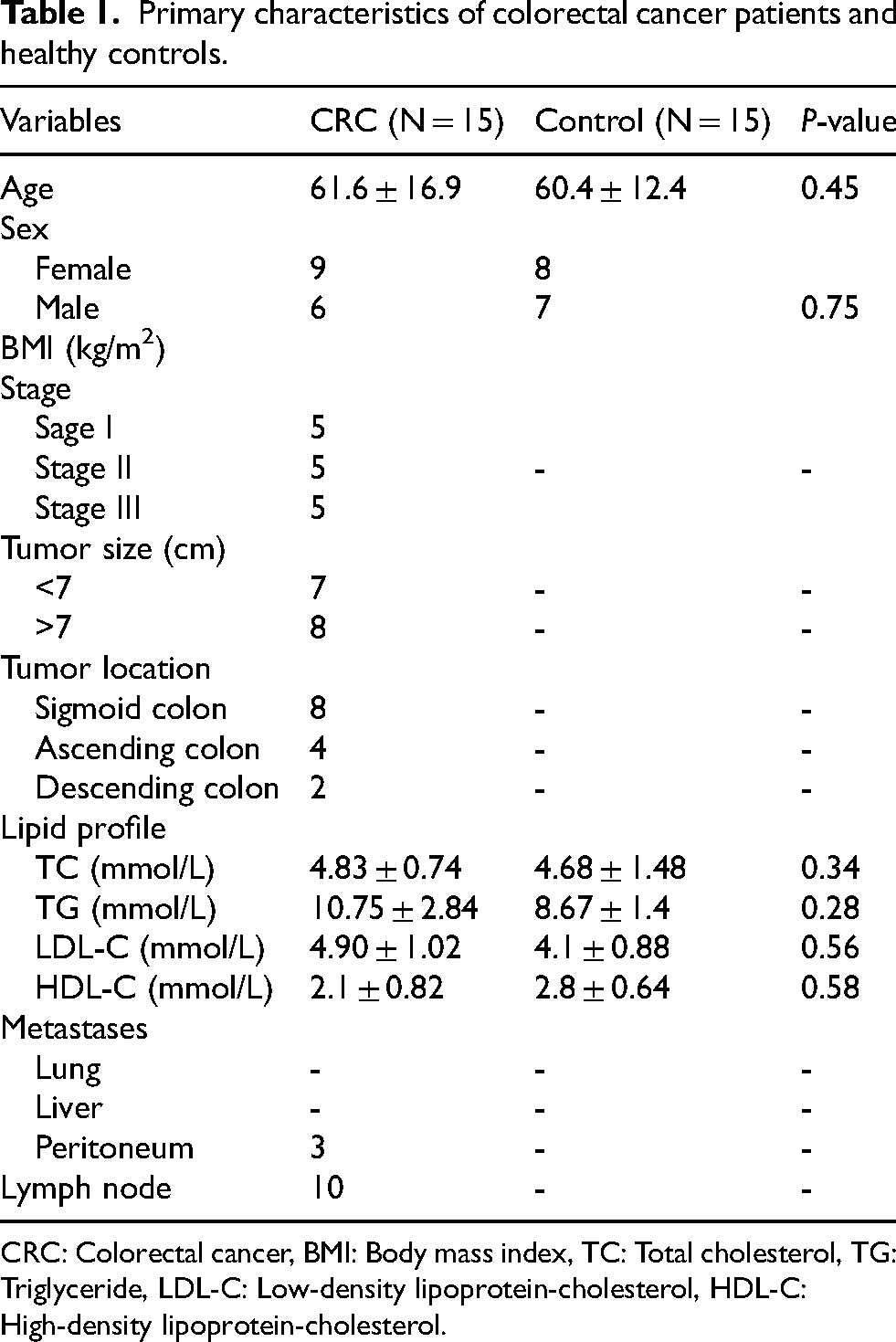
CRC: Colorectal cancer, BMI: Body mass index, TC: Total cholesterol, TG: Triglyceride, LDL-C: Low-density lipoprotein-cholesterol, HDL-C: High-density lipoprotein-cholesterol.
Selection of candidate miRNAs and their corresponding target genes
We preferred miRNAs and their target genes that indicate a dysregulation among CRC patients and individuals without CRC according to the following criteria: (1) at least 2 studies presented the identical dysregulation orientations (increase or decrease in expression); (2) the plenty of dysregulated orientations in serum/plasma and tissue miRNAs; and (3) the miR- colon cancer and target mRNA-colon cancer interaction were verified in more than 2 databases. Consequently, miR-34a with the specific target gene of SIRT-1 and miR-135b with target gene of DAPK-1 were detected as candidates for conducting a more in-depth analysis.
RNA extraction and qRT-PCR analysis
The extraction of total RNA was conducted from CRC tissue and serum using Trizol reagent (RNA extraction kit, Pars Tous kit) in accordance with the manufacturer's guidelines. To manage the variability in extracted RNA from the serum samples, miR-16 was incorporated into each sample. RNA was isolated utilizing the FAVORGEN miRNA Isolation Kit (FAMIK002) based on the manufacturer's instructions. Extracted total RNA was utilized as template for cDNA synthesis using the cDNA synthesis kit (Yekta Tajhiz YT4500). The analysis of gene expression was conducted through quantitative reverse transcription polymerase chain reaction (qRT-PCR) utilizing SYBR Green RealQ Plus 2x Master Mix Green (Ampliqon) on a StepOnePlus Real-Time PCR System (Applied Biosystems, Foster City, CA). The expression data was normalized using β-actin and U6 as reference genes. The expression level of each miRNA in the sample was determined using the formula 2−Δ CT. All reactions were performed in duplicate. The primer pairs utilized in qRT-PCR are presented in Table 2. The coefficients of variation (CVs) and interassay SDs of the CT values of duplicate samples were determined.
Primer sequences used in the study.

Statistical analysis
The results are expressed as mean ± standard deviation (SD). The difference between the expression levels of two groups were analyzed using student's t-test. The relationships between miRNAs and the risk of CRC were assessed using logistic regression, with adjustments made for age and sex. Linear regression analysis was utilized to identify the correlation between the expression of miRNAs and their target genes. Therefore, correlation analysis was conducted with Pearson's test. Receiver operating characteristic (ROC) curves were developed, and the area under the ROC curve (AUC) was estimated to evaluate the separate impact of candidate miRNAs as potential biomarker. P values were two-sided, and P < 0.05 showing significant difference. All of the statistical analyses were conducted using SPSS 24 (SPSS, Chicago, IL, USA) and GraphPad prism (Version 8).
Results
The demographic information of age, sex and stage of CRC were not statistically significant between patients diagnosed with colorectal cancer and their matched control group. The levels of miRNA-34a and miRNA-135b expression was measured in tissue and serum of colorectal patients compared to health control. The findings indicated that the expression levels of tissue and serum miR-34a were significantly reduced in patients with CRC compared to matched control, while, tissue and serum expression levels of miR-135b was sufficiently higher in CRC patients compared to control (Figure 1). In addition, the analysis of logistic regression indicated that miRNA-34a and miRNA-135b were considerably associated with CRC risk in patients with CRC compared to individuals without CRC (Table 3). In the following, we also studied the expression levels of SIRT-1 and DAPK-1 as specific target genes of miRNA-34a and miRNA-135b respectively, in the tissue sample of patients with CRC relative to control without CRC. As shown in Figure 2, an increase in SIRT-1 expression was observed in patients with CRC compared with control group. While, the expression of DAPK1 in patients with CRC was significantly lower than those without CRC.

The expression analysis of miR-34a and miR-135b in CRC tissue and serum samples compared to the individuals without CRC. Statistical testing was done by student t-test (mean ± SD). *P < 0.05, **P < 0.01 and ***P < 0.001 in comparison with normal control.
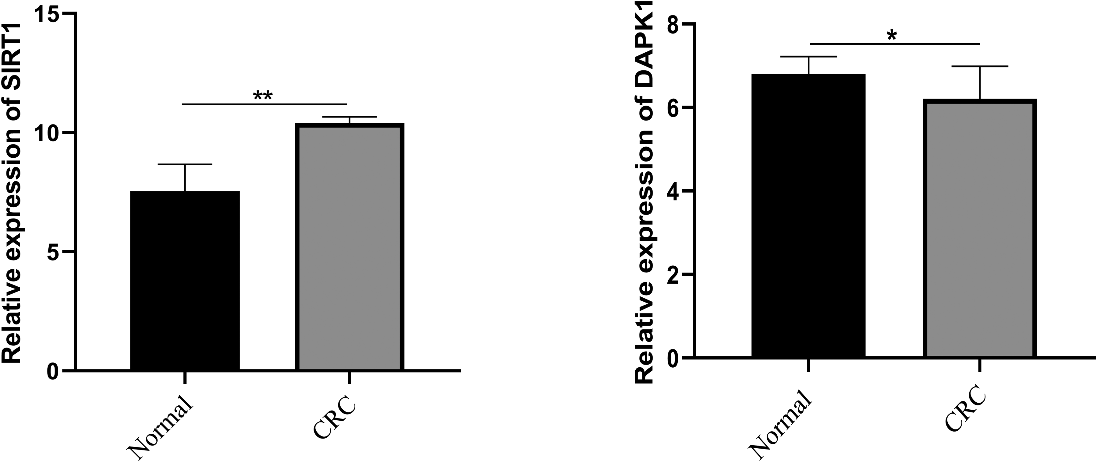
The mRNA expression of SIRT1 and DAPK1 in CRC and adjacent normal tissue. Statistical analysis was done by student t-test (mean ± SD). *P < 0.05 and **P < 0.01 in comparison with adjacent normal tissue.
The association between different miRNAs (miR-34a and miR-135b) and the risk of colorectal cancer.
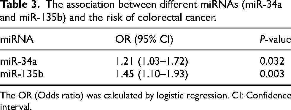
The OR (Odds ratio) was calculated by logistic regression. CI: Confidence interval.
The analysis of correlation was conducted based on Pearson's test. The results showed a significant inverse correlation between the expression of miR-34a and miR-135b with their specific target genes of SIRT1 and DAPK1 in patients with CRC (Figure 3).
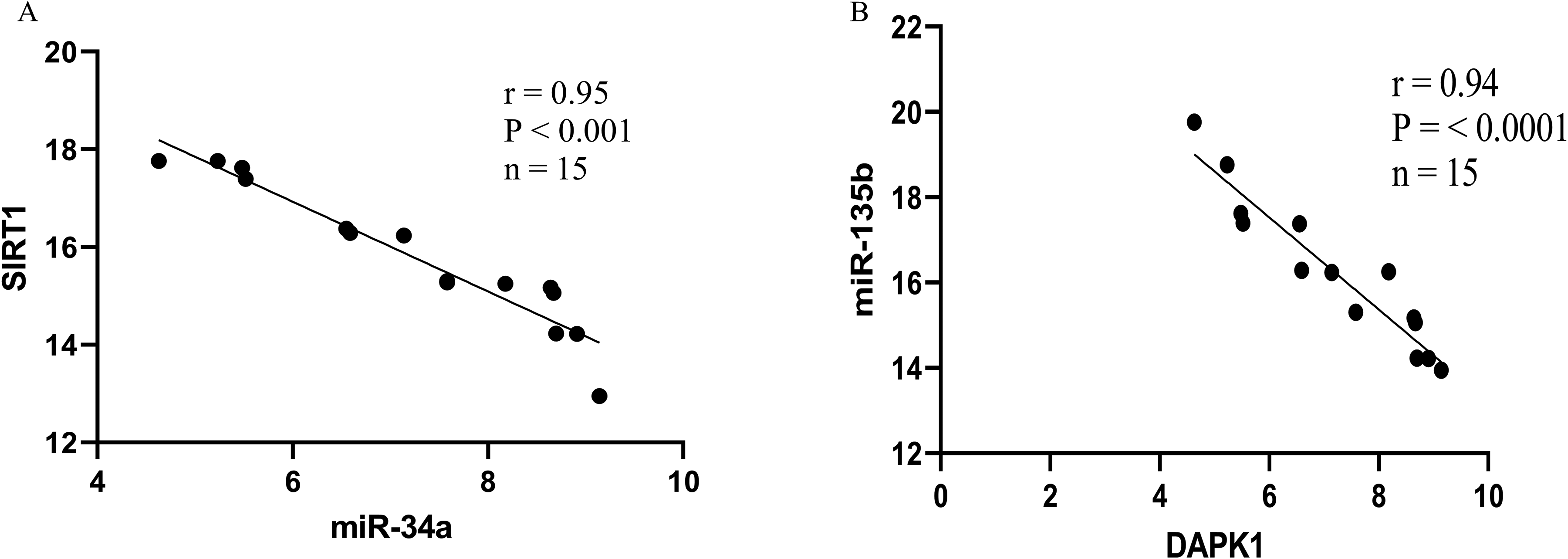
The analysis of regression of the expression of miR-34a and miR-135b and target genes of SIRT1 and DAPK1 in CRC tissue.
To find the diagnostic value of miRNAs in the identification of CRC patients from individuals without CRC as control, we employed the ROC curve to evaluate the levels of cut-off point, sensitivity, and specificity of the two previously mentioned miRNAs. The cut-off value was established as the point at which the combined sensitivity and specificity attained their maximum level. The cut-off value for miRNA-34a was 6.75 (sensitivity = 0.68, specificity = 0.70; for miRNA-135b, 9.45 (sensitivity = 1, specificity = 1). Besides, The AUC value of the miR-34a in the tissue sample of CRC was 0.90 ± 0.054, P = 0.002, and for miR-135b equal to 1 ± 0.00, P < 0.0001, recommend that the model including the introduced miRNAs can substantially enhance the diagnostic accuracy of the disease. Detailed of the ROC curve and AUC are shown in Figure 4.
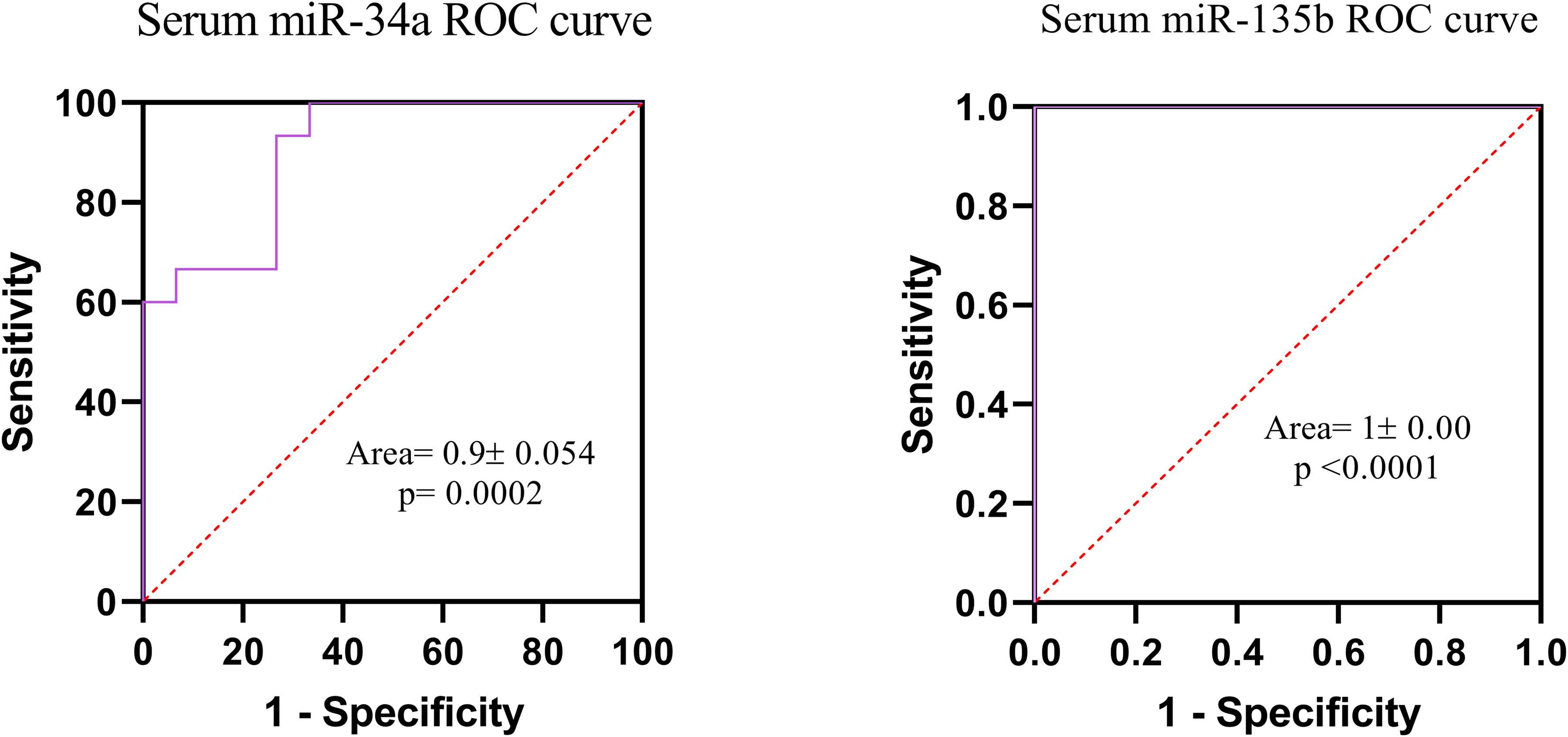
ROC curve of miR-34a and miR-135b in serum sample of patients with CRC.
Discussion
The present research evaluated the predictive significance of miRNAs (miR-34a and miR-135b) concerning the occurrence of colorectal cancer (CRC) within a population affected by the disease. Two miRNAs (miRN-34a and miRNA-135b) were significantly associated with to the risk of developing incident CRC. The sensitivity of the presented miRNAs in this study varied from 0.90 to 1. Our research provides population-level evidence indicating that miRNA panels may function as promising biomarkers for colorectal cancer (CRC). The study's case group consists of newly diagnosed colorectal cancer patients, candidate for surgery from the general population, who are paired with a control group that has no history of cancer.
Our findings revealed that the reduction of miR-34a level and elevated level of miR-135b in tissue and serum have a considerable detection precision for CRC. Additionally, the expression level of SIRT1 as specific target gene of miR-34a was sufficiently higher in tissue sample of patients with CRC compared to healthy control. In accordance with our findings, many studies reported that the expression of miR-34a were remarkably reduced in CRC in comparison with their levels in normal tissues and also miR-34a suppress both of the migration and invasion of colon cancer cell through targeting some of genes including fra-1.15,23 Zhang et al. demonstrated that the expression values of miR-34a in CRC cell lines and human samples (tissue or serum) was reduced in comparison to normal tissues, and the expression levels of miR-34a were found to be negatively correlated with metastasis of tumor. Besides, it was also reported that the increased survival rate was linked to elevated levels of miR-34a expression. 15 Therefore, miR-34a serves as a potent tumor suppressor gene that is regulated by the tumor suppressor protein p53 and directly leads to inhibition of migration, invasion and metastasis of several cancers such as CRC.24,25 In this study, we observed that the SIRT-1 expression significantly elevated in CRC tissue in comparison with normal tissue. For this reason, the findings of the research conducted by Meng et al. have indicated that miR-34a overexpression results in immunosuppression in CRC via the inhibition of SIRT1, which subsequently activates the NF-κB/B7-H3/TNF-α pathway. 16 The main benefit of therapy utilizing miR-34a lies in its ability to concurrently and comprehensively inhibit multiple targets associated with apoptosis, including BCL2, SIRT1, and BIRC5.26,27 miR-34a has been identified as an critical modulator of the immune system response in cancerous conditions, thus, there is a correlation between miR-34a and immune response through SIRT1/NF-κB signaling pathway.28,29 Additionally, multiple studies have indicated that SIRT1 typically suppresses NF-κB signaling through the deacetylation of the NF-κB subunit p65. 30 because of SIRT1, one of the best target gene is miR-34a and demonstrated that miR-34a enhances immune responses induced by NF-κB through the inhibition of SIRT1.31,32
Another result this study is that we reported the increased level of miR-135b in CRC tissue and serum samples along with downregulation of DAPK1 leads to reduced apoptosis process and enhanced cell proliferation. DAPK1 as a serine/threonine protein kinase promotes apoptosis and also is recognized as a tumor suppressor. The expression of DAPK1 is altered in various forms of cancer such as CRC. It plays a role in inhibiting metastasis by promoting programmed cell death.21,33 Towards this end, to gain a new insight into the selected microRNAs, we followed other miRNAs in some of studies. There is much evidence indicating that the dysregulation of the expression of miRNA has a main impact in the CRC advancement and also its metastasis.9,34 These miRNAs may act as either tumor suppressors or oncogenes, thereby affecting the expression of their corresponding mRNA targets. 35 Given the significant stability of miRNAs, they have been regarded and explored as a novel category of important biosignatures. The study performed by Raonic et al. demonstrated that high miRNA-29a expression associated with better progression-free survival of CRC patients. 36 on the other hand, some studies have shown that miRNA 29a involves in CRC by targeting matrix metalloproteinase 2 and E-cadherin through KLF428. 37 Furthermore, miR-125b was introduced to stimulate cetuximab resistance in CRC. 38 Taken together, these findings suggest that these miRNAs could be potential predictive biomarkers for incident CRC in contrast with healthy control.
Many methods are presented for the diagnosis of CRC such as colonoscopy. Not only colonoscopy is the gold standard in CRC screening but also the frequency of re-screening may be required only once every 10 years, contingent upon the outcomes obtained. 39 Moreover, the diagnostic yield in population-based colonoscopy screening is suboptimal due to the comparatively low rate of participation. 39 Faecal-based detection techniques, including faecal immunochemical tests (FITs), have recently introduced as a promising procedure in the realm of cancer treatment. Despite, FITs are characterized by being less invasive; however, the ability of these methods to specifically identify precancerous colorectal adenomas is limited.40,41 That is why, differently expressed of miRNAs in the early stages of conversion precancerous lesions to cancerous tissues can be able to the prediction in the most of cancers. 42 Moreover, miRNAs exhibit a considerable degree of stability in bio-fluids and can be readily identified in blood. Consequently, they present an opportunity for disease monitoring with non-invasive approaches. 42
Although, the use of two different samples to predicting the risk of colorectal cancer is novel and promising, there were several limitations to this study. Firstly, the sample size was small and the rate of higher stage of disease was low. Secondly, lack of complementary techniques for validation of findings. For example, RIN (RNA integrity number) along with the 260/280 or 260/230 ratios in measurement of RNA quality. Thirdly, the use of miRNA profile instead of two microRNAs and fourthly, the AUC of our regression model was almost one that showing the small sample size in our study. Given the limitations, the results of the present study should be interpreted with caution.
Conclusion
The present study revealed that the miR-34a and miR-135b provide a predictive ability with the high sensitivity for the prompt identification of colorectal cancer. The importance of miRNAs in cancer pathogenesis as potential targets, through regulation of their target genes suggests further investigations on their role in diagnosis and prognosis.
Footnotes
Abbreviations
Acknowledgment
We thank the Deputy of Research, Zanjan University of Medical Sciences that supported this study.
Ethics approval and consent to participate
This study was approved by the Research Ethics Committee of Zanjan University of Medical Sciences (IR.ZUMS.REC.1402.229). All participants signed written informed consent prior to being enrolled in the study.
Consent for publication
All the authors have consented to the publication of this paper.
Authors’ contributions
H. Kh and M. K contributed to the conception, and design and critically reviewed and approved the final manuscript as submitted. R. K, E. N, N. AD and B. M contributed to experimental work, data collection, statistical analyses, and interpretation and wrote the paper draft. A. B and E. Sh contributed to collection of tissue and serum samples from colorectal patients and healthy control. A. Sh as a General surgeon in the hospital contributed to the delivery samples. All authors approved the final version for submission.
Funding
The authors disclosed receipt of the following financial support for the research, authorship, and/or publication of this article: The present study was financially supported by grants (A-12-1636-4 and A-12-1320-5) from the Deputy of Research, Zanjan University of Medical Sciences.
Declaration of conflicting interests
The authors declared no potential conflicts of interest with respect to the research, authorship, and/or publication of this article.
Data availability
The data associated with this project are presented within the article and row data can obtain through contacting with corresponding author.