






Abstract
Background
The association between Epstein-Barr virus (EBV) and lung cancer risk remains controversial. Here, we used a two-sample Mendelian randomization (MR) analysis to test the causal relationship between EBV and lung cancer.
Methods
Data regarding lung cancer (outcomes) were collected from the Finnish database; the Genome-Wide Association Study (GWAS) summary-level dataset for EBV was obtained from the Open Forum Infect Dis. The inverse-variance weighted (IVW) method was used as a primary analytical approach; weighted median, MR-Egger, and weighted mode methods were used to ensure the robustness of the data. The MR-Egger regression assessed horizontal pleiotropy, and the MR pleiotropy residual sum and outlier (MR-PRESSO) method identified potential outliers. Cochran’s Q test evaluated heterogeneity among instrumental variables (IVs).
Results
IVW analysis indicated several significant causal effects. Genetically elevated levels of EBV ZEBRA and EBNA-1 antibodies increased the risk of SQC (OR=1.26 and OR=1.33, respectively). Increased EBNA-1 antibodies also raised the risk of overall lung cancer and small cell lung cancer. Conversely, higher VCA p18 antibody levels were associated with a decreased risk of lung adenocarcinoma (OR=0.70). Sensitivity analyses suggested these findings were robust, with no significant evidence of horizontal pleiotropy or heterogeneity.
Conclusion
Our data suggests a causal effect between EBV and the progression of lung cancer.
Introduction
Lung cancer (LC) is the leading cause of cancer deaths globally. 1 LC is characterized by significant pathological heterogeneity and is commonly classified into small-cell lung cancer (SCLC) and non-small-cell lung cancer (NSCLC). 2 SCLC has a poor prognosis due to its highly invasive and neuroendocrine cell-derived characteristics and poor prognosis, 3 whereas NSCLC predominates and encompasses three main subtypes: squamous cell carcinoma (SQC), adenocarcinoma (AdC) and large cell carcinoma (LCC). 4 Although advances in treatment strategies have been made, the treatment of SCLC and NSCLC remains challenging. The prognosis remains distressingly poor, with a low 5-year survival rate. 5
In addition to the well-known tobacco smoke as a major risk factor, air pollution, genetic susceptibility, specific occupational exposures, and certain viral infections are also strongly associated with the development of lung cancer, such as the high-risk human papillomavirus (HPV), Merkel cell polyomavirus (MCPyV), and EBV.6,7 EBV belongs to the group of enveloped gamma herpesviruses. EBV first invades cells within the Waldeyer’s ring structure and then infects immature B cells, prompting latent infection and transformation into memory B cells, which then disseminate throughout the systemic circulatory system. 8 Globally, EBV infection is highly prevalent, with the majority of people infected in childhood or adolescence entering a long-term latent state; the vast majority of infected individuals are asymptomatic. A meta-analysis integrating data on 886 lung cancer patients from 14 studies revealed that EBV infection was significantly associated with a more than fourfold increased risk of lung cancer, especially the rarer pulmonary lymphoepithelioma-like carcinoma subtype in non-small cell lung cancer. 9 Numerous studies have also confirmed the presence of EBV in lung cancer through diverse detection techniques. For example, a study in Spain used the Epstein-Barr encoding region in situ hybridization (EBER ISH) technique to detect 19 cases of AdC and SQC, of which 12 (63.2%) were positive. 10 In contrast, in an Italian study, EBV was detected in 26% (i.e.,21/65 cases) of breath condensate samples from 65 patients with NSCLC, as well as in 20% (i.e.,1/5 cases) of 5 patients with SCLC, as detected by real-time PCR. 11 Some studies, however, did not detect EBV by ISH in lung AdC and mesothelioma samples. 12 Also, studies on SCLC did not detect EBV gene expression even after the gold standard assay analysis of EBER-ISH. 13 Therefore, the causality of whether EBV directly causes lung cancer is still insufficiently confirmed and subject to some controversy and uncertainty.
To overcome the limitations of traditional observational studies, such as cross-sectional design, small sample size, insufficient follow-up time, differences in control group selection, and the effects of possible reverse causality and confounding factors, we used MR design to assess the association between EBV infection and lung cancer risk. The MR design utilizes genetic variants as instrumental variables for exposure because these genetic variants are randomly assigned to individuals at embryonic developmental stages and are theoretically independent of other traits (potential confounders or environmental factors), thereby reducing residual confounding effects and minimizing the impact of reverse causation. 14 The aim of this study was to investigate whether EBV infection increases the risk of lung cancer. Based on the existing accumulation of evidence, we hypothesized that there is an association between EBV infection and the development and progression of lung cancer.
Methods
Study design
The MR analysis followed the Strengthening the Reporting of Observational Studies in Epidemiology using Mendelian Randomization (STROBE-MR) guidelines.
15
The analysis adhered to three assumptions of MR studies (Figure 1)
16
: (1) IVs are strongly associated with exposure; (2) selected IVs are not associated with potential confounders; (3) IVs have a dependent, but not directly related, effect on the outcome through exposure (EMDIN C A 2017). Data used in this analysis came from approved studies that obtained informed consent from all participants. Description of the study design in MR study.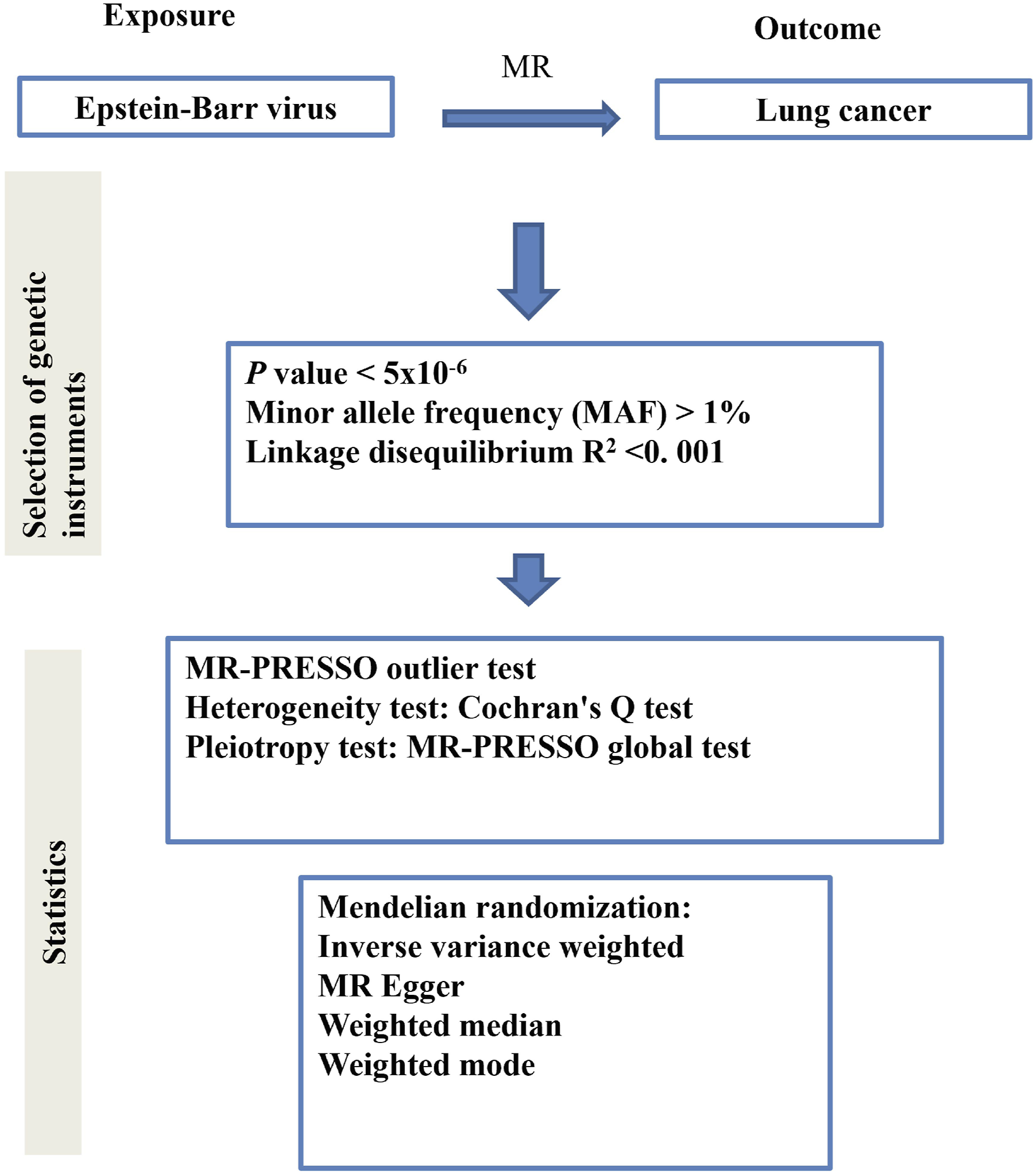
Data sources
Data regarding lung cancer (outcomes) were collected from the Finnish database (see Table S1). The Genome-Wide Association Study (GWAS) summary-level dataset for EBV, including anti-EBV IgG, VCA p18, ZEBRA, EBNA-1, and EA-D antibodies, was obtained from the Open Forum Infect Dis. 17 (see Table S2). The dataset is derived from the UK Biobank cohort of serologic measurements and genome-wide genotyping for up to 10,000 infectious diseases.
IVs selection
We calculated the variation in EBV explained by IVs, and the IVs included in this study were required to meet the following criteria: (i) autosomal duplex sequence SNPs significantly associated with the whole genome of EBV antibody were screened, i.e., meeting the criterion of P < 5 x 10⁻⁶; (ii) SNPs with a minimum allele frequency (MAF) > 0.01 were screened; (iii) SNPs with an R2 < 0.001 and window size = 10,000kb were excluded. (iv) according to the criteria of R2 < 0.001 and window size = 10,000kb, the linkage disequilibrium (LD) effect among SNPs was eliminated; (v) when the screened IV did not exist in the summary data of the ending, SNPs with high LD (R2 > 0.8) with the IV were searched for as surrogate SNPs, which were replaced; (vi) the F value of each SNP in the IV was calculated to assess the IV strength and to exclude possible weak instrumental variable bias between the IV and the exposure factors, which was calculated by the following formula: F = R 2 x(N-2)/(1-R 2 ), with R2 being the proportion of the variance of the exposure that can be explained by SNPs in the IV, and an F value of >10 was considered as a strong genetic IV. 18
MR analysis
We utilized MR analysis in order to determine a non-zero causal relationship between EBV and lung cancer. IVW was used as the primary analytical method in this analysis, and dominance ratios (odds ratios, ORs) and 95% confidence intervals (CIs) were calculated to assess the relationship between EBV and lung cancer. IVW is the predominant method for interpreting MR results and is considered the most robust indicator without evidence of directional multi-directionality among the selected IVs, as it calculates a weighted average of effect sizes by computing the inverse variance of each SNP as weights. 19 The robustness of the results was additionally tested using the MR-Egger, weighted median, and weighted mode methods. 20 The weighted median method assumes that half of the instrumental variables are valid and analyzes the causal association between exposure and outcome.
All analyses in this study were performed using the “TwoSampleMR” package (version R 4.2.3). Scatter plots, forest plots and funnel plots were used for visualization. Leave-one-out analysis (LOO analysis) was used to estimate the effect of the remaining SNPs on the results by sequentially removing individual SNPs and then performing IVW analysis again to determine whether a single SNP drove causality.
Sensitivity analysis
Sensitivity analysis was used to detect potential pleiotropy that may exist in MR studies. In this study, heterogeneity between IVs was assessed by Cochran’s Q test, and heterogeneity was considered low when P > 0.05, i.e., the valuation between instrumental variables varied randomly and exerted little influence on the IVW results. 20 If there was no significant heterogeneity, a fixed-effects model was used; otherwise, a random-effects model was applied. Considering also the influence of pleiotropy of genetic variation on the estimation of the association effect, this study used MR-Egger regression to explore the existence of horizontal pleiotropy, and when the intercept term of MR-Egger regression tends to zero or is not statistically significant, it suggests that there is no pleiotropy, and the exclusion assumption can be regarded as valid. 21 In addition, this study used the MR pleiotropy residual sum and outlier (MR-PRESSO) method to detect possible outliers (i.e.,SNPs with P < 0.05) and re-estimated the causal associations after eliminating them, thus correcting for horizontal pleiotropy. 22 All statistical tests were two-sided and were performed using the TwoSampleMR and MR-PRESSO packages in R software (version 4.2.3).
Results
IVs selection
In our study, when conducting MR analyses with exposure defined by seropositivity for anti-EBV IgG, EBV EA-D antibody, EBV EBNA-1 antibody levels, EBV VCA p18 antibody, ZEBRA antibody levels, EBNA-1 antibody levels, and EA-D antibody levels, we respectively identified 25, 5, 6, 18, and 18 relevant IVs, respectively (the mean F-statistics for these IVs was 22.84, 43.75, 107.62, 27.89, and 49.3, reflecting their overall predictive strengths; the minimum F-statistics was 20.84, 30.06, 31.14, 87.77, and 20.86, while and the maximum values reaching 31.4, 66.74, 342.35, 87.77, and 336.72 respectively. More data are shown in Table S3-S7.
MR data and sensitivity analysis
A causal association between EBV and lung cancer.

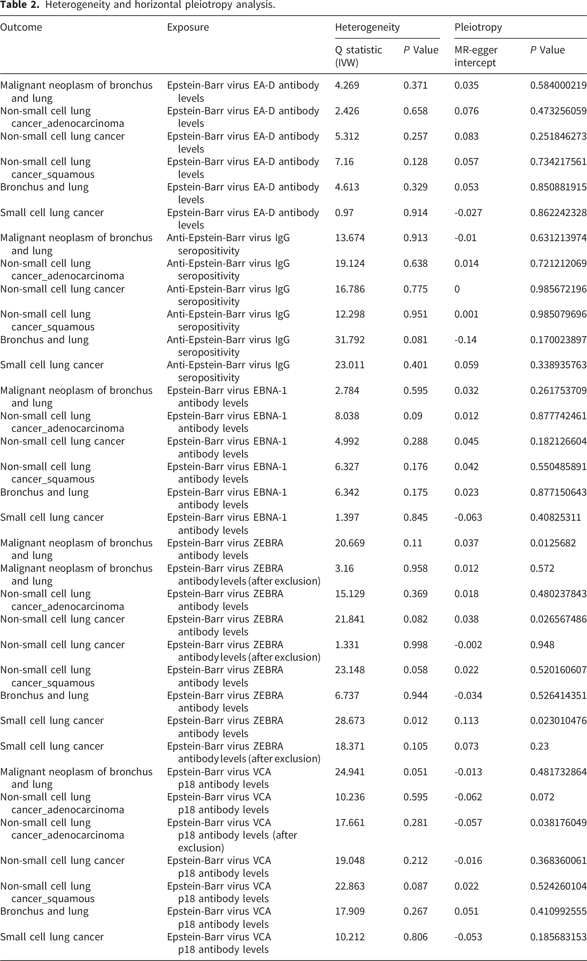
Heterogeneity and horizontal pleiotropy analysis.
MR-PRESSO analysis.
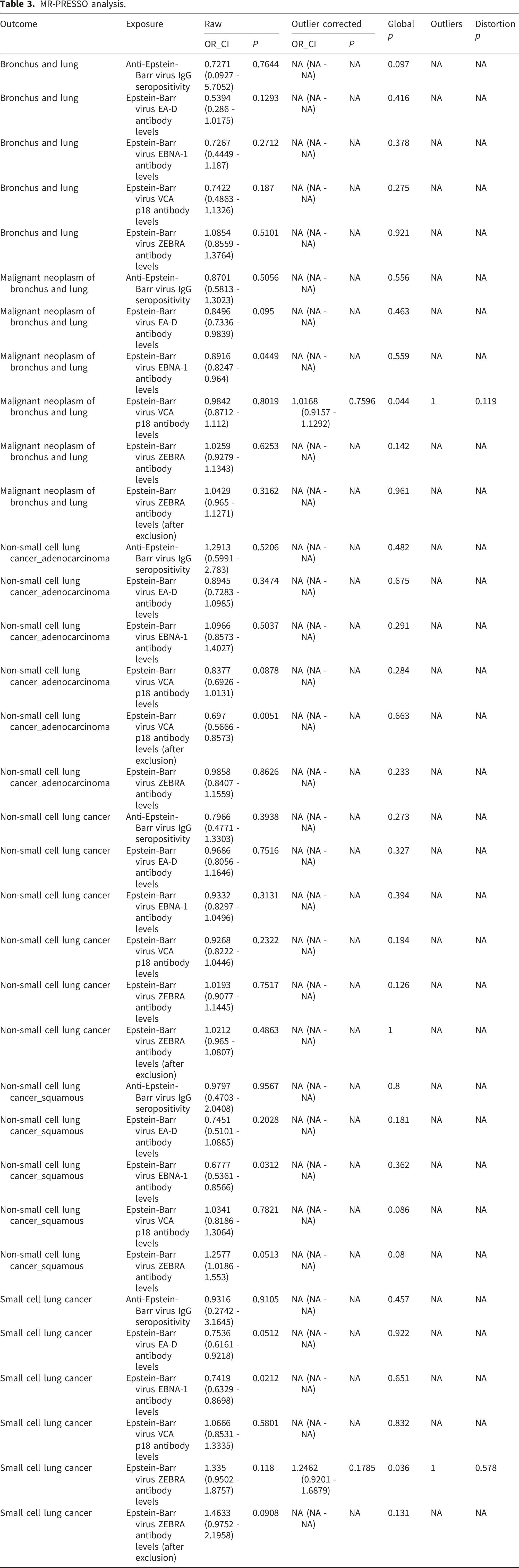
We also found that EBV ZEBRA antibody levels are a risk factor for non-small cell lung cancer (SQC) (OR=1.26, 95% CI=1.02-1.55, P=0.0331, Table 1, Figure S5). MR-Egger regression results and MR-PRESSO analysis confirmed the robustness of these results (Tables 2 and 3). IVW also suggested that EBV ZEBRA antibody levels had no effect on other outcomes (all P>0.05). Yet, MR-Egger regression analysis showed some horizontal pleiotropy for EBV ZEBRA antibody levels and malignant neoplasm of bronchus and lung (P=0.572), non-small cell lung cancer (P=0.026), and small cell lung cancer (P=0.023). However, these results become insignificant after correction (removal of one SNP) (see Table 2). Also, after correction, no heterogeneity was found between EBV ZEBRA antibody levels and outcomes (Table 3).
Also, IVW suggested that EBV VCA p18 antibody levels had no effect on outcomes (all P>0.05). Yet, after correction, EBV VCA p18 antibody levels had a causal effect on non-small cell lung cancer (adenocarcinoma) (OR=0.7, 95% CI=0.56 - 0.87, P=0.0016, Table 1, Figure S6). The MR-Egger regression indicated some effect of horizontal pleiotropy on the results. However, these results become insignificant after correction (removal of one SNP) (see Table 2, P=0.072). Also, after correction, no heterogeneity was found between EBV ZEBRA antibody levels and non-small cell lung cancer (adenocarcinoma) (Table 3).
No association was found between other exposure factors and outcomes (all P>0.05).
Discussion
In this study, we employed MR analysis to investigate a causal relationship between EBV and lung cancer. Our data suggests that EBV ZEBRA antibody levels may be a risk factor for non-small cell lung cancer (specifically, SQC). We also found some causal effects between EBV EA-D antibody levels, EBV EBNA-1 antibody levels, and EBV VCA p18 and malignant neoplasm of the lung. These results may open new ways for diagnosis and treatment of lung cancer.
After primary infection, EBV can persist in the host throughout their lifetime in a latent form, from which it can reactivate specific stimuli. The ZEBRA protein plays a crucial role in switching the virus from a latent to a productive mode. 23 Studies have suggested that modulation of host gene expression by ZEBRA can deregulate the immune surveillance to favor tumor progression. 24 For example, Joab et al. detected IgG anti-ZEBRA antibodies in 87% of nasopharyngeal carcinoma patients. 25 Moreover, Huang and colleagues suggested that sporadic lytic EBV ZEBRA infection may contribute to the progression of breast cancer. 26 Yet, the effect of ZEBRA protein on lung carcinoma has still not been fully explored. This is the first study reporting that ZEBRA antibody levels may be a risk factor for lung cancer, although this effect was seen only for non-small cell carcinoma (SQC). SQC of the lungs is less common than adenocarcinoma, but it still makes up roughly one-third of all lung cancer diagnoses. This data may open new ways to diagnose SQC. Yet, the mechanism of action needs to be further explored. Although great progress has been made in understanding the link between ZEBRA protein and cancers, 27 many aspects of EBV ZEBRA-related oncogenesis are still unknown and represent a major challenge in cancer research.
Interestingly, in this study, we found that EBV EA-D, VCA p18, and EBNA-1 antibody levels negatively affect lung cancer, which is not consistent with most studies showing a positive association between the two. However, reports on the association between these antibodies and EBV are still sparse and show inconclusive results, which suggests there is a need for research in different groups in the future. For example, one study of lung AdC and mesothelioma did not detect EBV by ISH. 12 In another study, 23 Asian SCLCs were analyzed by EBER-ISH and EBV gene expression was not detected. 13 In another study from Singapore, EBV was not detected by ISH in 110 lung AdCs. 28 Also, it should be considered that most EBV-associated diseases are represented by cancers occurring both in immunocompetent hosts and in patients with primary or acquired immunodeficiency. Also, for 30 to 40% of EBV genes, very little is known concerning their specific function. 29 In addition, some studies have suggested that EBV may interact with specific environmental factors (e.g., air pollutants, heavy metals, and certain chemicals) and, thus, indirectly affect lung cancer development. 30 For example, arsenic exposure can activate pathogenic gene expression of EBV, while certain pesticides and environmental pollutants may contribute to EBV activity and its association with lung carcinogenesis by inducing oxidative stress or interacting with EBV. 31 Therefore, the associations between EBV infection and lung cancer development may need to be further explored.
The strengths of this study are that it is the first attempt to explore the causal relationship between EBV and lung cancer by two-sample MR analysis using GWAS summary-level statistics. The two-sample approach has much summary-level genetic data to minimize potential confounders and reverse causality. Next, the findings were validated through various sensitivity analyses, applying multiple MR methods and different modeling assumptions, and the effects of outliers and multidirectionality were comprehensively assessed. All the analyses showed that the results of the study were consistent with good stability and reliability. However, this study is limited by the low proportion of EBV variants explained by validated SNPs, which makes it difficult to capture weak effects. In addition, this study is based on data from European populations, which may not accurately reflect populations in other regions, such as China or other Asian countries where EBV is prevalent. In specific lung cancer types such as pulmonary lymphoepithelioma-like carcinoma, the virus’s infection shows a more pronounced correlation with disease progression, particularly with high detection rates in Asian populations.32,33 This suggests that EBV’s role may be significantly influenced by tumor type and the genetic background of the population, highlighting the need for further elucidation of the mechanisms by which EBV operates within subtypes of lung cancer.10,34 Future research should delve deeper into these mechanisms, especially within the context of geographic and ethnic disparities, aiming to inform more precise strategies for preventing, diagnosing, and treating lung cancer. Additionally, establishing unified detection standards and enhancing the sensitivity and specificity of diagnostic techniques are crucial for accurately delineating the true nature of the relationship between EBV and lung cancer. 35
The present study demonstrated through rigorous genetic evidence that there is a significant causal association between EBV and lung cancer. This study provides new insights into the role of EBV in the pathogenesis of lung cancer and emphasizes the need for more experimental studies in the future to clarify the specific mechanisms of EBV’s role in lung cancer development in order to better guide the prevention, early diagnosis, and development of therapeutic strategies for lung cancer.
Supplemental material
Supplemental material - A causal association between epstein-barr virus infection and lung cancer: A two-sample mendelian randomization study
Supplemental material for A causal association between epstein-barr virus infection and lung cancer: A two-sample mendelian randomization study by Jia Wang, Kai Wang and Zhishang Wang in Cancer Biomarkers.
Supplemental material
Supplemental material - A causal association between epstein-barr virus infection and lung cancer: A two-sample mendelian randomization study
Supplemental material for A causal association between epstein-barr virus infection and lung cancer: A two-sample mendelian randomization study by Jia Wang, Kai Wang and Zhishang Wang in Cancer Biomarkers.
Footnotes

Ethical considerations
This article is a Mendelian Randomization study. The data for this study were obtained from publicly available databases and published literature data and do not require ethical approval and written informed consent.
Author contributions
Jia Wang carried out the studies, participated in collecting data, and drafted the manuscript. Jia Wang and Zhishang Wang performed the statistical analysis and participated in its design. Jia Wang and Kai Wang participated in acquisition, analysis, or interpretation of data and draft the manuscript. All authors read and approved the final manuscript.
Funding
The authors received no financial support for the research, authorship, and/or publication of this article.
Declaration of Conflicting Interests
The authors declared no potential conflicts of interest with respect to the research, authorship, and/or publication of this article.
Data Availability Statement
All data generated or analyzed during this study are included in this article and supplementary information files.
Supplemental material
Supplemental material for this article is available online.
References
Supplementary Material
Please find the following supplemental material available below.
For Open Access articles published under a Creative Commons License, all supplemental material carries the same license as the article it is associated with.
For non-Open Access articles published, all supplemental material carries a non-exclusive license, and permission requests for re-use of supplemental material or any part of supplemental material shall be sent directly to the copyright owner as specified in the copyright notice associated with the article.
