






Abstract
Introduction:
Regarding male breast cancer, a rare disease comprising ∼1% of breast cancers, data are generally scant. The present study aimed to quantify the imaging detected breast cancer in male gender corpses, determining in this way the prevalence of silent breast cancer in male gender.
Methodology:
The population target has been male corpses without clinical expression of breast cancer. Seventy-four male corpses have been submitted to bilateral subcutaneous radical mastectomy. Samples have been submitted to echography and mammography imaging and every lesion superior to BI-RADS 4a has been excised.
Results:
One excisional biopsy has been performed and no case of breast cancer has been identified.
Discussion:
Our findings suggest that screening of the general population for male breast cancer is not necessary.
Introduction
Male breast cancer (MBC) is a rare disease, comprising ∼1% of breast cancers. Therefore, data are generally scant on this issue. One national study (1) reported the diagnosis and treatment of 166 cases of male patients with breast cancer in Portugal between 1970 and 2013. Portugal is a participant in the International Male Breast Cancer Program, coordinated by the European Organization for Research and Treatment of Cancer (EORTC) and runs in conjunction with the Breast International Group (BIG) and the North American Breast Cancer Group (NABCG) networks.
The aim of the present study, the first one to appraise breast tissue via imaging by means of orienting the biopsy incision, is to quantify the actual number of cases of breast cancer present in male gender by calculating the prevalence of silent breast cancer in corpses. The intention was to quantify the cases of existing cancers that had not clinically manifested themselves.
In the international literature, there are only five publications (2 -7) based on medicolegal autopsies that were designed to define the “natural reservoir” of the disease, all of them regarding female gender.
Literature Review
In the present study, a thorough MEDLINE database search (from 1953 to 2016) was performed using the medical subject heading (MeSH) terms of “breast” AND “autopsy/ies”. After excluding case reports, hospital autopsies, breast benign disease, and series over autopsies in patients with breast cancer, five publications (2 -7) were identified, one of them being a meta-analysis, four of the papers were published between 1966 and 1997. None of them included male corpses.
Table 1 summarizes the five relevant studies that were identified. The most recent of the studies, published in 2015 by Stalsberg et al. (7) did not enable an improved evaluation of the “silent breast cancer” phenomenon because it was not designed to characterize the disease “reservoir” in the study population: The sample size remained small, the age limits were outside the target population, and the biopsy technique was neither oriented nor extensive.
Literature Review.
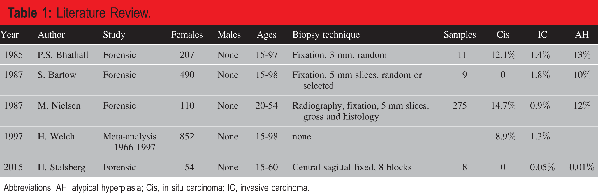
Abbreviations: AH, atypical hyperplasia; Cis, in situ carcinoma; IC, invasive carcinoma.
Objectives
The present study aimed: to determine silent breast cancer prevalence in male gender, to identify the specific profiles that influence the clinical manifestation of the disease, and to characterize the age distribution of the silent breast cancer in the population under study.
Study Design
The samples comprising the study population were obtained from the National Institute of Legal Medicine and Forensic Science in Lisbon, following a proper tissue collection authorization procedure.
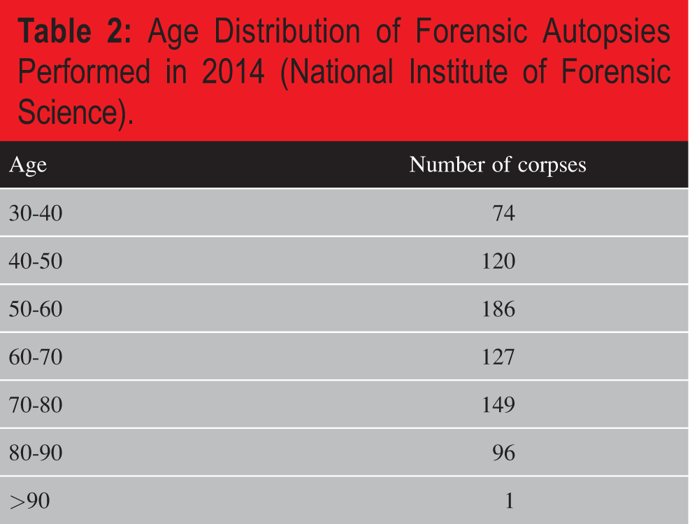
The advantage of forensic autopsies stems from two major factors: unexpected deaths and the relatively uniform age distribution of the population under study, as opposed to hospital samples ( Table 2 ):
Age Distribution of Forensic Autopsies Performed in 2014 (National Institute of Forensic Science).
Quantitative studies are very important because they allow one to determine the relationship between independent and dependent variables and achieve the study’s goal. This type of research also seeks to determine or establish facts, test hypotheses, and make study predictions. According to Vanderstoep and Johnson (8), quantitative research is advantageous because it yields accurate results that can be used to reflect the general population from which the sample was drawn. Furthermore, the result of the quantitative study is objective because the researcher remains detached when gaining, analyzing, and interpreting the research data. Therefore, the present study employed a quantitative research design to achieve its goals.
Sampling Procedure
Probability and nonprobability are the two main sampling techniques; the first one is known as choosing a random sample from a large population, while the latter one is regarded as a purposive sample created by targeted members of the population (9). This study employed the random (probability) sampling approach. The random sample can represent the whole researched population since it does not focus on the particular group of the population.
Sample Size
The study employed Cochran’s (10) sample size estimation procedure where the target population is infinite. The sample size formula is:
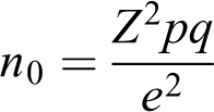
Where n 0 is the sample size, z is c interval (CI; 95%), p is the estimated proportion of an attribute that is present, q = 1 − p, and e is the desired level of precision (0.05). For instance, the approximate overall incidence of breast cancer in the Portuguese population is 0.12% (118.5 in 100 000 women).
Therefore, the sample size calculated at 95% CI, 0.12 proportion, and precision level of 0.05 will be:

Thus, the estimated population size = 27 cadavers are needed to achieve the null hypothesis in the male gender.
Data Collection
The data collection process of the cadavers included patients’ profile, gland characterization, lesion size, histological type, and molecular surrogates. Cadavers profile included age, ethnicity, comorbidities, medications, cause of death, breast screening adhesion, and breast cancer risk factors. Gland’s characteristics included dimensions, weight, and size.
Data Analysis
The quantitative statistical method of analysis was based on the overall multidimension constructs measurements for every factor, descriptive statistics, regression, and parametric as well as nonparametric tests. The regression statistics will be used to determine the correlation between the multidimension construct assessment and each factor, as well as the actual percentage of people with breast cancer. Moreover, the line fit plot will be employed to obtain illustrations based on the correlation and to provide the relationship between each factor. Further, the descriptive statistical analysis can be performed to provide the comparisons of age, ethnicity, and risks of breast cancer. The predicting factors of breast cancer will be determined using logistic regression.
Methodology
The study group consisted of a series of consecutive medicolegal autopsies on fresh Portuguese cadaver performed from July 2016 to December 2019 at the National Institute of Legal Medicine and Forensic Science, Lisbon, Portugal. The criteria for exclusion were age younger than 40 years, the autopsy performed in less than 48 hours after death, extensive injury to one or both breasts, and known or clinically evident breast cancer. Once the eligibility criteria were met, and the sample collection authorization was obtained, a bilateral subcutaneous modified radical mastectomy (bsMRM) was performed through a Douformentel incision (allowing the subsequent reconstruction, previous to corpses release) in each fresh cadaver at the National Institute of Legal Medicine and Forensic Science.
General information, such as age, height, weight, and body mass index (BMI), was obtained from the cadaver’s referring file when available, while past medical history data was not included due to inadequate collection. Each specimen was properly identified in means of spatial orientation and, after conditioning in sealed bags ( Figure 1 ), was transported within an appropriate container to the Hospital São Francisco Xavier (Lisbon, Portugal), and submitted to measuring (three-dimensions), waiting, inspection, palpation, ultrasound, and mammography by breast radiologists and breast surgeon.
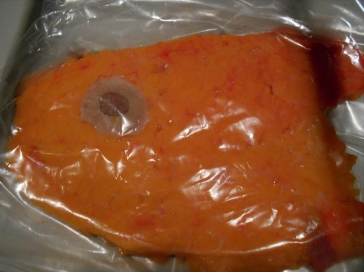
Left male breast sample.
The collected tissues were imaged using the G.E. Healthcare digital mammography system, Senographe Essential™ (G.E. Healthcare Bio Sciences, Pittsburgh, PA, USA) with an X-ray beam of 27 kV (range, 60-70 mA) and 10-15 dekanewtons (daN) compression, depending on tissue density and size ( Figure 2 ). The visualization screen had a resolution of five megapixels (G.E. Healthcare LOGIQ™ S7 Expert ultrasound system, with a medium frequency of 9-15 MHz; G.E. Healthcare Bio Sciences).
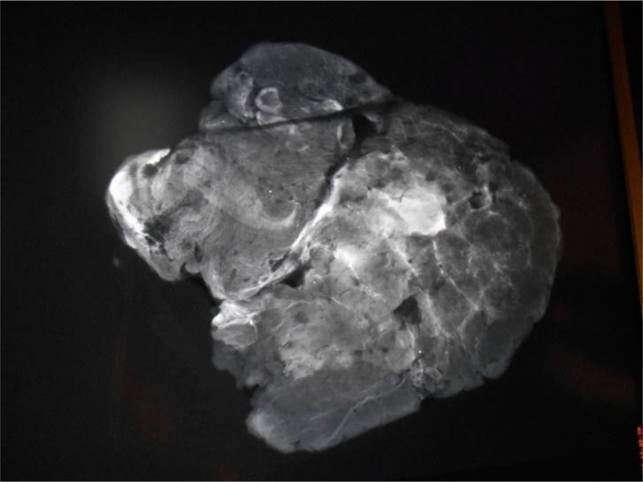
Sample’s mammogram.
Breast tissue, classified as Breast Imaging Reporting and Data System (BI-RADS) category three or higher, was submitted to wire-guided or direct excisional surgical biopsy by the author. According to the 5th edition of the ACR BI-RADS Atlas (11) system used: 0: Incomplete 1: Negative 2: Benign 3: Probably benign 4: Suspicious o 4A: low suspicion for malignancy, about 2% o 4B: intermediate suspicion of malignancy, about 10% o 4C: moderate concern, but not classic for malignancy, about 50% 5: Highly suggestive of malignancy 6: Known biopsy—proven malignancy
The samples were subsequently analyzed in the pathology department by an experienced breast pathologist.
Samples
In the preanalytical phase, breast biopsies were fixed in 10% buffered formalin (JTBaker) for 24 hours, and lumpectomy specimens were fixed for 48 to 72 hours at room temperature (20°C). Formalin-fixed, paraffin (VWR International, EUA) embedded tissues were processed in Sakura’s “Tissue-Tek VIP” and cut into 3 µm sections, one cut per adhesive slide (Superfrost Plus Gold—Thermo Scientific, EUA), with respective positive control. Tissue section adhesion time and temperature were held constant for 1 hour at 70°C. Following these procedures, the slide was subjected to labeling by the immunocytochemistry (ICC) method.
Immunocytochemistry Procedure
The ICC panel of primary antibodies used against Ki67 (clone 30-9, Cat. 790-4286), ER (clone SP1, Cat. 790-4324), and PR (clone 1E2, Cat.790-2223) were performed in the BenchMark ULTRA using Optiview DAB IHC Detection Kit (Cat. 760-700) for Ki67, and Ultraview Universal DAB Detection Kit (Cat. 760-500) for Estrogen Receptor (ER) and Progesterone Receptor (PR), all from Ventana Medical Systems, Tucson, USA. The slides were observed by a surgical pathologist under an optical microscope.
Results—Silent Male Breast Cancer
Results
All 74 cases were submitted to bsMRM and proceeded to tissue evaluation. The average postmortem to biopsy duration was 18 hours. Age at death ranged from 40 to 91 years, with a mean age of 63.9 years ( Figure 3 ).
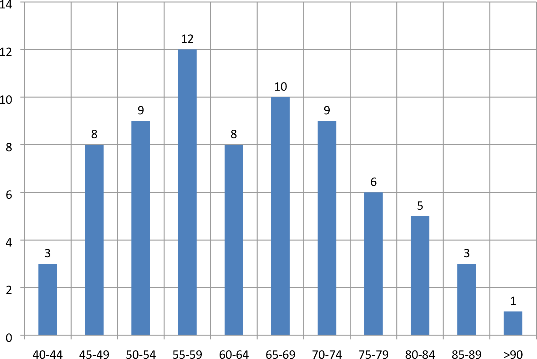
Age distribution of the male corpses. Age groups are presented on x-axis while y-axis denotes the number of corpses.
The mean BMI was 28.63 kg/m2; out of 74 cadavers, 90.54% were Caucasoid, six of Negroid, and one of Asiatic ethnicity. Of the 74 cases, 23 (31.08%) died suddenly from acute heart failure (myocardial infarction; Table 3 ; Figure 4 ), while the most interesting data is the diagnosis of four gastrointestinal tract silent adenocarcinomas (two colons, one gastric, and one pancreatic) and one lymphatic system neoplasm.
Autopsy Findings of the Cases.a
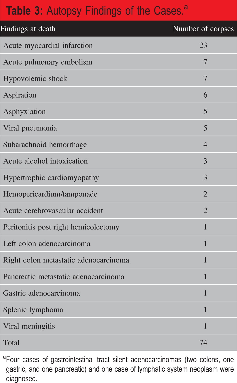
a Four cases of gastrointestinal tract silent adenocarcinomas (two colons, one gastric, and one pancreatic) and one case of lymphatic system neoplasm were diagnosed.
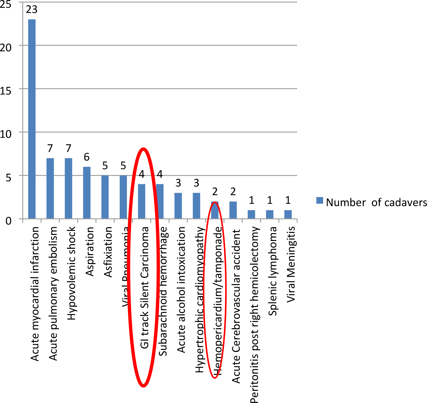
Autopsy findings of the cases. The x-axis presents the causes of death, while the y-axis shows the number of cases in each cause of death category.
No case of breast cancer was detected among the analyzed cadavers. None of the corpses had a history or scars of breast surgery, nor did they have a confirmed diagnosis or clinical signs of breast cancer.
The mean weight of processed breast tissue was 842.10 g/cadaver, with mean dimensions of mediolateral 23.46 cm, superoinferior 16.37 cm, and anteroposterior 0.83 cm per tissue ( Figure 5 ). There appeared to be no relationship between BMI and breast tissue weight.
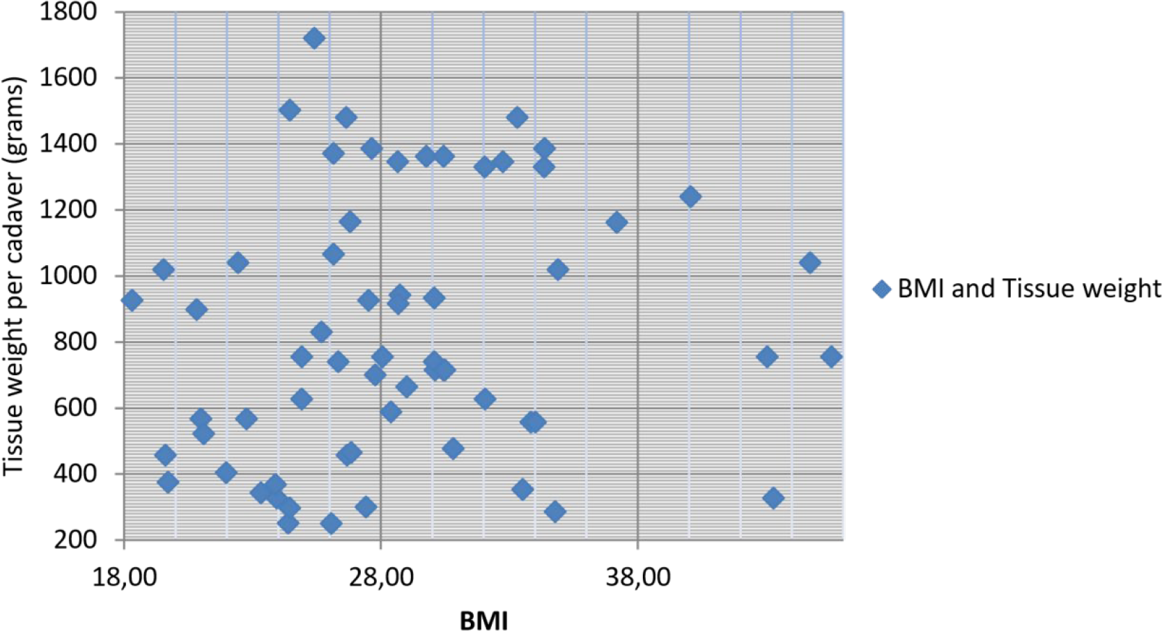
Body mass index (BMI) and mean tissue weight/cadaver. X-axis presents BMI values versus tissue weight per cadaver on the y-axis. There appeared to be no relationship between these two traits.
In volumetric terms, the breast tissue was submitted to imaging, and the following calculus was used to approximate its shape to a hemi-ellipsoid:
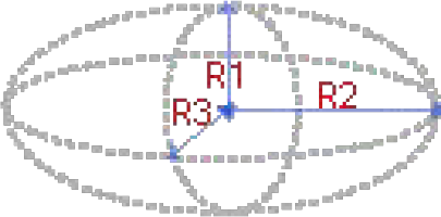 Ellipsoid dimensions and ellipsoid Formula (Knud Thomsen):

The total breast tissue volume elaborated was 102774,08 cm3 (102.77 L). The correlation between BMI and total breast volume (TBV) is depicted in Figure 6 . There appears to be no correlation (correlation index of 0.176279) between TBV and BMI.
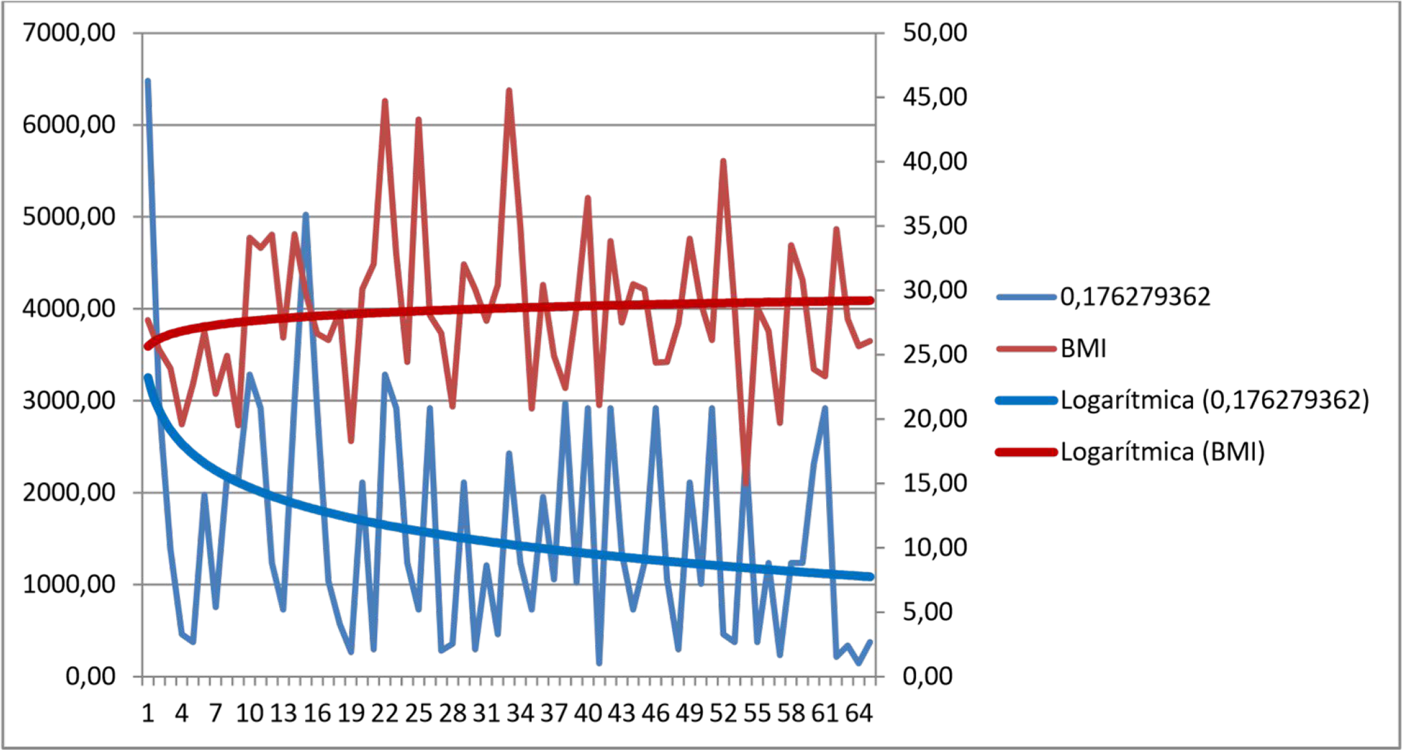
Body mass index (BMI) and total breast volume (TBV). A correlation index of 0.176279 was noticed between the two parameters.
Breast density and BMI also do not appear to have any correlation, presenting a negative correlation index of −0.13 ( Figure 7 ).
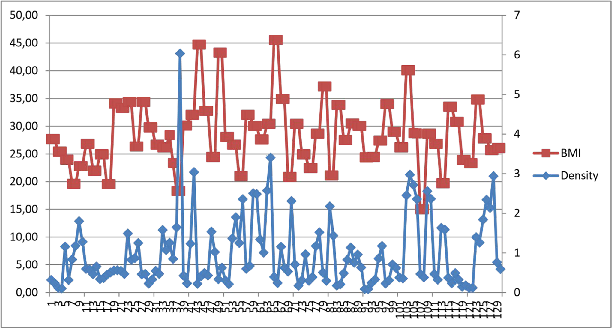
Body mass index (BMI) and breast density. Red line denotes BMI values, while blue line shows density. A negative correlation index of −0.13 was noticed between the two traits.
Breast Imaging Reporting and Data System classification indicated 1 in 129 breast samples (87.16%), 2 in 18 breast samples (12.16%), and 4a in 1 (0.67%) breast sample ( Figure 8 ).
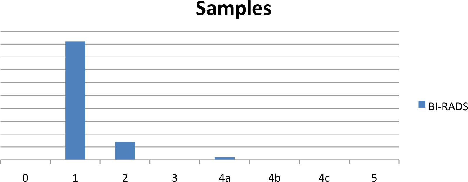
Breast Imaging Reporting and Data System (BI-RADS) classification of the samples.
Benign microcalcifications were detected in nine glands, dispersed in six cases, and localized in the remaining three. Benign macrocalcifications were detected in only five cases, localized in upper quadrants, while three cases had both types of benign calcifications. Moreover, intramammary lymph nodes were found in six cases (five cadavers, one biopsied included), while benign multiple axillary lymph nodes were present in seven cases (six cadavers).
The biopsied cadaver was a 47-year-old Caucasoid male who died from viral meningitis associated with a respiratory infection. The left breast echography revealed three intramammary nodules in the upper quadrants transition, classified as BI-RADS 4a (imaging of lymph nodes with thickened cortical).
The pathology report confirmed “reactive intramammary lymph nodes with no neoplastic lesion.” No other biopsy was performed.
Correlation Analyses
Correlation analysis was conducted on SPSS to determine the relationship of the gland’s classification (BI-RADS system) with age, weight, height, and BMI of the male corpses. The correlation was tested at a 95% CI, and a significant value (2-tailed) was used as a criterion to decide the significance of the relationship between the two variables. If the significance value is 0.05 or less, it indicates a significant relationship, and a greater value than 0.05 implies an insignificant relationship. Nonetheless, the Pearson correlation value was used to determine whether the relationship was negative or positive (based on the presence or absence of the negative sign).
The correlation matrix shown in Table 4 presents the correlation of glands (BI-RADS) with age, weight, height, and BMI of male corpses. The results indicated the glands (BI-RADS) have a significant relationship with the weight and BMI of male corpses as their respective significance values were 0.020 and 0.028, which were less than 0.05. However, based on negative signs of their Pearson correlation values, the relationship was significantly negative. The results also illustrated that glands (BI-RADS) have an insignificant relationship with the age and height of male corpses as their respective significance values were 0.170 and 0.346, which were more than 0.05. Thus, the gland (BI-RADS) found in male corpses increases as their weight and BMI decrease. Hence, higher BI-RADS is found in thinner individuals, which is probably due to the low sampling volume.
Correlation of Glands (BI-RADS) With Age, Weight, Height, and BMI of Male Corpses.
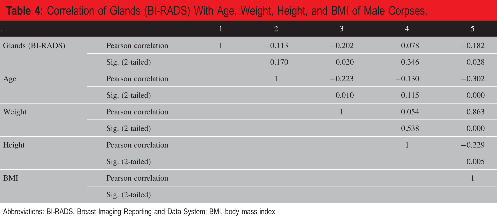
Abbreviations: BI-RADS, Breast Imaging Reporting and Data System; BMI, body mass index.
Moreover, a second correlation analysis was conducted to determine the relationship of the cause of death with the results of mammography, ecography, and glands (BI-RADS) on male corpses, as depicted in Table 5 . The results indicated the cause of death has a significant positive relationship with mammography, with a significance value of 0.027 and a positive Pearson correlation value of 0.568. However, the cause of death has an insignificant relationship with echography and glands (BI-RADS) of male corpses as their respective significance values were 0.732 and 0.085 which were greater than 0.05. The results depicted that the cause of death was insignificantly related to echography and gland’s BI-RADS of male corpses. Thus, the echography findings (calcifications) and gland’s BI-RADS did not correlate with the cause of death of the male corpses examined in this research. The cause of death and mammography findings might imply vascular calcification and consequent ischemic strokes or heart ischemic disease, data not uniformly supported by other studies.
Correlation of Cause of Death With the Results of Mammography, Ecography, and Gland’s BIRADS of Male Corpses.
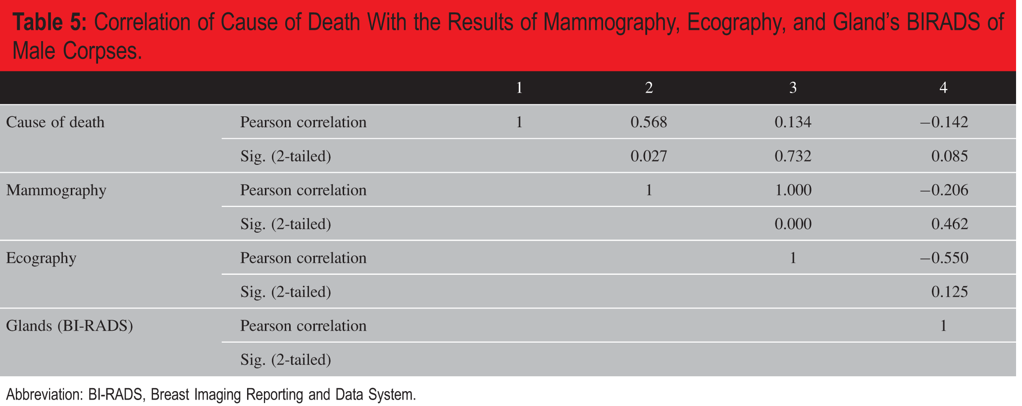
Abbreviation: BI-RADS, Breast Imaging Reporting and Data System.
Hypothesis Testing
The study’s goal was to quantify the number of male silent breast cancers that are not clinically manifested but can be identified through imaging analysis. The null hypothesis stated that the natural reservoir of silent breast cancer is not superior to the actual incidence of the disease. The alternative hypothesis stated that the natural reservoir of silent breast cancer is superior to the actual incidence of the disease.
The hypothesis was tested in the first period with 27 recruited male gender cadavers (12). The findings did not identify any silent breast cancer despite the fact that MBC’s molecular surrogate (usually ER, PR, and Androgen Receptor (AR) positive, Luminal B-like/HER2-negative, and 56% patients of T1 tumors) generally has a good prognosis; its late detection and consequent treatment dictates the disease course (5.1% with metastatic disease [M1] and Overall Survival (OS) 2.6 years).
Cross tabulation analysis was conducted on SPSS to test the null hypothesis. The glands’ results were expressed in seven BI-RADS categories. BI-RADS 1 shows negative examination, while BI-RADS 2 is consistent with benign findings. BI-RADS 3 is probably benign and should have shortened interval follow-up to determine stability; the risk of malignancy is below 2%. BI-RADS 4 is a suspicious abnormality, which can represent the chance of being malignant (in percent). The BI-RADS category 4 is subdivided into a, b, and c. The subcategory of (a) has a low probability of malignancy with a 2% to 10% chance of malignancy. The (b) subcategory has an intermediate risk of malignancy ranging from 10% to 50%. In comparison, the subcategory of (c) has a high probability of malignancy ranging from 50% to 95%. BI-RADS 5 is highly suggestive of malignancy more than 95%. In the cross tabulation analysis, the BI-RADS 4a: probably benign, has a low probability of malignancy, indication for biopsy was only one observation (0.67%); that is, less than 1%. Therefore, it was automatically ignored by the SPSS while performing the cross tabulation analysis. The cross tabulation results of the gland’s BI-RADS against the male corpses’ mammography are given in Table 6 .
Gland’s (BI-RADS) × Mammography Cross Tabulation.a
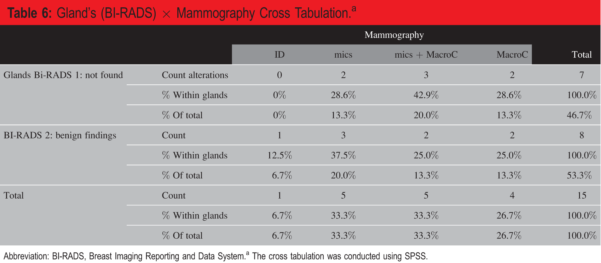
Abbreviation: BI-RADS, Breast Imaging Reporting and Data System.a The cross tabulation was conducted using SPSS.
The mammography results showed that male corpses samples had “microcalcifications,” “both microcalcifications and macrocalcifications,” and “macrocalcifications.” The majority, viz. 37.5% of the male corpses were found to have “microcalcifications,” 25% had “macrocalcifications,” and 25% had “microcalcifications and macrocalcifications.”
The cross tabulation results of the gland’s BI-RADS against the echography of the male corpses are presented in Table 7 . According to the echography results and BI-RADS classifications of 1 or 2, 25% of the male corpses had “Intramammary lymph nodes,” while the majority, 75%, had “Axillary lymph nodes.” In BI-RADS 2 cases, 80% of the male corpses were found to have “Intramammary lymph nodes,” and 20% were found to have “Intramammary lymph nodes” based on echography results.
Glands (BI-RADS) × Echography Cross Tabulation Among the Male Corpses.
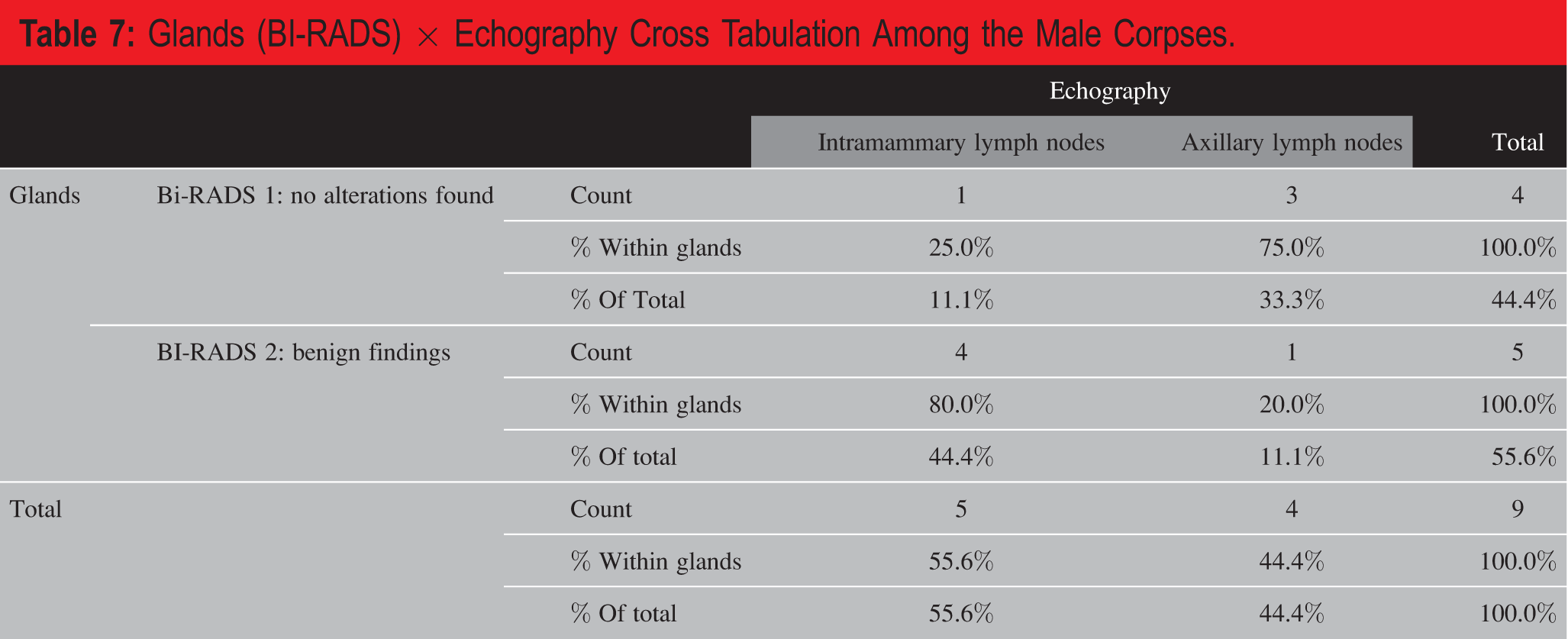
Abbreviation: BI-RADS, Breast Imaging Reporting and Data System.Based on cross tabulation results, it was evident that no malignancy signs were found by breast echography and mammography in the male corpses. However, to statically validate these findings, the level of significance was evaluated by correlation analysis. The correlations of the gland’s BI-RADS with the results of echography and mammography are shown in Table 8 . The correlations of gland’s BI-RADS with the results of echography and mammography were insignificant as their respective (2-tailed) significance values were 0.125 and 0.462, viz. greater than 0.05. Consequently, in male breast evaluation, BI-RADS classification obtained by echography and mammography cannot be used as a screening method (as suspicious findings are so scant) in the general population, as expected.
Correlations of Glands BI-RADS With the Results of Echography and Mammography.
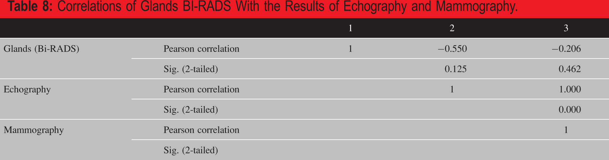
The statistical analysis found no significant incidence or suspicion of breast cancer in the male corpses, implying that the rate of MBC is not superior to the actual incidence in the general population.
Therefore:
We can conclude that the actual cases of MBC manifest themselves, and thus, we accept the null hypothesis that the natural reservoir of silent breast cancer is not superior to the actual incidence of the disease.
Discussion
Portuguese National Data
Thorough research on available national public databases was conducted yet leading to scant data. Male breast cancer incidence as recorded by the National Oncology Registry (RON; a national platform where all malignancies are individually registered) allowed collecting some data for 2001 to 2010. Male breast cancer has been registered for 477 cases (data for the years 2002-2004 is missing). Male breast cancer mortality was assessed using the National Statistics Institute data. In the following graphic ( Figure 9 ), data show that 1) the incidence of MBC in the Portuguese population is of a medium of 68.14 new cases/year, with no discernible trend (rising, decreasing, or plateau) and 2) the mortality rate due to MBC in the same population is 22.8 individuals/year, presenting a slight decrease over the last two and half decades (R2 = 0.0196).
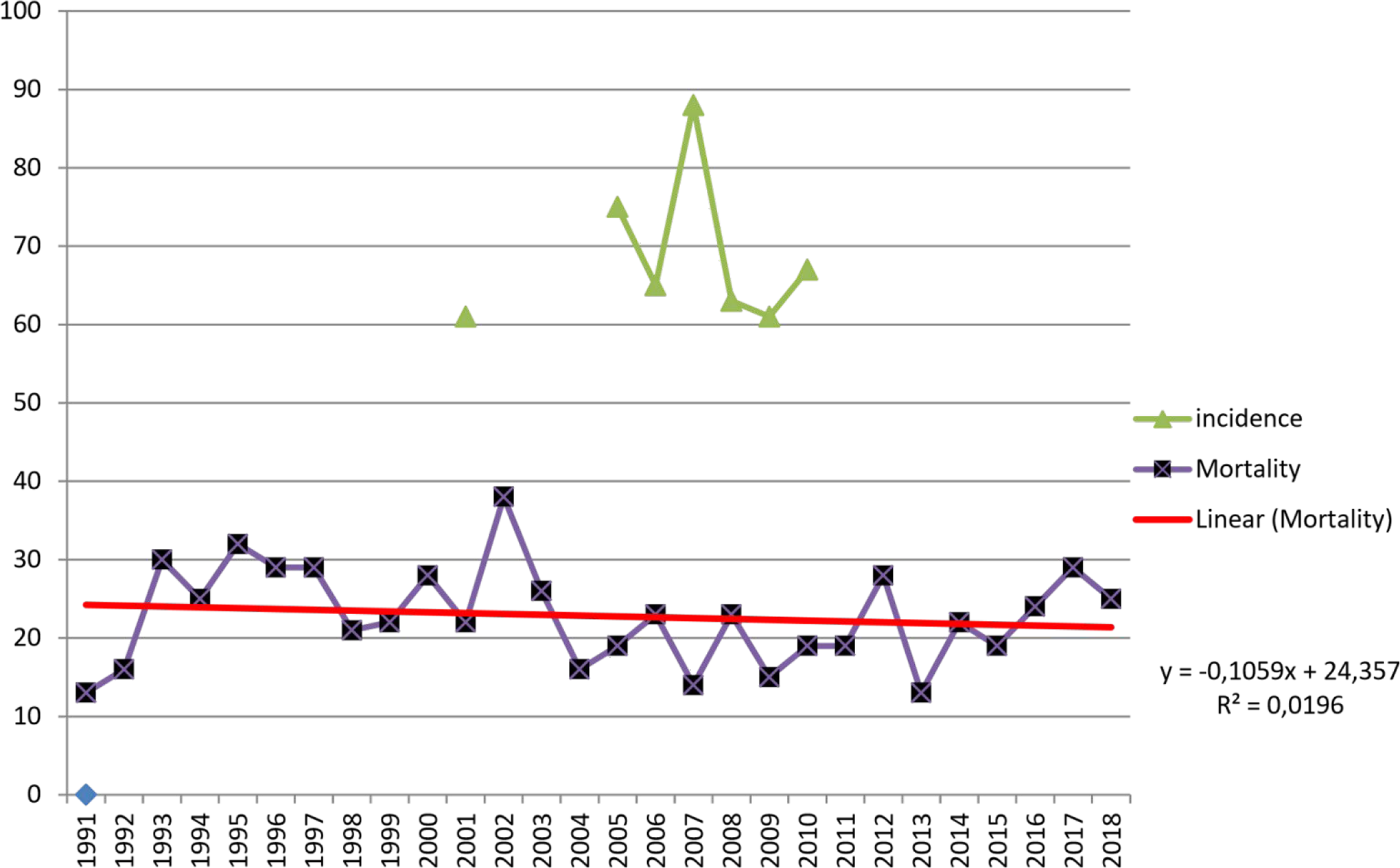
Portuguese MBC data incidence and mortality.
Male Breast Cancer and the State of Art
Male breast cancer (MBC) is a rare disease that has received little attention in terms of transcriptional profiling or genomic role players. The foundation of male cancer is usually based on the concepts and understanding of female breast cancer (FBC) and the existing literature on female assessment. Over the years, a great deal of effort has been expended in order to answer the question: Is MBC different and more aggressive than FBC? The current review gathered data from the literature on female and MBC from 2000 to 2018 to produce some conclusive results (13).
Male breast cancer, just as FBC, is a heterogeneous disease (14 -16). According to recent literature and Centre for Disease Control findings, MBC may differ from FBC at the molecular level (17). Various studies have established that there are two subgroups of MBC: the luminal M1 and the luminal M2, which are distinct from the currently known subtypes of FBC. Therefore, the novel subsets of the disease vary in males and females, and they have a unique characterization (17). Other studies have found that males with breast cancer have a lower chance of survival than females. A survey carried out in the United States between 2004 and 2014 indicated that even though MBC cases are few, the mortality rate is higher than that of FBC cases (18).
Women’s breast cancer has similarities to the one in males in some aspects. The similarities include the occurrence of invasive ductal carcinoma—frequently of positive hormone receptors ER and PR—as the most common histological type, which is often detected as a subareolar lamp, usually painless, with nipple retraction and bleeding (19, 20). Another significant similarity is that, in both male and FBC, a family history of breast and ovarian cancer is a risk factor of breast cancer development.
As far as differences between male and FBC are concerned, MBC is less common in males than females, accounting for nearly 1% of the total number of FBC (21 -23). Secondly, MBC is diagnosed in older men. The mean age of breast cancer diagnosis in men is 67 years, compared to 52 years in women. Moreover, lobular carcinoma is also relatively less common in men (24, 25). Furthermore, the prevalence of germline BRCA2 mutation in MBC patients is 14%, while BRCA1 is less common, occurring at a rate of 4% (15). To summarize, it seems that MBC affected patients are older, get diagnosed with a substantial delay (more than one year in almost half of cases), have a slightly less 5 year survival in stages I and II, and even worse in stages III and IV. Male breast cancer presents with a higher incidence in advanced stages and axillary evolvement. However, some studies suggest an equal overall disease-specific survival for both sexes, attributing the increased mortality rate in males to other noncancerspecific mortality (26).
Gender differences are significant at the molecular level, with 95% of MBC being luminal A or B (27 -29), compared to 73% of FBC (30). Both the HER2 and basal phenotypes are uncommon in men. Genetically, approximately 10% of MBC cases have BRCA2 mutations, while BRCA1 mutations are associated with less than 1%. Male breast cancer has been more linked with BRCA2 mutations, which account for 4% to 40% of hereditary compared to 5% to 10% in FBCs (31).
Cardoso et al. (32) reported that while MBC is distinct from FBC, the actual treatment of MBC is based on FBC protocols, with poor outcomes. Their study enrolled 1483 patients, at various participating institutions, with confirmed breast cancer diagnosed between 1990 and 2010. Biological material was handled and analyzed centrally. The findings suggested that most MBC cases were invasive ductal carcinomas, grade 2, and almost always ER+, PR+, and AR+. A trend toward higher OS was observed in patients with highly ER+ disease, highly PR+ disease, and highly AR+ compared with the low expression of the receptor (Allred scores 3-6). HER-2 expression was uncommon, and no association between outcome and HER-2 status was seen. High (≥20%) Ki67 expression was observed in only 24.9% of cases. The majority of patients had a Luminal B-like/HER-2-negative (48.6%) or a Luminal A-like (41.9%) disease. A small number of HER-2+ and triple-negative breast cancer was detected. Although 48.5% of patients had T1 tumors, only 4% had BCs. Sentinel Lymph Node Biopsy (SLNB) has seen a significant trend toward less aggressive axillary nodal management over the years.
Adjuvant radiotherapy (RT) was not delivered to 45% of patients treated with, nor to a significant proportion of patients (30.7%) with node-positive tumors treated with mastectomy. Since current recommendations suggest the use of similar algorithms for RT decision-making in males as in FBC patients, the low rates of adjuvant RT are a major concern as male patients usually have a higher stage at diagnosis. A significant trend toward increased chemotherapy (anthracycline) has been observed over time, with adjuvant Endocrine Therapy (ET) being administered to only 76.8% of patients. “The reasons for this under-use of an effective and low toxicity therapy are unknown” fortunately increasing the latest years (32).
Another large study by Wang et al. (33) pointed out that male patients with breast cancer significantly differed from their female counterparts by older age at diagnosis, a higher proportion of ER-positive subtype or advanced disease, and less likelihood of receiving conventional treatment. Men had higher mortality than women overall and across disease stages, particularly for ER-positive breast cancer. Clinical and treatment characteristics were the most common factors in sex-based disparity in mortality, but the differences persisted even after adjustment for age, race/ethnicity, clinical and treatment characteristics, and access to care.
Conclusions
To the best of our knowledge, this is the first study that explored the silent breast cancer incidence among men. We noticed that MBC is a rare disease, and its natural reservoir is extremely low, just like its incidence. Nevertheless, the few cases that exist need to be treated appropriately.
The screening of the general population for MBC is unnecessary; however, it should be targeted for men at elevated risk for breast cancer (34).
There is a need for consensus, and for that, clinical trials should be a priority. Fortunately, MBC dedicated guidelines have started to appear (35). Even though we have started to decode, we are still “lost in translation!” (36).
Limitations
The present research is subject to several limitations. First of all, the sampling number question is allocated. Since it was hypothesized that the prevalence of silent breast cancer is unknown and the actual disease incidence is low, finding a case of silent MBC would be quite unusual.
Another limitation of this study is that medical data from the analyzed corpses could not be collected, leaving out potentially harmful or protective factors that would have been very interesting to investigate. The third and perhaps the most obvious limitation of this study is that specimens were not examined through systematic histology. This “limitation” stems from the study’s somewhat unique design that aimed to identify imaging-detected silent breast cancer.
Footnotes
Acknowledgments
To the Hospital São Francisco Xavier, Instituto Nacional de Medicina Legal e Ciências Forenses, Universitat de Barcelona Medicine Faculty, and all the huge team that made this project work.
Author Contributions
Zacharoula Sidiropoulou, Ana Virginia Araujo, Inês Alegre, Claudia Santos, Filipa Costa, Diogo Cardoso, and Vasco Cardoso have been responsible for specimen collection, biopsies, and literature review.
Zacharoula Sidiropoulou has elaborated the protocol and supervised all trial.
Ana Paula Vasconcelos and Cristiana Couceiro have performed the breast imaging of the specimens.
Carlos Dos Santos has been responsible for inclusion/exclusion criteria of each cadaver and obtaining the authorization for each collection.
Rita Sampaio has performed the pathology analysis of specimens.
Fátima Cardoso and Pére Gascón have supervised and tutored the trial.
Ethical Approval
Autopsy authorizations have been cleared according to the Portuguese Law.
Statement of Human and Animal Rights
N/A
Statement of Informed Consent
Legal authorizations cleared in the national REENDA platform.
Disclosures & Declaration of Conflicts of Interest
The authors, reviewers, editors, and publication staff do not report any relevant conflicts of interest.
Financial Disclosure
No external funding to declare, all costs have been supported by the first author’s proper hospital, Hospital São Francisco Xavier-CHLO
