






Abstract
Coronavirus disease 19 (COVID-19) vaccination is considered an important part in improving health outcomes globally. While various adverse events following vaccination against COVID-19 have been reported, eosinophilic diseases have rarely been documented in the literature and are poorly understood. Although vaccination is lauded as being “safe,” it has become apparent that adverse reactions related to the vaccines can have detrimental health effects for certain individuals. We present a case of a death related to multiple severe preexisting comorbidities, complicated by new-onset gastrointestinal complaints which were temporally associated with recent COVID-19 vaccination and did not subside, but worsened prior to death. Autopsy revealed evidence of eosinophilic enteritis, associated with ascites, as well as eosinophilic inflammation elsewhere, including the lungs and heart. Histological examination revealed abundant eosinophils in tissues, including the small intestines, epicardium, and lungs. Whether or not the eosinophilic inflammatory process was caused by the recent vaccination cannot be stated with certainty; however, the temporal association between vaccination, symptom onset/progression, and death, and the literature which suggests a possible association between coronavirus vaccination and eosinophilic reactions leads to the conclusion that this death might have been related to an adverse reaction to COVID-19 vaccination.
Introduction
During the severe acute respiratory syndrome coronavirus 2 (SARS-CoV-2) worldwide pandemic, coronavirus disease 19 (COVID-19) vaccination has been recognized by many experts as playing an important part in improving health outcomes globally. Therefore, recognizing the adverse effects related to COVID-19 vaccination is essential. While various adverse events following vaccination against COVID-19 have been reported (1), eosinophilic diseases have rarely been documented in the literature and are poorly understood (2, 3). Although vaccination is considered to be “safe” from an overall public health perspective, it has become apparent that adverse reactions related to the vaccines can have detrimental health effects for certain individuals. Gathering information on potential adverse vaccine reactions is important to gain a better overall understanding of the potential risks versus benefits associated with the vaccinations. Here, we present a case of a death related to generalized eosinophilia presenting with symptoms of enteritis following Moderna COVID-19 vaccination.
Case Description
A 76-year-old man with a medical history significant for obesity died at home while sitting on the couch, witnessed by his wife. The death occurred indoors during late winter and there were no sudden changes to his status in the hours preceding his death. Notably, the patient received the Moderna COVID-19 vaccine 9 days prior to his death and experienced abdominal distension soon after vaccination on the same day. The abdominal distension continued to worsen and progressed to include shortness of breath in the days prior to death. He did not seek medical care for these symptoms. He became unresponsive while watching television with his wife. Cardiopulmonary resuscitation (CPR) was initiated following emergency medical services (EMS) personnel arrival. Resuscitation efforts were unsuccessful, and the patient was pronounced dead. Because of the temporal relationship between the patient’s symptoms, COVID-19 vaccination, and death, the decision was made to proceed with a medicolegal autopsy for this case.
The patient’s medical history included class 3 obesity, atherosclerotic cardiovascular disease, hypertension, hyperlipidemia, controlled type 2 diabetes mellitus, and prostate cancer. He had no history of significant gastrointestinal or respiratory disease. Medications included atenolol-chlorthalidone (50 mg-25 mg daily), fenofibrate (134 mg daily), glipizide (5 mg daily), lisinopril (10 mg daily), metformin (1000 mg twice daily), and pioglitazone (45 mg daily).
External examination at autopsy revealed the body of a well-developed, well-nourished, morbidly obese white male whose appearance was compatible with the stated age of 76 years. The body was 71 inches long and weighed 325 pounds (body mass index of 45.3). The heart weighed 680 grams, while the left lung was 530 grams, and the right lung was 720 grams. Brain examination revealed an unremarkable adult brain weighing 1300 grams, and the cerebral arteries contained focal areas of mild atherosclerosis. Cardiovascular examination revealed cardiomegaly, mild to severe coronary artery atherosclerosis, and mild to severe aortic atherosclerosis. Respiratory examination revealed a congested and mottled parenchyma. Gastrointestinal system examination revealed grossly unremarkable esophageal mucosa, gastric mucosa, pancreas, liver, small intestines, and large intestines. There were adhesions within the abdominal cavity and 1500 mL of serosanguinous fluid within the peritoneal cavity. Genitourinary system examination revealed granular subcapsular surfaces of the kidneys with thinned cortices.
Histological examination revealed abundant eosinophils in several tissues, including the small intestines ( Figure 1 ), the epicardium ( Figure 2 ), and the lungs ( Figure 3 ). Within the small intestine, there was an intense lymphoplasmacytic inflammatory infiltrate with numerous eosinophils; eosinophil counts ranged from “normal” to up >30 eosinophils per high-power field, consistent with a diagnosis of eosinophilic enteritis. The presence of numerous eosinophils in other organs (heart and lungs) was concerning for a systemic eosinophilic process. Histologic examination confirmed the presence of hypertensive and atherosclerotic cardiovascular disease and pulmonary emphysema. Liver examination was normal.
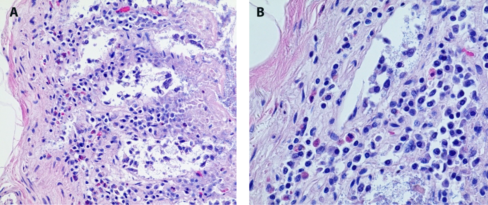
Small intestine inflammation. A – Numerous eosinophils within the mucosa of the small intestine (H&E, 200x). B – Higher magnification showing numerous eosinophils (H&E, 400x).

Epicardial inflammation. A – Chronic inflammatory cells within the epicardium, near the conduction system (H&E, 200X). B – Focus of inflammation with numerous eosinophils (H&E, 400X)

Bronchial inflammation. A – Chronic bronchial inflammation with numerous eosinophils (H&E, 200x). B – Higher magnification showing numerous eosinophils (H&E, 400x).
A polymerase chain reaction (PCR)-based test of nasopharyngeal and lung swabs was negative for viral infectious agents, including COVID-19. An independent Centers for Disease Control and Prevention consultation examination concurred with the autopsy findings of eosinophilic enteritis. The cause of death was ruled as hypertensive and atherosclerotic cardiovascular disease, with contributing causes of pulmonary emphysema, diabetes mellitus, obesity, and eosinophilic enteritis with possible generalized eosinophilia. The temporal relationship between vaccination and onset of gastrointestinal symptoms suggested a possible association between the vaccine and the eosinophilic inflammatory process.
Discussion
Eosinophilic gastroenteritis (EGE) is a disease under the broader category of eosinophilic gastrointestinal disorders (4). Its pathophysiology is not clear but appears to be associated with a history of allergic reactions (5). Symptoms depend on the subtype of EGE, with the mucosal subtype primarily presenting with nausea, vomiting, abdominal pain, and diarrhea (4). The muscular subtype may present as intestinal obstruction and perforation. The subserosal subtype can present as ascites and abdominal distension, both of which were evident in the case presented. Diagnosis frequently involves endoscopy with mucosal biopsy after establishing clinical suspicion (6). Although primary EGE is a chronic condition with a relapsing course, our patient did not have a known history of any form of gastrointestinal disorder, let alone EGE or allergic reactions. Because of the temporal relationship of vaccination and onset of abdominal symptoms, it is possible that patient developed EGE secondary to vaccination rather than presenting with new onset primary disease or a flare of preexisting disease.
First granted emergency use authorization in December 2020, the Moderna COVID-19 vaccine contains a SARS-CoV-2 spike mRNA encapsulated within a lipid nanoparticle (7). Although vaccine adjuvants have been linked with eosinophilic responses in the past, the Moderna vaccine does not contain adjuvants previously linked with systemic eosinophilia (8). Instead, the mRNA in the vaccine contains properties that causes immunostimulation through pattern recognition receptors, such as toll-like receptors, that detect cytosolic viral nucleic acids (9, 10). Additionally, the Moderna vaccine contains polyethylene glycol to help with stability (4). Polyethylene glycol has been implicated as the possible culprit for allergic reactions to the vaccines, including anaphylaxis. At the time of this writing, eosinophilic reactions to Moderna vaccine have not been reported in the English language literature; however, eosinophilic reactions have been reported with other COVID-19 vaccines (2, 4).
Authors in the past have noted that some coronavirus antigens may induce an eosinophilic response. Eosinophilic responses occurred in severe acute respiratory syndrome coronavirus 1 (SARS-CoV-1) vaccinated mice, ferrets, and monkeys following virus reexposure (10, 11). SARS CoV-1 shares approximately 80% of its nucleic acid sequence and with SARS CoV-2 and a similar pathophysiology (12). Several SARS-CoV-1 murine vaccine studies have noted eosinophilic reactions as well, including one study that found the presence or absence of alum did not affect eosinophilic responses (3, 6). Although our patient’s symptoms began soon after vaccination, there was no evidence of SARS-CoV-2 infection in our patient at the time of death. Additionally, in the studies above, eosinophilic disease was noted only in the pulmonary system rather than in multiple organs, as was found in our patient.
Treatment of EGE includes conservative measures, such as avoiding food allergens as determined by allergen testing (6). Systemic corticosteroids have been a mainstay for induction of disease flares, and frequently used in low doses to maintain remission. Budesonide, a steroid that primarily works locally in the gastrointestinal tract, has been shown to be effective in remission induction and maintenance. Budesonide carries a lower side effect burden compared to systemic corticosteroids. Corticosteroid-sparing therapy includes azathioprine and montelukast. Azathioprine is preferentially used in patients with steroid-dependent or refractory disease due to its significant immunosuppression, and not typically as a first line agent. Montelukast was shown to improve remission rates as monotherapy and as an adjunct to corticosteroids. Surgery is a last-line treatment reserved for severe disease complicated by perforation, intussusception, or intestinal obstruction, with estimates that 40% of patients with EGE requiring surgery during the course of their disease (6).
In recent years, eosinophils have been recognized for their role in antiviral responses, potentially attenuating viral illness severity, including COVID-19 (13). However, Lindsley et al., highlights the fact that eosinophils have the potential to become dysregulated under certain conditions, such as during allergic disease, resulting in an exaggerated response that could lead to tissue damage (14). While rare, eosinophilic dysregulation could lead to inflammation following COVID, causing significant morbidity and mortality in certain individuals.
The presence of eosinophilic inflammation in a person with no previous history of eosinophilic hyperactivation (i.e., allergic reaction) suggests that the potential eosinophilic response to the vaccine may have played a role in death. Performing autopsies on persons suspected of having an adverse reaction to vaccination represents an important but uncommon practice. Gaining a better understanding of why some people develop eosinophilia while others do not can help target who is most at risk for these adverse effects. More autopsies in the offices of medical examiners, coroners, and hospitals should be performed on patients who may have experienced such a reaction. Unfortunately, these cases tend to be overlooked due to reasons such as, but not limited to, a shortage of forensic pathologists, the ongoing and worsening opioid epidemic, and the COVID-19 pandemic itself causing excess deaths than would have normally been expected (15, 16). One solution would be to train more forensic pathologists to accommodate for the shortages seen across the United States.
Conclusion
This case represents a death related to multiple severe preexisting comorbidities, complicated by a history of new-onset gastrointestinal complaints which were temporally associated with recent COVID-19 vaccination and did not subside, but actually worsened prior to death. Autopsy revealed evidence of eosinophilic enteritis, associated with ascites, as well as eosinophilic inflammation elsewhere, including the lungs and heart. It is conceivable that the ascites and associated abdominal distention contributed to the decedent’s complaints of increased shortness of breath in the days leading up to his death. Whether or not the eosinophilic inflammatory process was caused by the recent vaccination cannot be stated with certainty; however, the temporal association between vaccination, symptom onset/progression, and death, and in-light of the literature which suggests a possible association between coronavirus vaccination and eosinophilic reactions leads to the conclusion that this death might have been related to an adverse reaction to COVID-19 vaccination. Further study is warranted, and clinicians and pathologists should be aware of the potential association between COVID-19 vaccinations and eosinophilic inflammatory reactions.
Footnotes
Ethical Approval
This case report was reviewed and deemed nonresearch by the WMU Homer Stryker MD School of Medicine Institutional Review Board. No protected health information or other uniquely identifying information is included in this manuscript.
Statement of Human and Animal Rights
This article does not contain any studies conducted with animals or on living human subjects.
Statement of Informed Consent
No identifiable personal data were presented in this manuscript.
Disclosures & Declaration of Conflicts of Interest
The authors have no conflicts of interest to declare.
Financial Disclosure
This study did not receive any funds.