
Abstract
Introduction
One of the intensively studied causes of male infertility in recent times is the integrity of DNA in the nucleus of mature ejaculated spermatozoa. 1 The incidence of sperm cells with fragmented DNA is higher in the ejaculate of participants with varicocele as compared to that in fertile donors. 2,3 This phenomenon may be correlated with an increase in reactive oxygen species (ROS), resulting in oxidative stress (OS), which causes the peroxidation of sperm plasma membrane and nuclear DNA damage. 4 Nitric oxide (NO) is probably an important source of such oxidative damage. 3,4 Nitric oxide is a potent ROS synthesized by 3 isoforms of NO synthase (NOS): nNOS (neuronal), eNOS (endothelial), and iNOS (inducible). Under pathological conditions, for example, varicocele, iNOS expression is upregulated, which is a possible cause of NO overproduction. 5,6 Varicocele has been accepted to be one of the main causes of infertility. 7 –10 In varicocele males, high NO production can induce the production of a highly toxic anion, peroxynitrite, as a result of peroxidation. 11 NO and peroxynitrite, which is a potent ROS, are produced in high concentrations in the dilated spermatic veins; therefore, these molecules could be the contributors to the high OS level in varicocele. 12
Normally, sperm chromatin is a highly organized and compact structure consisting of DNA and heterogeneous nucleoproteins. 1,13 Sperm DNA integrity is essential for the accurate transmission of genetic information. Oxidative stress and poor chromatin packaging can affect the integrity of sperm chromatin and cause sperm DNA damage. 14 Any form of sperm chromatin abnormalities or DNA damage may result in male infertility. 1,15 Chromomycin A3 (CMA3) assay, toluidine blue (TB) test, acridine orange (AO), and aniline blue (AB) staining are used to assess sperm chromatin/DNA. 14,16 –18
Some researchers have suggested that the administration of antioxidants reduces the extent of DNA damage 19,20 ; therefore, antioxidant supplementation may be used to treat infertile patients with varicocele. 15 Aminoguanidine (AG) is a specific iNOS inhibitor that is presumed to reduce NO content in tissues. 21 –23 Moreover, AG possesses antioxidant and free radical scavenging properties, which are especially observed in peroxynitrite production. 24 –26 This is the first report on the effects of AG on sperm chromatin integrity in varicocelized rats.
Material and Methods
Animals
Thirty adult Wistar male rats (mean weight, 300 ± 55 g) were maintained under standard laboratory conditions. The rats were acclimatized for 1 week under a 12 hour: 12 hour light: dark cycle at room temperature of 22 ± 2°C, and subsequently the male animals were mated with adult female rats and their fertility was evaluated (mating action was checked by vaginal plaque). Thirty male Wistar rats were divided into 5 groups: control, sham, varicocele, AG and placebo-treated groups. All chemicals were purchased from Sigma/Aldrich Chemical Co. (St. Louis, Missouri).
Surgical Procedure
The rats were weighed and then administered a general anesthesia with an intraperitoneal (ip) injection of ketamine (100 mg/kg) and xylazine (1 mg/kg). 9 Left varicocele was experimentally induced according to the method of Turner. 27 In brief, through a midline laparotomy incision, the upper left abdominal quadrant was approached. The left renal vein was carefully dissected medial to the insertion of the spermatic vein, and a 4.0-silk suture was tied around the renal vein over a 20-gauge needle. Then, the needle was carefully removed and approximately a 50% reduction in the diameter of the left renal vein was achieved. In the sham group, the rats underwent a similar procedure, but the ligation of the renal vein was not performed.
Treatment
At 10 weeks after the operation, the animals in treated groups received 50 mg/kg of AG (Sigma/Aldrich Chemical Co.) or placebo for 10 weeks. The drug was dissolved in distilled water just before ip administration.
Chromatin Structure of Sperm in the Caudal Epididymis
In order to assess the sperm DNA integrity, 4 different cytochemical staining methods including acridine orange (AO), chromomycin A3 (CMA3), aniline blue (AB), and toluidine blue (TB) assays were applied. For evaluation of protamine deficiency and to detect excessive presence of histones, we used CMA3 and AB staining, respectively. For assessing sperm chromatin structure and packaging were used TB and AO, respectively.
Sperm Collection
Animals were sacrificed by cervical dislocation and their left caudal epididymis was separated carefully from the testis and minced in 5 mL Hank's solution at 37°C. The sperm suspension was placed on a slide glass for chromatin assessment. 28
Assessment of Chromatin
Aniline blue AB staining
After sperm preparation, 5 µL of the prepared spermatozoa were spread onto glass slides, and the slides were allowed to dry. The smears were fixed in 3% buffered glutaraldehyde in 0.2 M phosphate buffer (pH 7.2) for 30 minutes. Then, the slides were stained with 5% aqueous AB mixed with 4% acetic acid (pH 3.5) for 5 minutes; 200 sperm cells per slide were examined and the percentage of stained sperm heads was calculated. Sperm cell heads with good chromatin integrity were nearly colorless, while those with diminished integrity were blue (Figure 1A ). 18
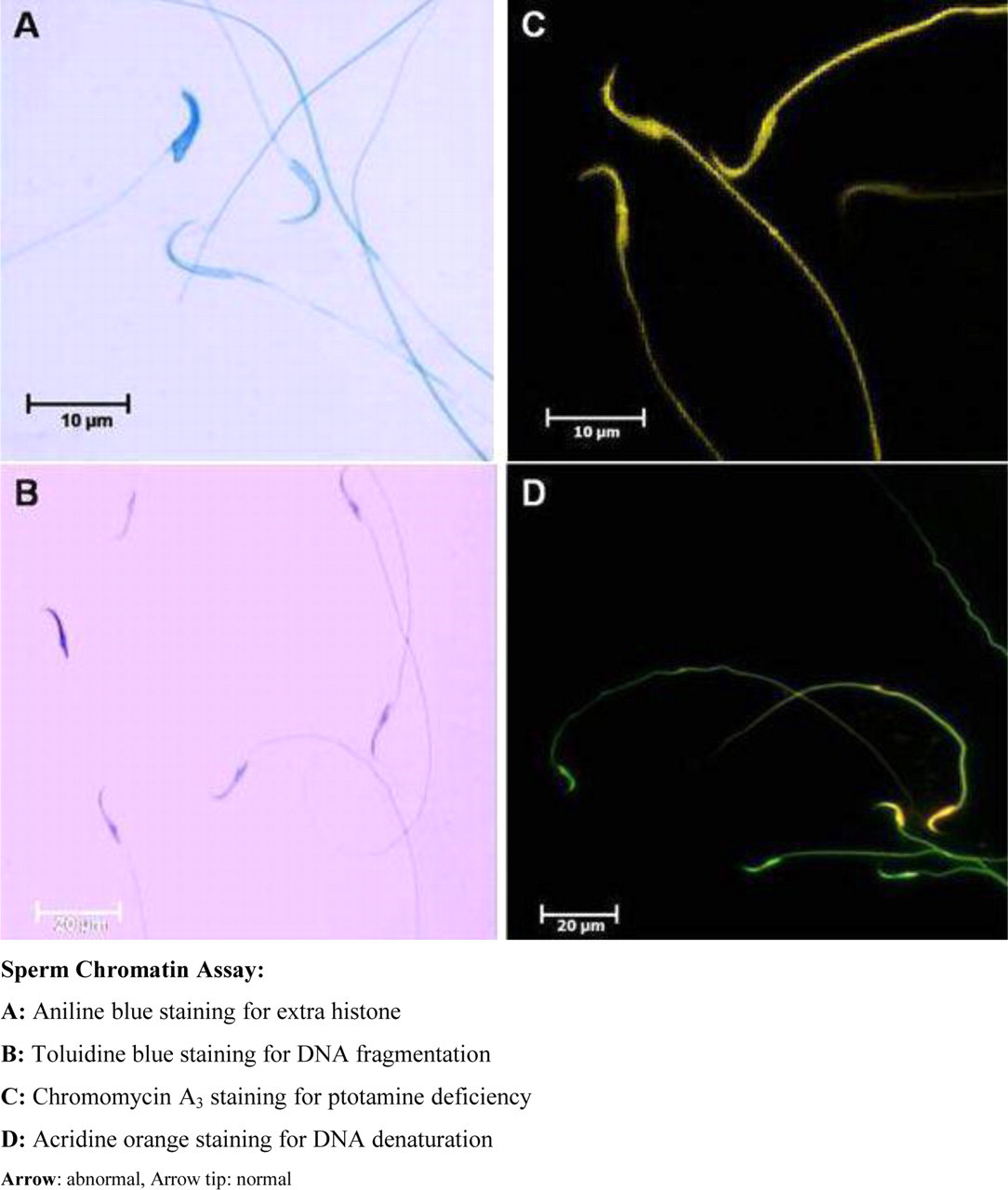
Characteristics of nuclear DNA integrity of spermatozoa retrieved from the caudal epididymis of rats. Sperm chromatin assay: (A) aniline blue staining for extra histone; (B) toluidine blue staining for DNA fragmentation; (C) chromomycin A3 staining for ptotamine deficiency; (D) acridine orange staining for DNA denaturation. Arrow, abnormal, Arrow tip, normal.
Toluidine blue TB staining
Dried smears were fixed in 96% ethanol and acetic acid (1:1) for 30 minutes, then hydrolyzed with 0.1 N, HCl for 5 minutes in 4°C. The slides were washed with distilled water 3 times for 2 minutes and stained with 0.05% TB for 5 minutes. Sperms with light blue heads were normal with good DNA integrity, while those with dark heads (purple) were abnormal with poor DNA integrity 16,17 (Figure 1B).
Chromomycine A3 CMA3 staining
Spermatozoa retrieved from twice-washed swim-up samples as well as from semen smears were fixed in methanol/glacial acetic (3:1) at 4 for 5 minutes and then spread on slides. Each slide was treated for 20 minutes with 100 µL of CMA3 (Sigma) solution (0.25 mg/mL in McIIvaline buffer [pH 7.0] containing 10 mmol/L MgCl2). The slides were then rinsed in the buffer and mounted with buffered glycerol. Fluorescence analysis was performed using an Olympus microscope (Germany) with appropriate filters. We randomly examined 200 spermatozoa on each slide. The results were evaluated on the basis of the color: bright yellow stained spermatozoa (CMA3 positive) and dull yellow stained spermatozoa (CMA3 negative) 29 (Figure 1C).
Acridine orange AO staining
A small aliquot (20 µL) was smeared on precleaned glass slides. The smeared slides were air-dried and later fixed overnight in freshly prepared Carnoy’s solution (methanol/acetic acid, 3:1). After fixation, the slides were air-dried and stained for 5 minutes with freshly prepared 0.19 mg/mL AO stain as follows:
Ten milliliter of 1% AO in distilled water was added to a mixture of 40 mL of 0.1 mol/L citric acid and 2.5 mL of 0.3 mol/L Na2HPO4.7H2O. The AO solution was stored in dark at 4°C for 4 weeks.
After staining, the slides were washed with distilled water, covered with glass cover slips, and immediately examined using a fluorescent microscope (Leitz, Germany; at the excitation wavelength of 450-490 nm). An average of 200 sperm cells were counted on each slide by the same examiner, and the duration of evaluation was not more than 40 seconds per field. Spermatozoa showing green fluorescence had normal DNA, whereas sperms showing a spectrum of yellow-orange to red fluorescence had damaged DNA 30 –32 (Figure 1D).
Statistical Analyses
All values are presented as mean ± standard deviation (SD). One-way analyses of variance (ANOVA) and post-hoc Duncan test were performed to determine the differences in the staining characteristics of all groups using the SPSS/PC computer program (version 13.OSS). A probability of P < .05 was considered to be statistically significant.
Results
The results of comparison of sperm nuclear integrity (CMA3, AB, AO, and TB staining) in the 5 groups namely, control, sham, varicocele, and treated groups are shown in Table 1 . The results for control and sham groups did not differ significantly, but the varicocele group showed a significant increase in comparison with the control group (P < .05). There was also a significant decrease between the AG-treated and varicocele groups in AO and TB tests (P < .01).
Characteristics of Nuclear DNA Integrity of Spermatozoa in the 4 Groups a
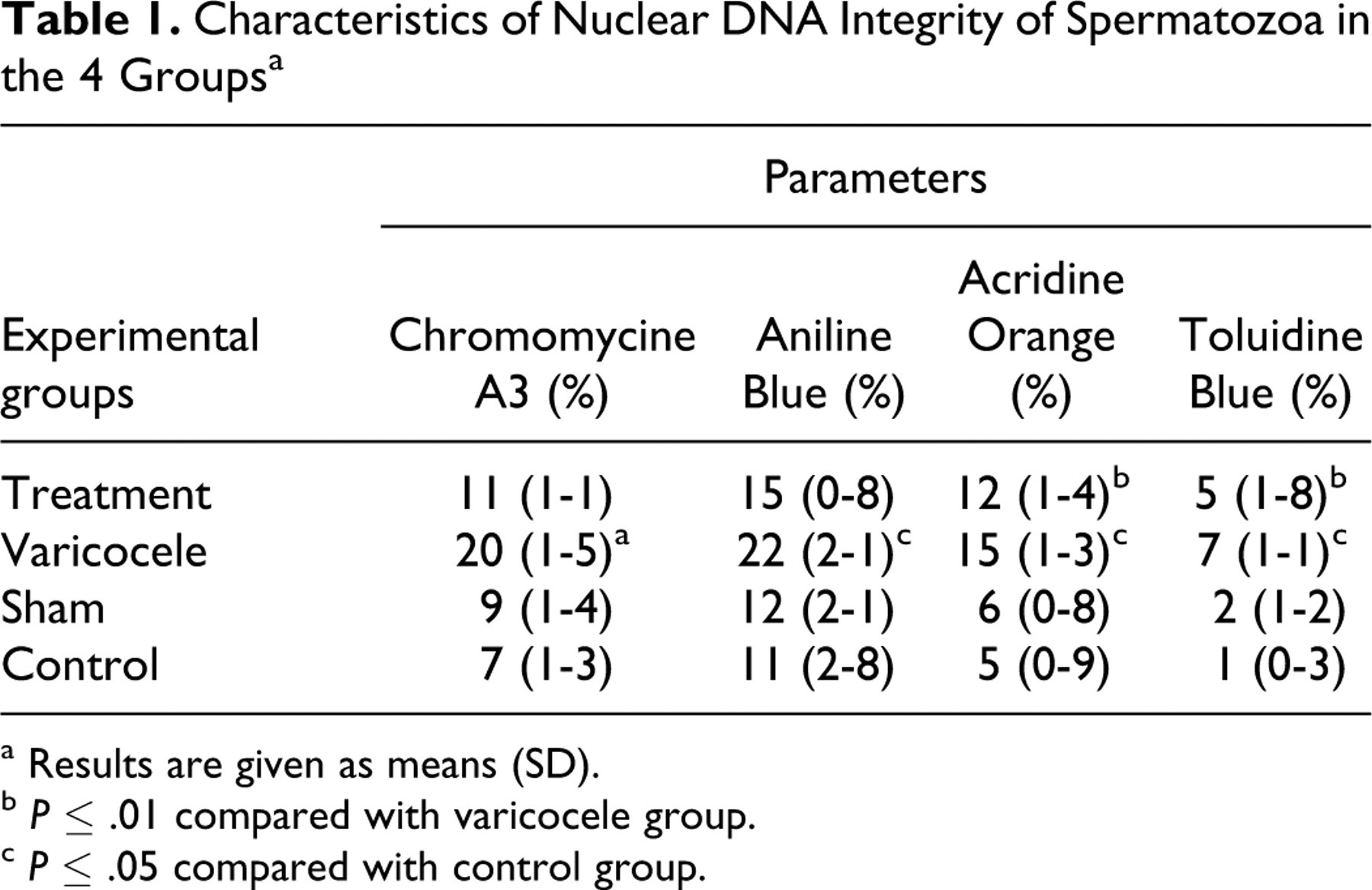
a Results are given as means (SD).
b P ≤ .01 compared with varicocele group.
c P ≤ .05 compared with control group.
Discussion
Varicocele is the abnormal dilatation of testicular veins that drain the testicle. 33 –35 Infertility-related to varicocele has multiple pathophysiological mechanisms. The effects of elevated seminal ROS and the role of NO in infertile patients with varicocele have been extensively studied. 36 Nitric oxide is considered as the causative factor of poor sperm function in varicocele. 5,33,37,38 The mean NO concentration in the seminal plasma of males with varicocele was significantly higher than that in the control. 20
Nitric oxide synthase activity is high in dilated veins and Leydig cells in patients with varicocele. 6,23 Varicocele has no significant effect on nNOS production 39 and both normal and varicocele patients express eNOS in blood vessels and Leydig cells 5 ; however, the expression of iNOS is markedly elevated in the testis of varicocele patients. Some authors reported that iNOS activity is upregulated in varicocele patients and that it is a possible cause of elevated NO levels. 5,6
According to one of the most important hypotheses, the molecular mechanism of sperm DNA damage involves ROS. 1,38 Sperm chromatin abnormalities, such as enhanced ROS production and high number of sperm cells with fragmented DNA were observed in rat models of varicocele. 4
In our experiment, varicocele rats exhibited a significant increase in chromatin abnormalities in the 4 tests (AO, TB, CMA3, and AB; P < .05). Recent studies have shown that varicocele patients have a significantly higher proportion of sperm cells with fragmented DNA than fertile men. 2,4,40 Smith et al reported that the number of spermatozoa with damaged DNA is markedly high in patients with varicocele. 41 Further, Talebi et al showed that varicocele patients have a higher proportion of spermatozoa with abnormal DNA and immature chromatin than fertile men as well as infertile men without varicocele. Therefore, infertility in varicocele patients may be attributed to the production of spermatozoa with poorly condensed chromatin. 35
Changes in sperm chromatin after the induction of varicocele may be mainly due to the direct effects of elevated NO levels in dilated veins. In adult varicocele patients, the NO levels in the varicose veins are 25 times higher than in those peripheral veins. 5,33 Oxidative stress affects the integrity of sperm genome by introducing single strand DNA breaks at high frequencies. 2,42,43 Because iNOS activity was markedly elevated in the Leydig cells of varicocele rats, some researchers suggest that iNOS activity may play an important and novel role in the testicular dysfunction associated with varicocele. 6
Guanidines such as
In our study, after AG administration to varicocele rats, DNA integrity (AO and TB) improved significantly (P < .01); it may be due to inhibition of iNOS and reduction in NO. 23,45,46 The positive changes observed in sperm chromatin integrity in this study may be attributed to the antioxidant effect of AG. Aminoguanidine directly scavenges hydroxyl radicals and inhibits DNA damage. Perhaps, the interference of AG with free-radical generation could be one of the causes of increased DNA repair in varicocele patients. In this study, we did not determine the amount of NO in rats.
However, the A in a competitive manner suppress the action of
Although we observed improvement in the results of AO and TB tests after AG administration, DNA maturation (AB and CMA3) did not show significant improvement in the treated groups. Our results indicated that in addition to NO, there might be other factors that influence DNA condensation and maturation. Immaturity is a consequence of defective spermatogenesis that could also lead to differences in disulfide cross-linking and insusceptibility toward DNA fragmentation. 4 Patients with male factor infertility and those with varicocele may produce spermatozoa with poorly condensed chromatin, 47 which allows ROS to have better access to the inside of the sperm nuclei and cause more damage. 4
Conclusion
The results of this study suggest that AG improves sperm DNA fragmentation that is associated with infertility in varicocelized rats. So some drugs such as AG may be useful for treatment of varicocele. Also, this study supports the hypothesis that AG exerts protective effects owing to its direct or indirect antioxidant activities, but it needs more experiments to determine the exact mechanisms of action.
Footnotes
The authors declared no potential conflicts of interests with respect to the authorship and/or publication of this article.
Acknowledgement
This research was supported by Tehran University of Medical Sciences, grant No 88-01-30-8370.
The authors received no financial support for the research and/or authorship of this article.