
Abstract
Ouabain can normalize the blood pressure of the adult intrauterine growth restriction (IUGR) offspring through retaining the number of glomeruli of the IUGR newborn. However, the melioration of hemodynamic features coinciding with the improvement in cardiac structure and function is poorly understood. Intrauterine growth restriction was induced in pregnant rats with protein intake restriction, and ouabain was administrated using osmotic mini pumps from the second gestational day. The male offspring of the mothers with normal diet, low-protein diet, and low-protein diet added with ouabain treatment were randomly divided into 2 groups, one of which received normal diet and the other was treated with isocaloric 8% high-salt diet. We found that maternal malnutrition caused fetal growth retardation. At the end of a 40-week research, the offspring of the IUGR group presented high blood pressure and deteriorative cardiac performance and even worse in the offspring fed with 8% high-salt diet. Ouabain can normalize the blood pressure and improve the cardiac performance, even if following 8% high-salt diet challenge. Pathological and molecular analyses showed IUGR following 8% high-salt diet significantly increased the cardiac hypertrophy, whereas the unfavorable effects were ameliorated in the offspring treated with ouabain. Results suggest that the effects of ouabain on restoration of glomerular number in newborn and normalization of blood pressure during adulthood in IUGR male offspring can benefit the cardiac structure and function, especially under high-salt diet challenge.
Introduction
Intrauterine growth restriction (IUGR), also known as fetal growth retardation, refers to the established fetal weight below the 10th percentile for the given gestational age. The incidence of IUGR is approximately 5% to 10%, and IUGR is the main cause of perinatal morbidity and mortality. 1 All these epidemiological studies on humans and animal models have revealed that IUGR is associated with the later development of hypertension and cardiovascular diseases in adulthood. 2 –4 Several rodent models of IUGR-induced hypertension have been developed, and hypertension has been hypothesized as a result of congenital deficiency in nephron number. 5 This congenital deficiency results in a reduced surface area for filtration and a subsequent reduction in glomerular filtration rate (GFR) as well as an increase in sodium retention and hypertension. Studies have also suggested that the incidence and severity of hypertension are higher in male offspring than in females. 6 –8
Systemic high blood pressure increases functional load on cardiomyocytes, thereby activating the signaling pathways and gene expression, upregulating protein synthesis and formation of novel sarcomeric units, and increasing the size of the heart and cardiomyocytes. 9 Left ventricular hypertrophy (LVH) is one of the main outcomes of chronic hypertension, which usually progresses to a decompensated stage of heart failure. The LVH is also an independent risk factor for myocardial infarction and arrhythmia. The pathogenesis of essential hypertension may most likely occur from the association of several pathophysiological stimuli with environmental factors and genetic background. High salt intake is an important environmental factor that may elevate blood pressure, promote hyperplasia and hypertrophy of myocardial cells, and facilitate collagen deposition, thereby resulting in cardiac hypertrophy and myocardial fibrosis. 10,11 Hypertension caused by IUGR together with high-salt diet challenge may enhance the cardiac insults. 12
Ouabain is a poisonous cardiac glycoside that inhibits the activity of sodium–potassium adenosine triphosphatase (Na-K-ATPase). Ouabain is used to treat heart failure, angina pectoris, and myocardial infarction by blocking the sodium transporter. Recent studies have indicated Na-K-ATPase as a signal transducer, and low concentrations (nanomolar or subnanomolar) of ouabain may stimulate Na-K-ATPase, triggering several complex signal pathways and exerting different biological effects. 13,14 Further investigation on cultured embryonic kidneys has suggested that low concentration of ouabain decreases the nephric apoptosis exerted by serum deprivation. 15 In vivo study has presented that ouabain restores nephron number, reversing the unfavorable consequences of maternal protein restriction. 16
Although ouabain can restore the glomerular number in newborn after IUGR, the long-term outcomes about blood pressure and cardiac function should be clarified. To assess the effects of ouabain treatment during pregnancy on the blood pressure and cardiac function of adult IUGR offspring, particularly during high-salt diet challenge, Sprague-Dawley (SD) rats were used, and IUGR was induced by maternal protein restriction, whereas ouabain was delivered using osmotic mini pump. The male offspring were fed with either normal diet (0.1% sodium chloride) or isocaloric high-salt diet (8% sodium chloride) after weaning and followed up for 40 weeks. Blood pressure, cardiac function, heart weight, histological features, and messenger RNA (mRNA) related to cardiac hypertrophy were studied.
Methods and Materials
Animals
All procedures on animals were approved by the Ethics Review Board for Animal Studies of Drum Tower Hospital affiliated to the Medical School of Nanjing University and performed in accordance with the guide for the care and use of laboratory animals published by the US National Institutes of Health (Publication No. 85-23, revised 1996). Fifteen female SD rats weighing between 250 and 265 g were used. The rats received standard chows and purified water before mating. Female rats were mated with male rats during estrus determined by daily vaginal smears. Gestational day 0 (GD0) was designated by the appearance of sperm in vaginal smears. Pregnant rats were individually housed in standard rat cages with water ad libitum. The food intake of each rat was recorded daily.
The 15 pregnant rats were randomly divided into 3 groups. In the control group (n = 5), all pregnant rats received standard laboratory rat chow (No. 20121023, Xietong Organism, Nanjing, Jiangsu, China; protein, 22.10%; fat, 5.28%; carbohydrate, 52.00%; metabolizable energy, 3520 kcal/kg) throughout pregnancy. In the IUGR group (n = 5), all pregnant rats were fed with 75% caloric low-protein diet (SY01, Slac Laboratory Animal, Shanghai, China; protein, 9.00%; fat, 3.20%; carbohydrate, 72.6%; metabolizable energy, 3530 kcal/kg) determined by quantification of the control group mean food intake starting at GD0 and continuing throughout pregnancy. In the ouabain group (n = 5), all pregnant rats received the same diet as that fed to the IUGR group, and an osmotic mini pump delivering ouabain (10 μg/kg body weight/d) was implanted in the abdominal cavity.
The newborn rats were weighed within 6 hours after birth, and the dams were returned to normal diet (22.1% protein). After weaning, 3-week-old male offspring from each group were randomly allocated to receive either a normal diet (0.1% sodium chloride, Xietong Organism) or an isocaloric high-salt diet (8% sodium chloride, Xietong Organism). Thus, the male offspring were divided into 6 groups as follows: control + normal diet (control/ND), IUGR + normal diet (IUGR/ND), ouabain + normal diet (ouabain/ND), control + high-salt diet (control/HS), IUGR + high-salt diet (IUGR/HS), and ouabain + high-salt diet (ouabain/HS). The male offspring were followed up for measurement of blood pressure and echocardiographic parameters then killed at the age of 40 weeks.
Measurement of Blood Pressure
A programmable tail-cuff sphygmomanometer (BP-2006A; Softron, Tokyo, Japan) was used to measure the systolic blood pressure (SBP), diastolic blood pressure (DBP), and heart rate. The male rats were first trained to acquaint with the measurement devices and then allowed to remain calm in the mental chamber during the inflation–deflation cycle. The measurement was conducted in a quiet room, and the male rats were kept in a 37°C mental chamber for 15 minutes warming to relatively eliminate stress during the blood pressure measurement. To eliminate the bias resulting from struggling or other physiological alterations, the readings were used only if the standard deviation of the measurement was <6 mm Hg. An average of 3 readings was recorded for each animal.
Echocardiographic Analysis
Rat cardiac function was assessed by transthoracic ultrasonic cardiogram using VisualSonic Vevo2100 (Fujifilm VisualSonics, Inc, Toronto, Canada) ultrasound system equipped with a 30-MHz transducer. The male rats were anesthetized by chloral hydrate. Their chests were shaved and hair removal gel was applied to minimize resistance to ultrasonic beam transmission. The male rats were then fixed on a heating pad with rectal temperature kept between 35°C and 37°C during the measurement. The paws were connected to the eletrocardiogram. Each echocardiographic examination was conducted by one experienced examiner and accomplished within 15 minutes after induction of anesthesia. M-mode measurements of the left ventricle (LV) dimensions were averaged from more than 3 cycles. Interventricular septal thickness in diastole (IVSd) and systole (IVSs), LV posterior wall thickness in diastole (LVPWd) and systole, and LV internal dimension in diastole (LVIDd) and systole (LVIDs) were measured. Percentage of LV ejection fraction (LVEF) was calculated as described previously.
Histological Study of Cardiomyocytes and Assessment of Fibrosis
At the age of 40 weeks, the male rats were anesthetized and their thoracic cavities were uncovered to expose their beating hearts. The hearts were rapidly harvested, rinsed in ice-cold 0.9% saline solution, blotted with liquid, weighed, and fixed with the neutral 4% formaldehyde for 48 hours. The hearts were then dehydrated in graded alcohols and embedded in paraffin. Sections of 2 μm thickness were sliced and stained with hematoxylin–eosin. Cross-sectional areas of the cardiomyocytes were measured at the nuclear level in longitudinally sectioned myocytes. Suitable cross-sections were defined as having nearly circular capillary profiles and nuclei. Five fields of LV sections were randomly selected, and captured images were analyzed by Image-Pro Plus 6.0 image analysis software. At least 50 cells per randomly chosen microscopic field were selected to calculate the mean cardiomyocyte cross-sectional area. Fibrosis was detected by Masson trichrome staining on paraffin-embedded sections.
Evaluation of Hypertrophic Marker Gene Expression in Cardiac Tissues
Hypertrophic marker gene expression was analyzed by quantitative real-time polymerase chain reaction (qPCR). RNA was isolated from LV samples with Trizol (Qiagen, Valencia, California). Complementary DNA (cDNA) was synthesized using iScript cDNA synthesis kit (Bio-Rad Laboratories, Hercules, California). The qPCR was performed using iQ SYBR Green supermix kit (Bio-Rad Laboratories) in a CFX96 real-time PCR detection system (Bio-Rad Laboratories). The following primers were used: glyceraldehyde 3-phosphate dehydrogenase (GAPDH) forward, 5′-TCTACATGTTCCAGTATGACTC-3′; GAPDH reverse, 5′-ACTCCACGACATACTCAGCACC-3′; atrial natriuretic peptide (ANP) forward, 5′-CTCCGATAGATCTGCCCTCTTGAA-3′; ANP reverse, 5′-GGTACCGGAAGCTGTTGCAGCCTA-3′; b-type natriuretic peptide (BNP) forward, 5′-TTGGGCAGAAGATAGACCGGAT-3′; BNP reverse, 5′-GGTCTTCCTAAAACAACCTCA-3′; beta-myosin heavy chain (β-MHC) forward, 5′-AACCTGTCCAAGTTCCGCAAGGTG-3′; β-MHC reverse, 5′-GAGCTGGGTAGCACAAGAGCTACT-3′. The expression level of GAPDH was used as internal control.
Statistical Analysis
The results are expressed as mean ± standard error of the mean. Differences in birth weight, blood pressure, echocardiographic elements, and cross-sectional areas of cardiomyocytes were assessed by 2-way analysis of variance. Significance was considered at P value <.05. Statistical analysis was performed using SPSS Statistics, Release 18 (IBM Software Group, Chicago, Illinois).
Results
Maternal Protein Restriction Caused Fetal Growth Restriction
Compared with the control group, low-protein diet significantly reduced the birth weights in the IUGR and ouabain groups (IUGR vs control, P < .001; ouabain vs control, P < .001; ouabain vs IUGR, P = .749; Figure 1A). At the age of 3 weeks, the body weights of the IUGR and ouabain groups remained significantly less than those of the control group. No significant difference existed between the IUGR and the ouabain groups (IUGR vs control, P < .001; ouabain vs control, P < .001; ouabain vs IUGR, P = .062; Figure 1B).
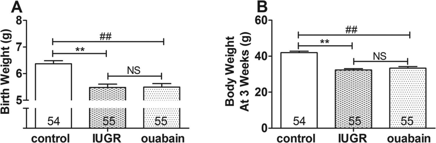
Body weights of offspring at birth and at 3 weeks of age. A, Birth weights. B, Body weights at 3 weeks of age. Results are presented as mean ± standard error of the mean (SEM). *P < .05, **P < .001, intrauterine growth restriction (IUGR) group versus control group; # P < .05, ## P < .001, ouabain group versus control group; & P < .05, && P < .001, ouabain group versus IUGR group.
Blood Pressure Significantly Increased in the Offspring of IUGR Group, Especially Under High-Salt Diet Challenge
Among the male offspring fed with normal diet, the IUGR/ND group exhibited significantly higher SBP than those of the control/ND and ouabain/ND groups (IUGR/ND vs control/ND, P < .001; ouabain/ND vs IUGR/ND, P < .001; ouabain/ND vs control/ND, P = .119; Figure 2A). The DBP of the IUGR/ND group was also significantly higher than those of control/ND and ouabain/ND groups (IUGR/ND vs control/ND, P = .001; ouabain/ND vs IUGR/ND, P = .032; ouabain/ND vs control/ND, P = .114; Figure 2C).
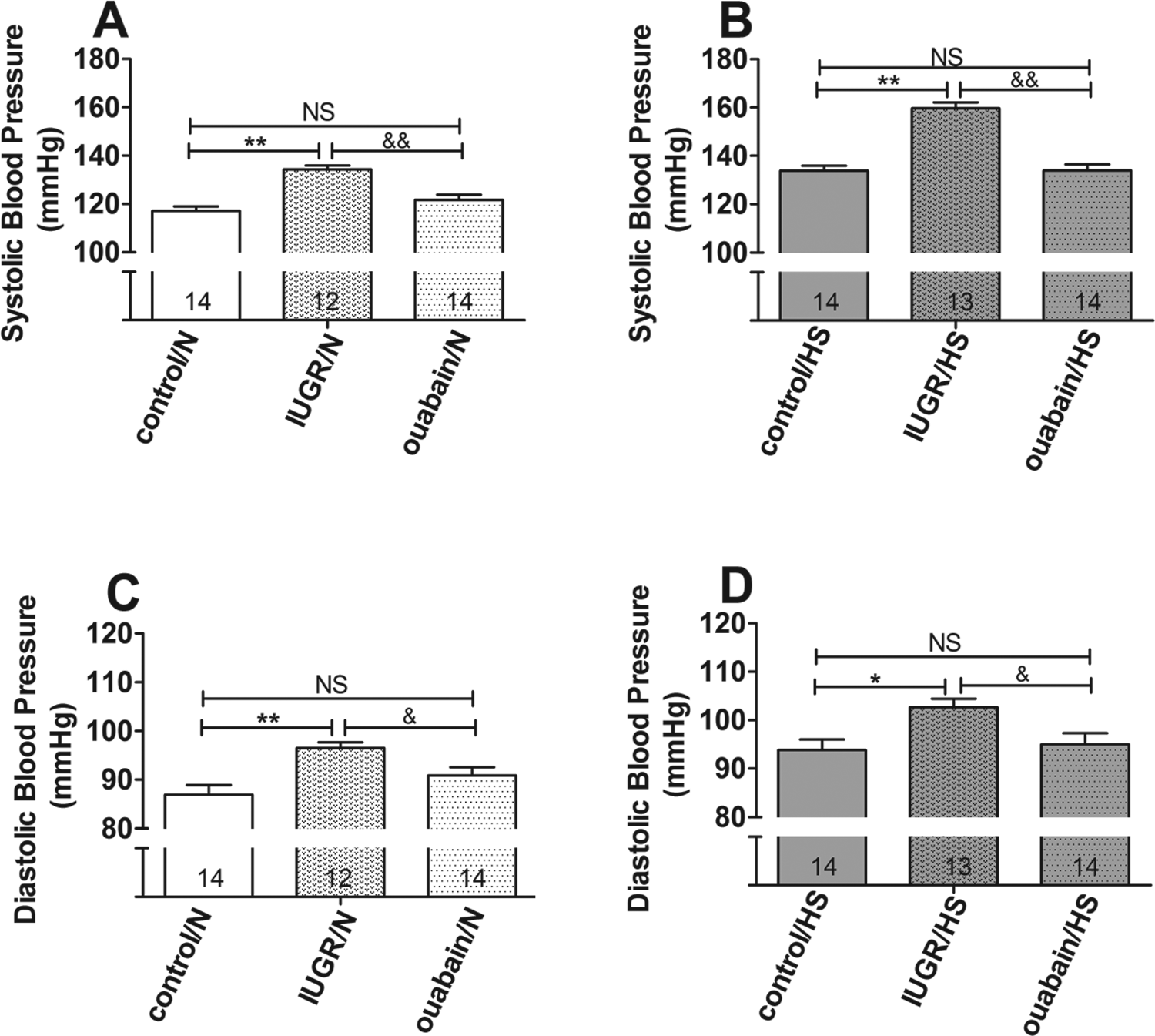
Systolic and diastolic blood pressures (SBP and DBP) in control + normal diet, IUGR + normal diet, ouabain + normal diet, control + high-salt diet, intrauterine growth restriction (IUGR) + high-salt diet, ouabain + high-salt diet groups. A, The SBP of male offspring receiving normal diet. B, The SBP of male offspring receiving 8% high-salt diet. C, The DBP of male offspring fed with normal diet. D, The DBP of male offspring fed with 8% high-salt diet. Results are mean ± standard error of the mean (SEM). *P < .05, **P < .001, IUGR group versus control group; # P < .05, ## P < .001, ouabain group versus control group; & P < .05, && P < .001, ouabain group versus IUGR group. NS indicates no significant difference.
The male offspring that received 8% high-salt diet presented higher SBP and DBP than those of the offspring treated with normal diet. In the IUGR/HS group, high-salt diet significantly increased the SBP, which was significantly higher than those of the control/HS and ouabain/HS groups (IUGR/HS vs control/HS, P < .001; ouabain/HS vs IUGR/HS, P < .001; ouabain/HS vs control/HS, P = .967; Figure 2B). Compared with those of the control/HS and ouabain/HS groups, the DBP of the IUGR/HS group also significantly increased (IUGR/HS vs control/HS, P = .008; ouabain/HS vs IUGR/HS, P = .019; ouabain/HS vs control/HS, P = .712; Figure 2D).
High-Salt Diet Impaired the Cardiac Performance and Increased the Heart Size of IUGR/HS Group
Figure 3 illustrates representative tracings of M-mode echocardiography at the age of 40 weeks. No significant difference existed in the IVSd, IVSs, LVPWd, LVIDd, LVIDs, and LVEF among the control/ND, IUGR/ND, and ouabain/ND groups, except for the IVSd of the IUGR/ND group, which significantly increased compared to that of control/ND group (Table 1). Compared to the groups fed with normal diet, the 8% high-salt diet increased the dimensions of IVSd, IVSs, LVPWd, LVIDd, and LVIDs. Compared to the control/HS and ouabain/HS groups, high-salt diet significantly increased the IVSd, LVPWd, and LVIDd in the IUGR/HS group, whereas no significant differences existed in the IVSd, IVSs, LVPWd, LVIDd, LVIDs, and LV EF between the control/HS and the ouabain/HS groups.

Representative M-mode echocardiograms of control, IUGR, and ouabain rat offspring fed with either normal diet or 8% high-salt diet. ND indicates normal diet; HS, 8% high-salt diet.
Echocardiographic Studies of Heart Dimensions and Functions at Age of 40 Weeks.
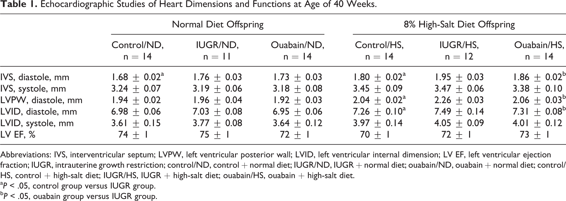
Abbreviations: IVS, interventricular septum; LVPW, left ventricular posterior wall; LVID, left ventricular internal dimension; LV EF, left ventricular ejection fraction; IUGR, intrauterine growth restriction; control/ND, control + normal diet; IUGR/ND, IUGR + normal diet; ouabain/ND, ouabain + normal diet; control/HS, control + high-salt diet; IUGR/HS, IUGR + high-salt diet; ouabain/HS, ouabain + high-salt diet.
a P < .05, control group versus IUGR group.
b P < .05, ouabain group versus IUGR group.
Maternal protein restriction significantly reduced the net heart weight and the heart weight–body weight ratio of the offspring fed with normal diet. No significant difference existed between the IUGR/ND and the ouabain/ND groups (heart weight: IUGR/ND vs control/ND, P = .007; ouabain/ND vs IUGR/ND, P = .962; ouabain/ND vs control/ND, P = .004; heart weight–body weight ratio: IUGR/ND vs control/ND, P = .020; ouabain/ND vs IUGR/ND, P = .669; ouabain/ND vs control/ND, P = .039; Figure 4A and C). The 8% high-salt diet significantly increased the heart weight and the heart weight–body weight ratio of the IUGR/HS group compared to those of the control/HS and ouabain/HS groups. No significant difference was observed between the control/HS and the ouabain/HS groups (heart weight: IUGR/HS vs control/HS, P = .044; ouabain/HS vs IUGR/HS, P = .035; ouabain/HS vs control/S, P = .941; heart weight–body weight ratio: IUGR/HS vs control/HS, P < .001; ouabain/HS vs IUGR/HS, P = .004; ouabain/HS vs control/HS, P = .291; Figure 4B and D).
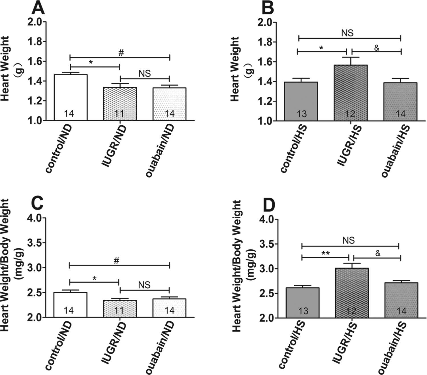
Heart weight and heart weight–body weight ratio. A, Heart weight of male offspring fed with normal diet. B, Heart weight of male offspring fed with 8% high-salt diet. C, Heart weight–body weight ratio of male offspring fed with normal diet. D, Heart weight–body weight ratio of male offspring fed with 8% high-salt diet. Results are mean ± standard error of the mean (SEM). *P < .05, **P < .001, intrauterine growth restriction (IUGR) group versus control group; # P < .05, ## P < .001, ouabain group versus control group; & P < .05, && P < .001, ouabain group versus IUGR group.
Histological Study Showed Significantly Increased Myocardial Cross-Sectional Area and Fibrosis in IUGR/HS Group
Figure 5A shows a representative cross-sectional area of cardiomyocytes in hematoxylin–eosin-stained transverse LV sections that were used to assess the minor diameter of myocytes. The cardiomyocytes cross-sectional area in the IUGR/ND group was slightly higher than those in the control/ND and ouabain/ND groups. No significant difference existed among these 3 groups (IUGR/ND vs control/ND, P = .085; ouabain/ND vs IUGR/ND, P = .236; ouabain/ND vs control/ND, P = .578; Figure 5B). Compared to the male offspring fed with normal diet, the 8% high-salt diet increased the cardiomyocyte cross-sectional area in IUGR/HS group, which was significantly higher than those in the control/HS and ouabain/HS groups (IUGR/HS vs control/HS, P = .001; ouabain/HS vs IUGR/HS, P = .021; ouabain/HS vs control/HS, P = .167; Figure 5C).
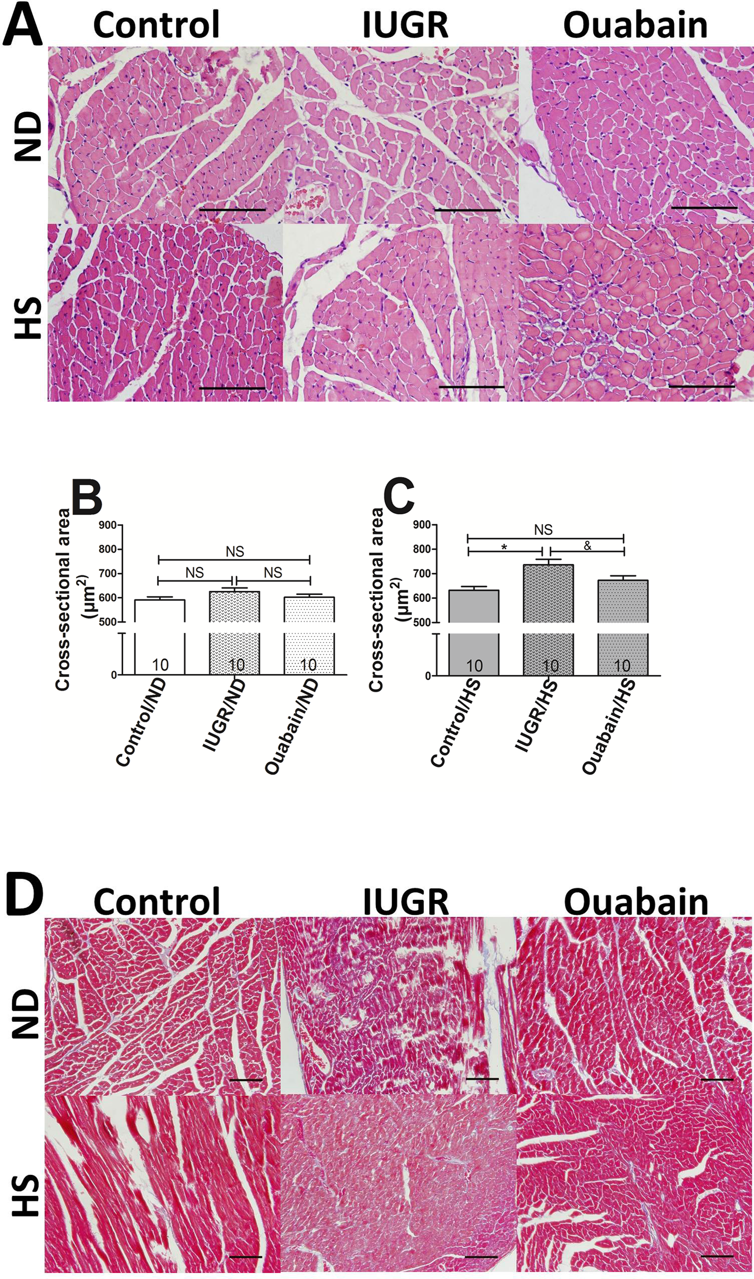
Effects of maternal protein restriction with or without ouabain treatment subsequent to normal diet or 8% high-salt diet. A, Sample slices of the hematoxylin–eosin-stained transverse sections of the left ventricle. B, Cross-sectional area of offspring fed with normal diet. C, Cross-sectional area of offspring fed with 8% high-salt diet. D, Histological analysis of heart section stained with Masson trichrome. Bars = 100 μm. Results are mean ± standard error of the mean (SEM). *P < .05, **P < .001, intrauterine growth restriction (IUGR) group versus control group; & P < .05, && P < .001, ouabain group versus IUGR group.
We also used Masson trichrome staining to evaluate the LV fibrosis. The results showed that fibrosis was positively related to blood pressure, which may lead to pathological cardiac hypertrophy.
Quantitative Real-Time PCR Showed 8% High-Salt Diet Increased the Expression of Hypertrophic Marker Genes, Especially in IUGR/HS Group
The mRNA expression of ANP, BNP, and β-MHC was induced by 8% high-salt diet, and the expression of these 3 genes was significantly higher in the IUGR/HS group than in the control/HS and ouabain/ HS groups (Figure 6A–C). No significant difference existed among the 3 groups, which all received normal diet.
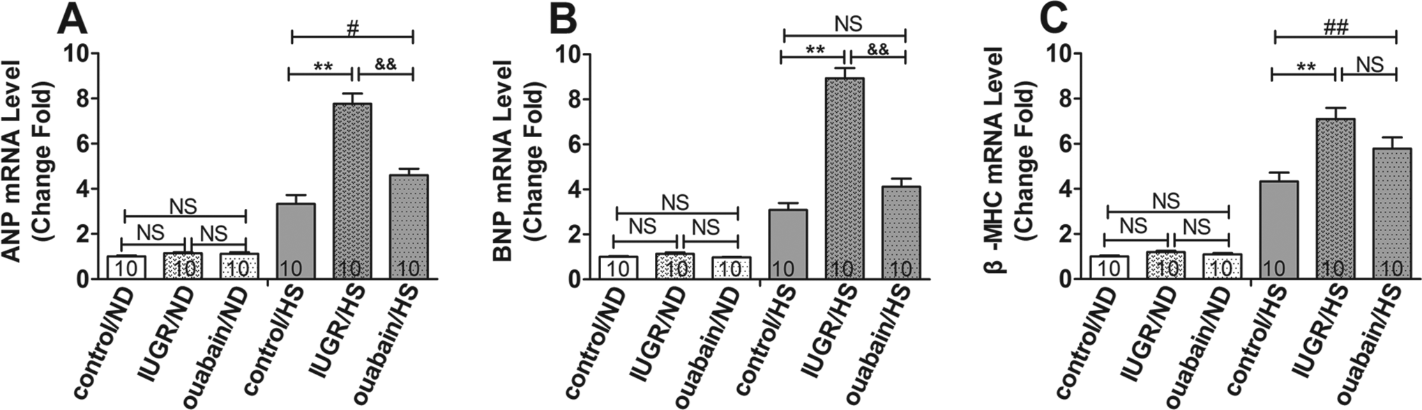
Expression of ANP, BNP, and β-MHC of left ventricular tissue. A, ANP expression level was induced by 8% high-salt diet and significantly higher in intrauterine growth restriction (IUGR) + high-salt diet (IUGR/HS) group. B, BNP expression was upregulated in offspring fed with high-salt diet and even higher in IUGR/HS group. C, β-MHC expression level increased in offspring treated with high-salt diet. Results are mean ± standard error of the mean (SEM). *P < .05, **P < .001, IUGR group versus control group; # P < .05, ## P < .001, ouabain group versus control group; & P < .05, && P < .001, ouabain group versus IUGR group.
Discussion
The present study mainly revealed that ouabain can prevent later development of hypertension induced by maternal malnutrition, especially following high-salt diet, thereby reducing hypertrophy of cardiomyocytes and benefiting cardiac function. Ouabain may also be a new therapeutic alternative for curing the unfavorable effects of IUGR on fetal kidney.
The concept of the “fetal programming” aims to explain the association of unfavorable perinatal events (eg, malnutrition, stress, and alcohol or tobacco abuse) and the susceptibility to certain diseases in adulthood (eg, metabolic syndrome, hypertension, and cardiovascular diseases). 17 –19 Several studies have demonstrated that maternal malnutrition will compromise the development of fetal kidney and reduce the kidney volume accompanied with significant decrease in glomerular number. 20 Long-term follow-ups after birth have shown an affected renal function and an increased incidence of hypertension in the IUGR group compared with the control group. 3 Interestingly, epidemiological studies have indicated that the IUGR male offspring increased susceptibility to hypertension and cardiovascular and renal risks in a sex-specific manner. 6 –8 Thus, in the current study, we used male rat offspring to explore the adverse outcomes of IUGR and the effects of ouabain. Our study showed that the male IUGR offspring exhibited higher SBP and DBP than those of the control group, whereas the female offspring were slightly affected, and no significant difference existed between the IUGR and the control groups (data not shown).
In addition to the function of Na-K-ATPase as an ion pump, this enzyme can also serve as a signal transducer. As a highly specific ligand of Na-K-ATPase, the classical mechanism of action of ouabain involves inhibition of the ion pump, especially at the high concentrations. Besides the typical mechanism, low (nanomolar or subnanomolar) concentrations of ouabain stimulate the Na-K-ATPase and act as signal transducer. 13 Studies have shown that low concentrations of ouabain can activate the calcium-dependent transcription factor nuclear factor κB and protect nephrogenesis from serum deprivation in vitro or maternal malnutrition in vivo. 15,16 Researches have attributed hypertension of IUGR offspring to reduction in glomerular number. Considering ouabain can rescue nephrogenesis during maternal protein restriction, whether ouabain can normalize the blood pressure becomes unclear. Our current study indicated that ouabain decreased the high blood pressure induced by IUGR. Although the SBP and DBP of the ouabain group remained higher than those of the control group, no significant difference existed between the ouabain and the control groups.
Renal sodium excretory function is intrinsically accountable for the long-term control of blood pressure. Maternal undernutrition decreases the number of nephron and renal hemodynamic alteration, thereby resulting in reduced GFR. 21 However, maternal undernourishment increases tubular sodium transporters and renal oxidative stress, thus increasing the reabsorption of sodium. 5 Decreased GFR and increased sodium reabsorption together contribute to hypertension. The kidney is an organ of strong reserve capacity. Thus, compromised nephron endowment encountering adverse event challenge (eg, high dietary sodium intake) will deteriorate hypertension. We applied isocaloric 8% high-salt diet to mimic the adverse challenge until 40 weeks old. Our results showed that long-term dietary sodium intake increased the SBP for 16, 26, and 13 mm Hg in the control, IUGR, and ouabain groups, respectively. The results suggested that IUGR decreased the reserve capacity of kidney, and exacerbated hypertension. Therefore, ouabain can partially reverse the undesirable effects.
High blood pressure increases the afterload that the heart needs to contract against, thereby resulting in adaption of the cardiac function and structure. Compensatory LV hypertrophy is a pathological reaction to long-term hypertension at the middle stage, and LV failure is the end-stage outcome. 22,23 Echocardiography is the principal method for assessing the cardiac function, with which the thickness of the muscle can be measured. The LVH showed increased IVSd, LVPWd, and LVIDd. Pathological analysis of LVH usually shows increased heart weight or heart weight–body weight ratio, enlarged cardiac fiber, and augment of fibrosis.
Our results showed that the cardiac function was slightly affected in the offspring fed with normal diet, except the IVSd of the IUGR/ND group, which was significantly higher than that of the control/ND group. However, the pathology indicated that maternal protein restriction may decrease the heart weight of the offspring, even if modified by body weight, and ouabain cannot reverse this effect. The cross-sectional areas of the cardiomyocytes also did not differ significantly among the 3 groups fed with normal diet, whereas fibrosis was more evident in the IUGR/ND group than in the control/ND and ouabain/ND groups. These findings suggested that IUGR may decrease the heart weight of the offspring, and ouabain cannot cure this effect. Fortunately, the heart can still function well with mild alteration in cardiac structure, but this condition will eventually lead to mild or moderate hypertension.
However, many changes will occur when the offspring were treated with 8% high-salt diet. The increased volume load of the heart resulted from increased dietary sodium intake and decreased sodium excretion, accompanied with disordered renin–angiotensin–aldosterone system, which significantly increased the blood pressure of the IUGR/HS group compared to those of the control/HS and ouabain/HS groups. 24,25 Therefore, the IUGR group exhibited poor reserve capacity to deal with the unfavorable events. Eventually, malignant hypertension will deteriorate the cardiac function. Our study showed that 8% high-salt diet affected the cardiac performance, and the IVSd, LVPWd, and LVIDd of the IUGR/HS group were even higher than those of the control/HS and ouabain/HS groups. Morphological and pathological studies were consistent with the echocardiographic measurements, which exhibited increased heart weight, heart weight–body weight ratio, cross-sectional area of cardiomyocyte, and collagen deposition. The results strongly suggested that ouabain can protect the cardiac function and structure of the IUGR offspring against high-salt diet challenge.
At pathological conditions, multiple signaling pathways are disturbed, leading to myocardial hypertrophy, vascular dysfunction, matrix remodeling, and ultimately heart failure. Cardiac hypertrophy has been associated with the reactivation of ANP, BNP, and β-MHC genes. 26,27 Hence, we also investigated the expression level of ANP, BNP, and β-MHC, and the results showed that the mRNA levels of ANP, BNP, and β-MHC were highly induced by 8% high-salt diet and even higher in the IUGR/HS group than in the control/HS and ouabain/HS groups.
Conclusion
Maternal protein restriction will result in hypertension in adult offspring, especially when the IUGR offspring underwent unfavorable environmental events. Ouabain administrated during pregnancy can normalize the blood pressure of the IUGR offspring, even if challenged by adverse events, through decreasing the apoptosis of the embryonic kidney and increasing the glomerular number in the newborn. This advantage may protect the cardiac function and structure against malignant hypertension resulting from the double whammy of impaired endowment of nephron and postnatal detrimental lifestyle. Low-sodium diet should be the conventional intervention for the IUGR offspring to prevent the injury on the renal and cardiac function resulting from high GFR and hypertension. This study is also our first proposal for a totally new alternative to treat dysplasia of a specific organ in the IUGR.
Footnotes
Authors’ Note
This research job was mainly conducted at the Nanjing Drum Tower Hospital affiliated to Medical School of Nanjing University, Nanjing City, Jiangsu Province, China.
Acknowledgments
The authors would like to thank Dr Kui Meng, Dr Qiang Zhou, and Dr Hong Sun of the Pathological Department of the Nanjing Drum Tower Hospital for their technical assistance in the histological study. We also appreciate Kairong Wu, Xiaokang Li, Liping Qian, and Yeqing Qu of the Animal Research Center of the Nanjing Drum Tower Hospital for the rats breeding.
Declaration of Conflicting Interests
The author(s) declared no potential conflicts of interest with respect to the research, authorship, and/or publication of this article.
Funding
The author(s) disclosed receipt of the following financial support for the research, authorship, and/or publication of this article: This study was financially supported by the National Natural Science Foundation of China (No. 31171386).