






Abstract
Purpose:
Labor is thought to positively influence immune system development in the offspring, but studies investigating the impact of different modes of delivery on maternal immune system cells are scarce. Therefore, the aim of this study was to investigate the effect of labor on maternal peripheral blood T-cell subsets and on the recently described regulatory T and B cells.
Methods:
Cross-sectional study comparing the absolute counts and percentages of peripheral blood T-cell subsets (maturation and activation profiles) and regulatory T and B cells between healthy pregnant women who delivered their newborns via elective cesarean (no labor; n = 14) and those who had a spontaneous vaginal delivery (after labor; n = 18). The cells were characterized using flow cytometry.
Results:
We found that compared to the women who had elective cesareans, those who had spontaneous vaginal deliveries had significantly (P < .05) lower absolute counts of B cells (median [cells/μL]: 146 [interquartile range, IQR = 49] vs 192 [IQR = 65]) and natural killer-like T (NKT-like) cells (median [cells/μL]: 154 [IQR = 125] vs 224 [IQR = 117]) in the peripheral blood. No further significant differences, particularly in regulatory T and B cells, were identified between the study groups.
Conclusion:
Labor does not seem to have a major impact on maternal peripheral blood T-cell subsets or regulatory T and B cells.
Background
The immune system of a healthy pregnant woman undergoes a transformation that allows it to tolerate fetal alloantigens, thus permitting the development of the fetus in the maternal uterus. It is plausible that several types of immune cells might contribute to this maternal–fetal immunotolerance. Physiological pregnancy has been compared to a state of quiescent systemic inflammation, and parturition has been compared to an immunological reaction that results in the recruitment of immune cells to the systemic circulation and the maternal–fetal interface. 1 This entire process may have an effect on the maturation and activation profiles of T cells in the peripheral blood (PB) of pregnant women. 2,3
Traditionally, this state of immunological fetal tolerance has been explained by the predominantly Th2-type immunity found in normal pregnancies; in contrast, a predominantly Th1-type immunity was used to explain pathological pregnancies, such as those characterized by recurrent spontaneous abortion, intrauterine growth restriction, and preeclampsia. 4 –9 Currently, this immunotolerance is explained by both Th2-type immunity and the roles of regulatory T cells (Tregs). Tregs are potent suppressors of inflammatory immune responses. Furthermore, they play a crucial role in immune tolerance, and their role in maintaining pregnancy has widely been reported in both humans and mice. 10,11 A decrease in maternal Tregs could cause the fetus to be rejected, and such decreases have been reported in conditions related to preterm labor and to immunological tolerance failure, such as miscarriage and preeclampsia. 9,12 –15
Recent reports have suggested that another heterogeneous population of T cells might also have an important function in fetal–maternal immunological tolerance and in the regulation of Th1/Th2 balance. The CD3+CD56+CD16+ natural killer-like T (NKT-like) cell subset simultaneously expresses T cell and natural killer cell surface markers and has been found in PB and in human decidua. 16 Published data regarding the role of the circulating levels of NKT-like cells in embryo implantation have been contradictory. 17,18
Recent studies 19,20 show that in addition to their humoral activity, specific subsets of B cells have a regulatory function (regulatory B cells [Bregs]). Their counts increase during the first trimester of pregnancy, and they have the capacity to inhibit proinflammatory responses through the secretion of the anti-inflammatory cytokine interleukin 10, which protects against pregnancy loss. 20 These findings suggest that these cells with regulatory functions may be important for better understanding the mechanisms characterizing pregnancy and delivery.
Natural labor involves physiological processes that may not occur in pregnant women who deliver their newborns via elective cesarean. For example, during labor, the maternal immune system is activated, resulting in increased absolute counts and percentages of PB leukocytes 21 –23 mainly because of the increase in the absolute counts and percentages of neutrophils and the decrease in the absolute counts and percentages of PB lymphocytes. 24
The number of cesarean deliveries has dramatically increased worldwide over the past decades. Although a cesarean delivery can benefit the well-being of the mother and the child in certain situations, it is not a risk-free procedure; potential risks include infection, thrombosis, abnormal placentation, hysterectomy, and even maternal death, and cesarean deliveries are associated with more maternal complications compared to vaginal deliveries. Earlier studies have shown that offspring delivered via prelabor cesarean have a higher risk of immune disorders later in life, 25,26 and early changes in the subpopulations of lymphocytes can be detected in neonates soon after birth. 27 Data related to the PB T-cell subsets in mothers have been scarce, 27,28 and neither of the published studies of this topic investigated the effect of labor on maternal Tregs and Bregs. The results of such investigations would be clinically important because they could provide further knowledge regarding the impact of different modes of delivery on maternal health and the maternal immune system. Therefore, the objective of our study was to investigate the effect of labor on the maternal circulating T-cell subsets, Tregs, and Bregs.
Materials and Methods
Study Population
This was a cross-sectional study comparing the characteristics (ie, cell quantification and phenotype identification) of the PB T-cell subsets, Tregs, and Bregs between women who gave birth vaginally (ie, normal spontaneous vaginal birth) and by elective cesarean (ie, any surgical operation for delivering a baby through the abdominal wall without labor and with a duration of ruptured membranes lasting 5 minutes or less). The details of these modes of delivery were recorded in the patients’ obstetric records.
An experienced practitioner decided whether to opt for vaginal delivery or elective cesarean. The hospital’s procedure for managing vaginal deliveries and elective cesareans did not change over the course of our study. As part of this procedure, each newborn was evaluated at birth by a pediatrician.
Our study included a sample of the healthy pregnant women (n = 32) attending the outpatient clinic of our hospital. Healthy pregnant women were defined as women with asymptomatic, uncomplicated third trimester singleton pregnancies. These women received regular antenatal care and had appropriate fetal growth (based on uterine fundal height and ultrasounds performed after 28 weeks of gestation).
We excluded all women who did not have a term delivery (≥37 weeks of gestation); did not give birth to a newborn whose birth weight was above the 10th percentile for gestational age; had a labor induction, diabetes, hypertension, atherosclerosis, autoimmune disease, vascular disease, or renal disease; had spontaneous rupture of the fetal membranes 48 hours before labor; had any clinical or laboratory signs of infection; used prenatal medications other than vitamins, folic acid, and iron supplements; or smoked during the 6 months prior to the PB sample collection.
All of the women were recruited between July 2013 and March 2014 at Hospital CUF Descobertas in Lisboa (Portugal). The ethics committee of this hospital approved the study protocol. All of the procedures involving human participants were performed in accordance with the ethical standards of the institutional and/or national research committee and with the 1964 Helsinki Declaration and its later amendments or comparable ethical standards. All of the women provided written informed consent prior to being included in the study.
Study Procedures
On the delivery day, PB samples were collected from all pregnant within 15 minutes after placental expulsion and oxytocin administration. Flow cytometry was then used to characterize the T-cell subsets, Tregs, and Bregs. Finally, these characterizations were compared between the pregnant who gave birth vaginally and those who gave birth via elective cesarean. The samples collected after the elective cesareans were used to represent a state with no labor, whereas the samples taken from vaginal deliveries were used to represent the state after the completion of labor. Labor was defined as the presence of regular uterine contractions that occurred at a frequency of at least 2 every 10 minutes and that were associated with cervical changes that led to vaginal delivery.
The following data were collected from all of the women: maternal age, ethnicity, body mass index (BMI), parity, systolic and diastolic blood pressure, gestational age at delivery, mode of delivery, and prolonged maternal hospital stay. The following data were collected from the newborns: weight at birth, sex, APGAR score, arterial cord pH <7.0, and intensive care unit admission.
Flow Cytometry Analysis and Laboratory Measurements
A BD FACS Calibur (BD Biosciences, San Jose, California) equipped with 2 lasers (a 488-nm air-cooled argon-ion laser and a 635-nm red-diode laser) was used to characterize the PB lymphocytes of the pregnant included in this study. To obtain absolute cell counts, PB was assayed using the BD Multitest IMK kit (with CD3, CD4, CD8, CD16, CD19, CD45, and CD56) in BD Trucount tubes (all from BD Biosciences) following the manufacturer’s instructions. Then, all of the samples were treated with a prevalidated panel of monoclonal antibodies (mAbs) that included the following fluorescence-conjugated mAbs acquired from Biolegend: CD3 FITC (clone SK7), CD3 PerCP (clone UCHT1), CD4 PerCP Cy5.5 and CD4 APC (clone SK3), CD8 PE Cy7 and CD8 APC (clone SK1), CD19 PerCP Cy5.5 (clone HIB19), CD24 PE (clone ML5), CD25 PE (clone BC96), CD27 FICT (clone O323), CD38 APC (clone HIT2), CD45RA FITC (clone HI100), CD62L PE (clone DREG-56), CD127 Alexa647 (clone A019D5), CD152 (CTLA-4; clone L3D10), and anti-HLA DR FITC (clone L243). For surface antigen characterization, PB was processed with a simple lyse-wash protocol using BD FACS lysing solution (BD Biosciences) to lyse the red blood cells. The Human FoxP3 Buffer Set (BD Pharmingen, San Jose, California) was used to evaluate Foxp3. Following the manufacturer’s recommendations, the cells were lysed with BD FACS lysing solution (BD Biosciences) and then incubated with anti-CD4 and -CD25 (see earlier) mAbs for surface antigen staining. After the fixation and permeabilization steps, the cells were incubated with Anti-Foxp3 Alexa488 (clone 259D/C7; BD Pharmingen). CellQuest software (BD Biosciences) was used for both acquisition and analysis. A minimum of 10 000 T cells was acquired in the T-cell panels, and a minimum of 2000 B cells was acquired in the B-cell panels. The data analysis and gating strategies are described in Figures 1 to 3.
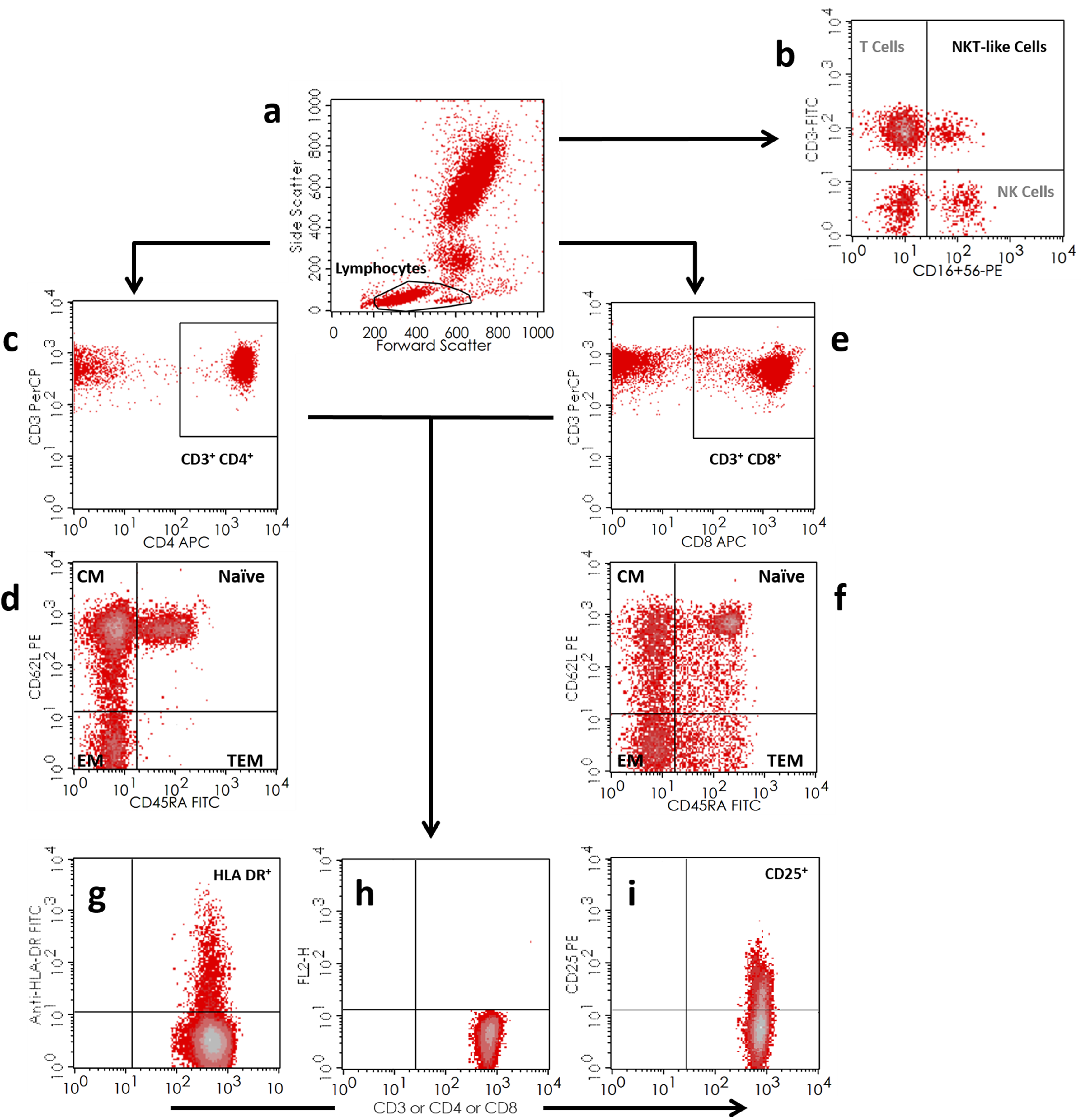
Gating strategy for the identification T-cell subsets. A, Lymphocyte gate (FSC/SSC). B, Identification of CD3+CD16+/CD56+ NKT-like cells. C-F, Naïve and memory subsets of CD4+ and CD8+ T cells: CD4+ T cells were identified within the lymphocyte gate by the coexpression of CD3 and CD4; CD8+ T cells were similarly identified by the coexpression of CD3 and CD8. CD4+ and CD8+ T-cell subsets were classified as represented in the CD45RA/CD62L dot plots: Naïve—CD45RA+CD62L+; Central Memory, CM—CD45RA−CD62L+; Effector Memory, EM—CD45RA−CD62L−; Terminally Differentiated Effector Memory, TEM—CD45RA+CD62L−. G-I, Activation markers (HLA DR and CD25) were evaluated in CD3+, CD3+CD4+ and CD3+CD8+ T cells. Fluorescence minus one (FMO) tubes (dot plot H) were used to assess CD25 positivity.
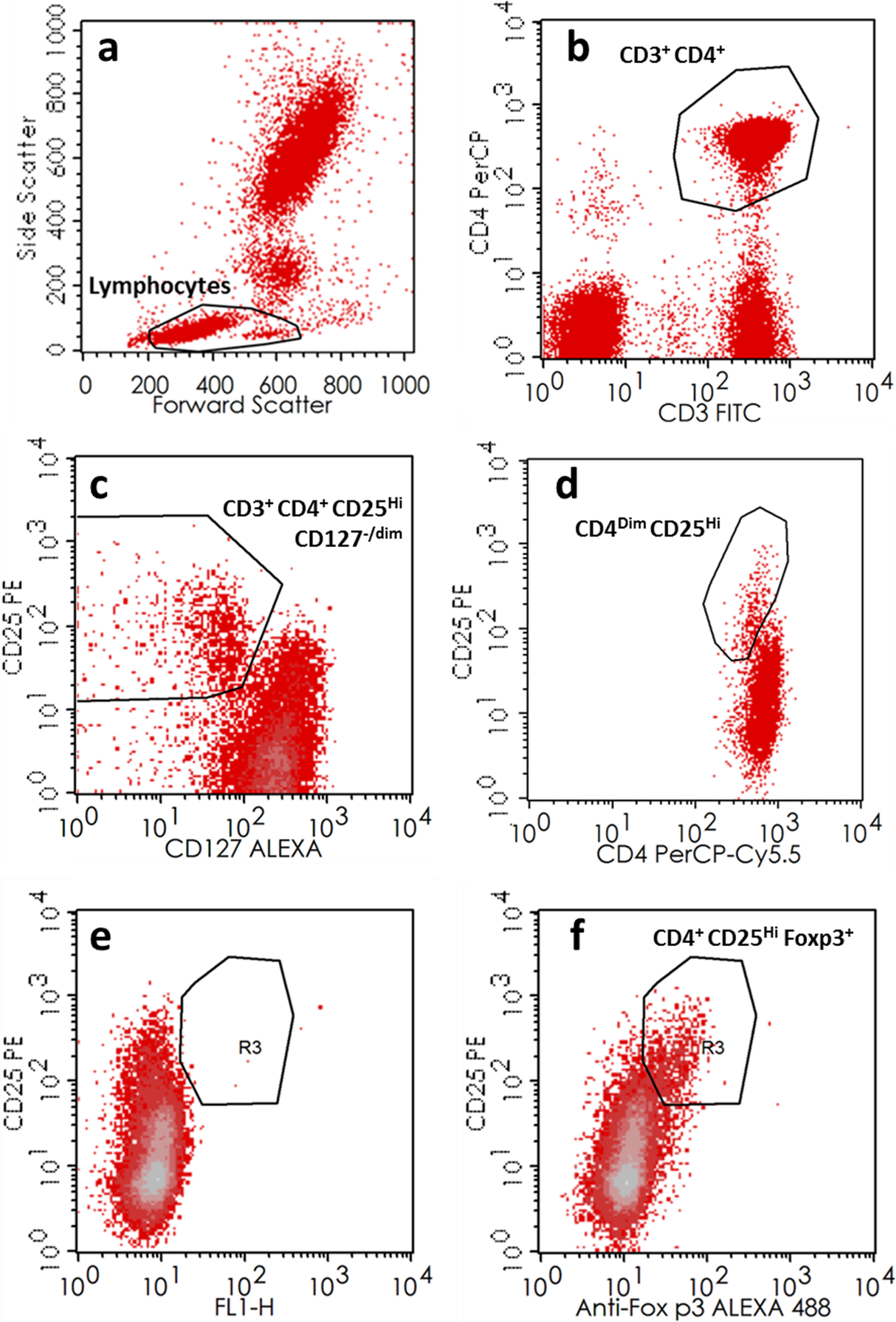
Gating strategy for the identification of regulatory T cells. Three analytical strategies were used to characterize Treg cells: CD4DimCD25Hi, CD4+CD25HiCD127−/dim , and CD4+CD25HiFoxp3+ T cells. A-B, CD4+ T cells were recognized as the CD3+ CD4+ (or CD4+) cells within the lymphocyte gate. C, Dot plot exemplifying the identification of CD4+CD25HiCD127−/dim regulatory T cells. D, CD4 versus CD25 dot plot showing the identification of CD4+(dim)CD25Hi regulatory T cells. E-F, Identification of CD4+CD25HiFoxp3+ regulatory T cells with dot plots of fluorescence minus one (FMO; E) and Foxp3 (F) tubes.
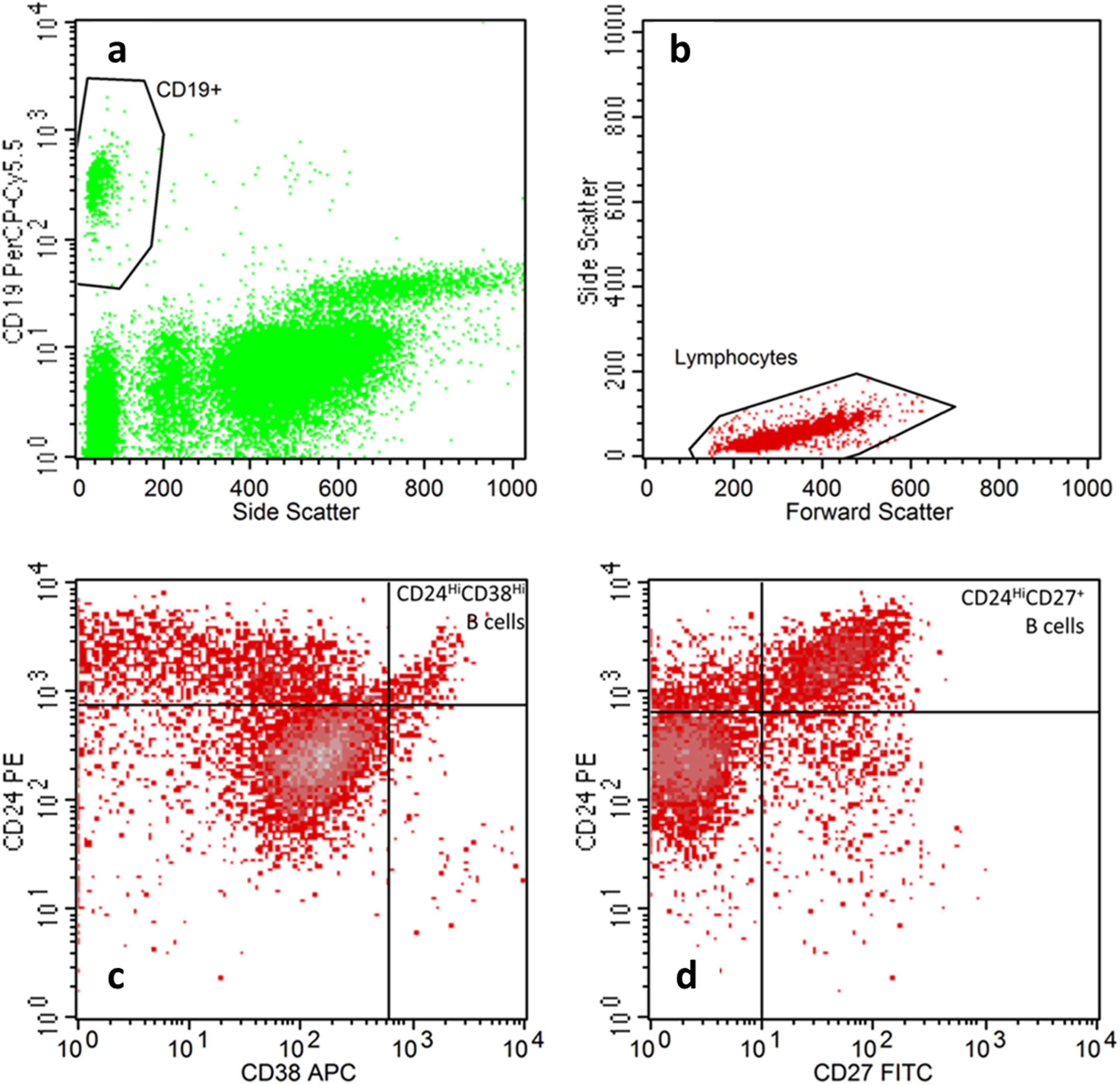
Gating strategy for the identification of regulatory B cells. Considering the lack of consensus regarding the Bregs phenotype, 2 analytical strategies were used to characterize Bregs: CD24hiCD27+ and CD24hiCD38hi. A-B, B cells were identified as CD19+ cells (a) in the lymphocyte population, gated according to forward and side scatter (B). C-D, Identification of regulatory B-cell subsets according to their expression of CD24, CD27, and CD38 (CD24HiCD38Hi Bregs and CD24HiCD27+ Bregs).
Statistics
The normality of data was assessed using the Shapiro-Wilk test. Normally distributed data were compared using t tests and summarized as mean and standard deviation, and nonnormally distributed data were compared using Mann-Whitney U tests and reported as the median with the interquartile range (IQR). Count data were compared using Fisher exact test. Statistical significance was defined by a P value <.05. All data were analyzed using R software, version 3.12 for Windows.
Results
Our study included 32 pregnant who gave birth to their newborns at term either via spontaneous vaginal delivery (n = 18) or via elective cesarean (n = 14). As Table 1 shows, these women did not differ significantly in terms of age, ethnicity, BMI, systolic and diastolic blood pressure, gestational age, and prolonged maternal hospital stay. However, there were significantly (P < .05) more nulliparous pregnant who delivered their newborns vaginally (n = 12) than via elective cesarean (n = 1). Among the spontaneous vaginal deliveries, the median time for dilation from 4 to 10 cm was 6 hours (95% confidence interval, 4.8-9.3 hours). Regardless of the mode of delivery, all of the pregnant received regional analgesia and/or anesthesia. None of the women received general anesthesia.
Demographics and Clinical Variables.a

Abbreviations: BMI, body mass index.
aSD, standard deviation.
b t test.
cFisher exact test for count data.
dWilcoxon rank test.
eMore than 2 days after vaginal delivery or more than 3 days after cesarean.
All 18 of the vaginal deliveries were spontaneous (without forceps or vacuum) and received epidural analgesia during labor. All elective cesareans (n = 14) were made with regional (combined spinal epidural) anesthesia. The indications for the elective cesareans were previous cesarean section (n = 12; 86%) and breech presentation (n = 2; 14%).
Tables 2 to 5 present a comparison of the PB T-cell subsets, Tregs, and Bregs between women who had spontaneous vaginal deliveries and those who had elective cesarean births. Compared to women who had elective cesareans (no labor), those who delivered spontaneously and vaginally had significantly (P < .05) lower circulating absolute counts of B cells (median [cells/μL]: 146 [IQR = 49] vs 192 [IQR = 65]) and NKT-like cells (median [cells/μL]: 154 [IQR = 125] vs 224 [IQR = 117]).
Comparison of Maternal Peripheral Blood Lymphocytes Subsets Between Women Who Gave Birth by Spontaneous Vaginal Delivery and by Elective Cesarean.a

aNormally distributed data expressed as mean (SD, standard deviation): group comparisons with Student t test; nonnormally distributed data expressed as median (IQR, interquartile range): group comparisons with Mann-Whitney U test.
Comparison of the Maturation Profiles of CD4+ and CD8+ T Cells Between Women Who Gave Birth by Spontaneous Vaginal Delivery and by Elective Cesarean.a

Abbreviations: CM, central memory; EM, effector memory; TEM, terminally differentiated effector memory.
aNormally distributed data expressed as mean (SD, standard deviation): group comparisons with Student t test; nonnormally distributed data expressed as median (IQR, interquartile range): group comparisons with Mann-Whitney U test.
Comparison of Activation Profiles of CD3+, CD4+, and CD8+ T cells Between Women Who Gave Birth by Spontaneous Vaginal Delivery and by Elective Cesarean.a

aNormally distributed data expressed as mean (SD, standard deviation): group comparisons with Student t test; nonnormally distributed data expressed as median (IQR, interquartile range): group comparisons with Mann-Whitney U test.
Comparison of Regulatory Subsets of T and B Cells Between Women Who Gave Birth by Spontaneous Vaginal Delivery and by Elective Cesarean.a

aNormally distributed data expressed as mean (SD, standard deviation): group comparisons with Student t test; nonnormally distributed data expressed as median (IQR, interquartile range): group comparisons with Mann-Whitney U test.
Furthermore, no significant differences in T cells, CD4+ and CD8+ T cells, the maturation profiles (naïve, central memory, effector memory, and terminally differentiated effector memory) of CD4+ and CD8+ T cells, and the activation profiles of CD3+, CD4+, and CD8+ T cells (markers for T-cell activation included HLA-DR and CD25) were identified between the study groups.
Additionally, no significant differences in Tregs and Bregs were identified between the pregnant with spontaneous vaginal deliveries and those who underwent elective cesareans.
Discussion
In this study, we investigated whether labor has an effect on maternal PB T-cell subsets, Tregs, and Bregs. We found that compared to elective cesareans (no labor), spontaneous vaginal deliveries (after the completion of labor) were associated with significantly lower median absolute counts of B cells and NKT-like cells. No further significant differences between spontaneous vaginal deliveries and elective cesareans were identified.
Variations in some of the subpopulations in PB may indicate that there is a selective migration of these cells out of the PB (into the sites of inflammation), a change in the survival of these cells, or an obstacle to their differentiation. The determination of these phenotypes in pregnancy and labor may contribute to our understanding of T-cell homeostasis during labor.
In our study, the finding of lower counts of B cell in spontaneous vaginal deliveries compared to elective cesareans could be explained by the migration of these cells into the decidua during labor, which would result in the decrease in the absolute counts of these cells in PB. Indeed, a recent study 29 demonstrated for the first time in humans that specific subtypes of leukocytes infiltrate the decidua during labor, suggesting that the inflammatory mediators produced by leukocytes play a role in decidual activation, a preliminary event in the labor process. Our results are consistent with those of a previous study 30 in an animal model that described immune cell migration from PB to the uterus, as one of the mechanisms responsible for the B lymphopenia that occurs late in pregnancy. We hypothesize that during labor, recruitment events are more intense.
We also found that compared to no labor, spontaneous vaginal deliveries were associated with significantly lower median absolute counts of NKT-like cells. To our knowledge, this finding has not been described previously in labor; however, 1 study found that NKT-like cells may be recruited from the PB immune cell pool after embryo implantation. 31 Similar to B cells, NKT-like cells may also migrate selectively to the decidua during labor, thus decreasing the absolute counts of these cells in the PB. The data pertaining to the function of NKT-like cells in pregnancy immune modulations are limited. 16,18 Further research is necessary to investigate the exact function of these cells during labor.
Nevertheless, spontaneous vaginal deliveries and elective cesareans did not seem to have significantly different impacts on PB absolute counts or the percentages of the majority of immune cell subsets, and this seems to be supported by the literature. First, it has been shown that in human term pregnancy, a significantly higher percentage of activated T cells are present in the decidua than in the maternal PB as a result of the local expansion of these cells. 32 Additionally, recent research in humans has shown that the absolute counts of Tregs and the estrogen levels in the PB increase progressively from early to late pregnancy 11 and decrease after delivery. 11,33 This suggests that Treg may play a role in the immunological changes that occur before delivery rather than during or after labor. Finally, Bregs were already shown to expand in normal early pregnancy when immunosuppression is necessary. 20 We hypothesize that, like Tregs, they are probably not directly involved in the immunological mechanisms in labor.
In our study, analgesia and/or anesthesia were administered in all cases of vaginal deliveries and elective cesareans. It has been reported that the use of analgesia and/or anesthesia at birth is associated with reduced maternal and fetal endocrine stress responses and that vaginal deliveries are associated with higher endocrine stress responses than elective cesareans are. 34 It is currently unknown whether the use of analgesia and/or anesthesia has any impact on the maternal immune response at delivery.
The main limitation of our study was that there were significantly more nulliparous who delivered their newborns vaginally than by elective cesarean. Nonetheless, our sample was homogenous, as we only included a pregnant woman who had singleton pregnancies between 37 and 41 weeks of gestation, and we used standardized protocols for the management of vaginal deliveries and elective cesareans, with no induction of labor or intravenous analgesia. Studies with larger sample sizes may be required to further investigate the impact of labor on maternal immune system cells. Furthermore, it is important that upcoming studies compare immune cell subsets in the PB and decidua at delivery from the same healthy pregnant women.
In conclusion, this study found that labor does not affect significantly the studied circulating lymphocytes subsets. During labor, these changes may indicate that what is happening in the PB does not reflect what is happening in the placenta. The failure to detect a significant or major difference in maternal PB T-cell subsets, especially in Tregs and Bregs, between the presence or the absence of labor is consistent with the previous studies that reported that these cell subsets have a more important functional role in early pregnancy for tolerating the fetus rather than during labor. In the future, it will be interesting to determine whether the presence or absence of labor has different effects on the immune system of pregnant patients with autoimmune disorders and might eventually affect their clinical outcomes.
Footnotes
Authors’ Note
All of the authors designed the study and created the study protocol; J Lima had the original research idea, recruited the patients, collected the data, and drafted the manuscript; C Martins and G Nunes analyzed the blood samples using flow cytometry; LM Borrego supervised all the work and the research protocol; all of the authors contributed to the data analysis and interpretation, revised it, intellectually contributed, and approved the final version of the manuscript.
Declaration of Conflicting Interests
The author(s) declared no potential conflicts of interest with respect to the research, authorship, and/or publication of this article.
Funding
The author(s) disclosed receipt of the following financial support for the research, authorship, and/or publication of this article: This study was partially funded by José de Mello Saúde.