
Abstract
We investigated whether serum osteopontin (OPN) levels are different according to specific phenotypes of adenomyosis and endometriosis. We conducted a prospective laboratory study in a university referral center for endometriosis between May 2005 and May 2013 and included 148 nonpregnant women, younger than 42 years, undergoing surgery for a benign gynecological condition and who had a preoperative pelvic magnetic resonance imaging (MRI). The presence of focal and/or diffuse adenomyosis was determined by pelvic MRI, and women were classified into 3 groups: no-adenomyosis (No-AM), isolated diffuse adenomyosis (DIF-AM), and focal adenomyosis with or without diffuse adenomyosis (FOC-AM). After complete surgical exploration of the pelvic cavity, the presence and type of endometriosis was surgically determined and histologically confirmed. We distinguished 4 phenotypes: no endometriosis, superficial peritoneal endometriosis (SUP), ovarian endometrioma, and deep infiltrating endometriosis (DIE). Osteopontin levels were measured by enzyme-linked immunosorbent assay in serum samples obtained in all participants in the month preceding surgery. Our results show lower OPN levels in women with focal adenomyosis compared to adenomyosis-free controls. Our results also show a decrease in OPN levels in women with associated DIE and focal adenomyosis compared to women with SUP. Various serum biomarkers have been studied in the context of endometriosis severity and subtypes, whereas data on serum markers of adenomyosis are scarce. Both entities are often associated, and adenomyosis could be a confounding factor influencing results. Future research on serum biomarkers should describe subtypes of adenomyosis and endometriosis and analyze results according to well-defined subtypes.
Introduction
Osteopontin (OPN) is a calcium-binding glycophosphoprotein of the small integrin binding ligand N-linked glycoprotein family implicated in various cell functions, which was originally isolated in the bone matrix. 1 In humans, OPN is secreted by epithelial cells in the gastrointestinal, urinary, and reproductive tracts, the gallbladder, pancreas, lung bronchi, lactating breast, salivary glands, and sweat ducts. 2 It is detected in body fluids and in extracellular matrix components. Osteopontin can function as a cell adhesion protein and as a cytokine for several integrins and cell differentiation antigen 44. 3 Osteopontin plays a key role in various physiological and pathological processes such as mineralization, blood vessel formation, and acute and chronic inflammation, where it acts through the recruitment of monocytes/macrophages and mediates cytokine secretion in leukocytes. 4 In the endometrium, it also plays a key role in the window of implantation (WOI) and in the blastocyst—endometrium adhesion process through its interaction with αvβ integrin. 5 -7 Numerous publications also describe its role in neoplastic transformation and cancer development in a variety of tumors through its implication in cell attachment, migration, invasion, and proliferation. 8 -10 Its use as a diagnostic, prognostic, or follow-up serum biomarker has been described for several neoplastic and nonneoplastic conditions such as ovarian cancer, hepatocellular carcinoma, breast cancer, multiple sclerosis, and alcoholic steatohepatitis. 11 -15
Since OPN is a secreted protein implicated in inflammation, cell adhesion, and migration, the authors have hypothesized that it could be involved in the pathogenesis of endometriosis by favoring cell migration, attachment, and invasion of the peritoneum and pelvic organs. 16 -18 Yang et al recently showed that the activation of OPN induced by estrogen could upregulate matrix metalloproteinase 9 (MMP-9) and favor migration of endometrial epithelial cells in endometriosis. 16 Several studies have analyzed OPN and αvβ integrin by immunohistochemistry, Western blot, and/or messenger RNA (mRNA) expression in the eutopic and ectopic endometrium of women with endometriosis showing conflicting results. 17,19 -21 Two studies report higher plasma OPN levels in women with endometriosis compared to endometriosis-free controls; OPN levels were, however, not related to the severity of the disease evaluated by revised American Fertility Society (r-AFS) scoring. 17,18,22
Adenomyosis, a frequent chronic gynecological condition defined by the benign ectopic invasion of endometrial tissue—glands and stroma—into the myometrium with hyperplasia of adjacent uterine smooth muscle cells, is often associated with endometriosis. 23,24 Both entities, endometriosis and adenomyosis, share clinical symptoms such as dysmenorrhea, noncyclic chronic pelvic pain (NCCPP), dyspareunia, and infertility. A single study has shown a lower expression of integrin β3 and OPN in the eutopic endometrium during the WOI in women with adenomyosis. 25 There are no studies to date on the expression of OPN by endometrial cells from adenomyosis foci located in the myometrium or on OPN as a serum biomarker of adenomyosis.
In the present study, we assayed OPN in serum from a large cohort of women operated for benign gynecological conditions to determine levels according to well-characterized phenotypes and severity of adenomyosis and endometriosis.
Materials and Methods
Since 2005, we conducted a cross-sectional study using data prospectively collected from all nonpregnant women younger than 42 years who underwent a surgical intervention by laparoscopy or laparotomy for a benign gynecological indication at our institution. The local ethics committee (Comité Consultatif de Protection des Personnes dans la Recherche Biomedicale) of Paris Cochin approved the study protocol. Excluded from this population were women with cancer or borderline tumors and those who did not consent to the study. Within the cross-sectional study, we performed a substudy to analyze serum OPN levels according to the presence of adenomyosis diagnosed by pelvic magnetic resonance imaging (MRI). We selected all women from the cohort from May 2005 to May 2013 who had a pelvic MRI performed by our senior radiologist during the preoperative workup. Women were allocated into 2 groups according to MRI findings: the adenomyosis group (n = 109) consisted of participants with MRI findings of diffuse adenomyosis according to the criteria published by Bazot et al (a maximal junctional zone thickness [JZmax] of ≥12 mm and/or a JZmax/myometrial thickness ratio of >40% and/or high signal intensity myometrial spots) and/or features of localized focal adenomyosis. 26 Focal adenomyosis was defined on T2-weighted MRI images as a localized, ill-defined, low signal intensity mass within the myometrium. 27 The control group consisted of women without any criteria for adenomyosis at MRI (n = 39). Women in the adenomyosis group were further subdivided into isolated diffuse adenomyosis (DIF-AM) and focal adenomyosis with or without diffuse adenomyosis (FOC-AM) according to MRI findings.
The diagnosis of endometriosis was based on surgical exploration and histological confirmation. Patients visually diagnosed with endometriosis but without histological confirmation were excluded from the study. When present, the extent of endometriosis was surgically staged and scored (total, implants, and adhesions scores) according to the r-AFS. In addition, endometriosis lesions were classified into 3 different subtypes—superficial peritoneal endometriosis (SUP), endometriomas (OMAs), and deep infiltrating endometriosis (DIE). Since these subtypes are frequently associated, according to the previously described classification, patients were assigned to the group corresponding to the most severe (worst) lesion, in the following order from the least to the most severe—SUP, OMA, and DIE. 28
We also analyzed data according to the following groups— group A, no endometriosis with either no adenomyosis or DIF-AM; group B, SUP with either no adenomyosis or DIF-AM; group C, DIE with FOC-AM.
For each patient, personal history data were obtained during face-to-face interviews conducted by the surgeon during the month preceding surgery. We used a highly structured previously published questionnaire. 28,29 The following data were recorded—age, parity, gravidity, height, weight, body mass index (BMI), existence of infertility, menorrhagia, gynecologic pain symptoms (dysmenorrhea, deep dyspareunia, NCCPP), and gastrointestinal and lower urinary tract symptoms. 30,31 Pain intensity was evaluated preoperatively using a 10-cm visual analog scale. Women without hormonal treatment were cycling women (in the proliferative or secretory phase of the menstrual cycle) without any hormonal treatment use in the last 6 months before the surgery. 32 We also recorded current hormonal treatment use (including progestogens, estroprogestatives, or gonadotrophin releasing hormone [GnRH] agonists) and previous endometriosis and/or uterine surgery.
Collection of Serum
Serum samples were collected from all participants during the month that preceded surgery. Briefly, samples of 5 to 10 mL of venous blood were collected through a peripheral venous catheter and then centrifuged at 800g for 12 minutes at 4°C. Serum supernatants were collected, aliquoted, and stored at −70°C within 2 hours after collection until use.
Measurement of OPN Concentrations
Osteopontin concentrations in serum were determined in duplicate using an enzyme-linked immunosorbent assay (ELISA) kit (Reference DY1433 Human OPN; R&D Systems, Inc, Minneapolis, Minnesota), according to the manufacturer’s recommendations. The range of determination was 30 to 4000 µg/L. Osteopontin levels <30 µg/L were undetectable and were considered as 0 µg/L for statistical analysis. Each sample was tested in duplicate and the mean value calculated. The intra-assay and interassay coefficient variability of the OPN ELISA kit were 7.8% and 9.8%, respectively.
Statistical Analysis
All data were collected in a computerized database and analyzed using Statistical Package for the Social Sciences software (IBM Corp, Released 2012, IBM SPSS Statistics for Windows, Version 22.0., Armonk, New York), and GraphPad Prism (GraphPad Prism version 6.04 for Windows, GraphPad software, San Diego, California) was used for the figures.
We compared women with DIF-AM and FOC-AM to control women using analysis of variance for quantitative variables and Pearson χ2 for qualitative variables, as appropriate. Data are presented as mean ± standard deviation (SD).
Considering the non-Gaussian distribution of serum OPN levels, statistical analysis between groups was performed with the Kruskal-Wallis test. Post hoc pairwise comparisons were performed using Dunn multiple comparison test. We performed a simultaneous multiple regression analysis to explore the relationship between serum OPN levels and the type of endometriosis, the type of adenomyosis, and the type of hormonal treatment.
According to the non-Gaussian distribution, correlations between detectable serum OPN levels and clinical, biological, and anatomical characteristics of disease severity, measured with semi-quantitative variables, were examined using the nonparametric Spearman rank correlation test excluding undetectable OPN levels. Throughout the article, P < .05 was considered statistically significant. When multiple comparisons were performed using the Dunn test, adjusted P values are reported.
Results
Patients and Controls
One hundred forty-eight women were recruited for this study, 109 adenomyosis-affected women and 39 adenomyosis-free controls (No-AM). Their major clinical, imagery, and surgical features are presented in Table 1. According to the MRI classification of adenomyosis, the 109 adenomyosis-affected women were classified as follows: 52 (47.7%) DIF-AM and 57 (52.3%) focal adenomyosis (FOC-AM; Figure 1).
Baseline Characteristics.a
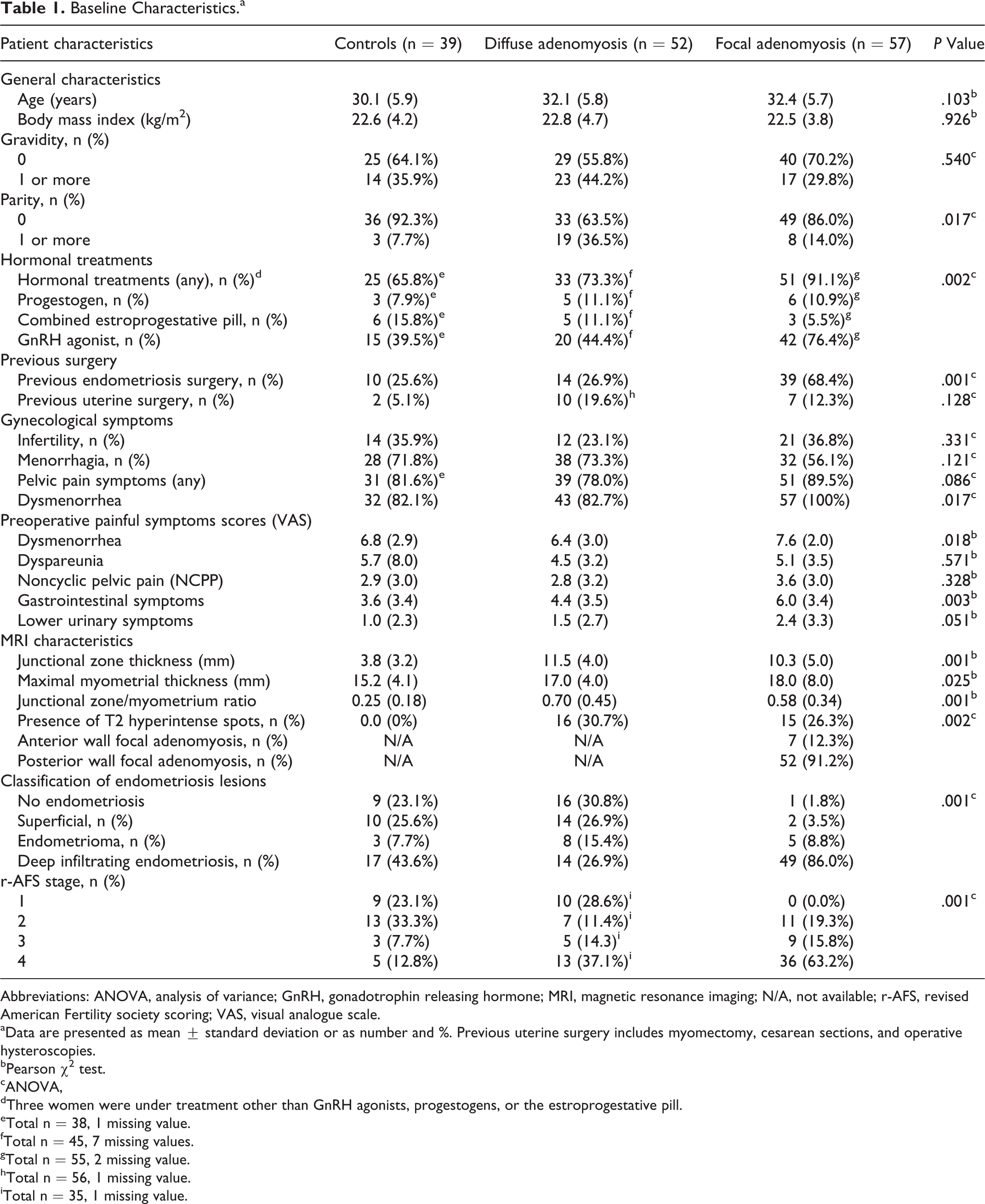
Abbreviations: ANOVA, analysis of variance; GnRH, gonadotrophin releasing hormone; MRI, magnetic resonance imaging; N/A, not available; r-AFS, revised American Fertility society scoring; VAS, visual analogue scale.
aData are presented as mean ± standard deviation or as number and %. Previous uterine surgery includes myomectomy, cesarean sections, and operative hysteroscopies.
bPearson χ2 test.
cANOVA,
dThree women were under treatment other than GnRH agonists, progestogens, or the estroprogestative pill.
eTotal n = 38, 1 missing value.
fTotal n = 45, 7 missing values.
gTotal n = 55, 2 missing value.
hTotal n = 56, 1 missing value.
iTotal n = 35, 1 missing value.
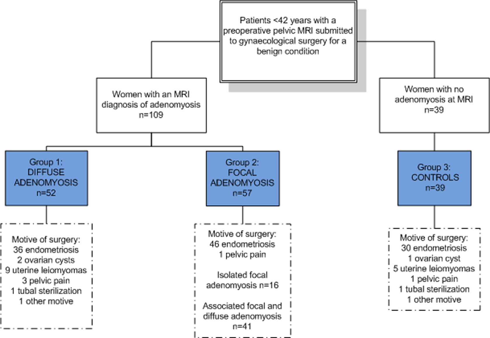
Patient inclusion flowchart.
In women with DIF-AM, 6 (11.5%) had the 3 diagnostic criteria, 22 (42.3%) had both JZmax ≥12 mm and JZmax/myometrial thickness >40%, 4 (7.7%) had both JZmax/myometrial thickness >40% and high signal intensity myometrial spots, 15 (28.8%) had JZmax/myometrial thickness >40% only, and 4 (7.7%) had only high signal intensity myometrial spots.
Women with FOC-AM had associated diffuse adenomyosis in 71.9% of cases; 9 (15.8%) had the 3 diagnostic criteria, 6 (10.5%) had both JZmax ≥12 mm and JZmax/myometrial thickness >40%, 3 (5.3%) had both JZmax/myometrial thickness >40% and high signal intensity myometrial spots, 14 (24.6%) had JZmax/myometrial thickness >40% only, and 1 (1.6%) had only high signal intensity myometrial spots. The focal lesion was located in the posterior wall of the myometrium in 91.2% of the cases with a mean lesion size of 15.2 (6.1) mm. The lesion was located in the anterior wall in 12.3% of the cases with a mean (SD) lesion size of 21.5 (8.5) mm. Only 3.3% of women had associated anterior and posterior focal lesions.
As shown in Table 1, women with FOC-AM had higher pain scores for dysmenorrhea, gastrointestinal, and urinary pain symptoms compared to No-AM and DIF-AM.
The distribution of the hormonal treatment use differed significantly between adenomyosis groups (P = .002). In a pairwise comparison, there was no difference in the type of hormonal treatment use between No-AM and DIF-AM (P = .497). There was a significant difference in the treatments used in women with FOC-AM versus DIF-AM and FOC-AM versus No-AM (P = .003 and P = .003, respectively). Women with FOC-AM were under GnRH agonist treatment in 76.4% of cases, whereas 44.4% of women with DIF-AM and 39.5% of women with No-AM used that treatment.
Serum OPN Levels
Serum OPN was measured in all 148 women studied. Considering 30 µg/L as the threshold for detection, OPN was detected in 106 women and undetectable in 42 women (28.4% of cases). Osteopontin was detected in 29 (74.4%) control women (No-AM) and in 77 (70.6%) women with adenomyosis (P = .80). Among the adenomyosis-affected women, OPN was detected in 45 (86.5%) women with DIF-AM and 32 (56.1%) with FOC-AM (P = .001).
Thirty women had menstrual cycles without hormonal treatments. Osteopontin levels did not differ between days 1 to 13 (290.1 [0.0-1702.7] µg/L) and days 14 to 35 (377.8 [0.0-2624.9] µg/L) of the menstrual cycles (P = .617).
Osteopontin levels were significantly different between women with hormonal treatments and women without treatment (180.5[0.0-4000.0] µg/L vs 517.2 [0-2228.7] µg/L; P = .039). As shown in Table 2, OPN levels were different according to the type of treatment (no treatment: 517.2 [0-2228.7] µg/L; GnRH agonist: 163.5 [0.0-4000.0] µg/L; estroprogestative: 783.7 [0.0-3566.6] µg/L; progestin 267.7 [0.0-4000.0] µg/L; P = .032). None of the pairwise comparisons reached statistical significance.
Serum Osteopontin (OPN) Levels Measured by ELISA According to the Presence or Absence and According to the Type of Hormonal Treatment at the Time of Surgery.
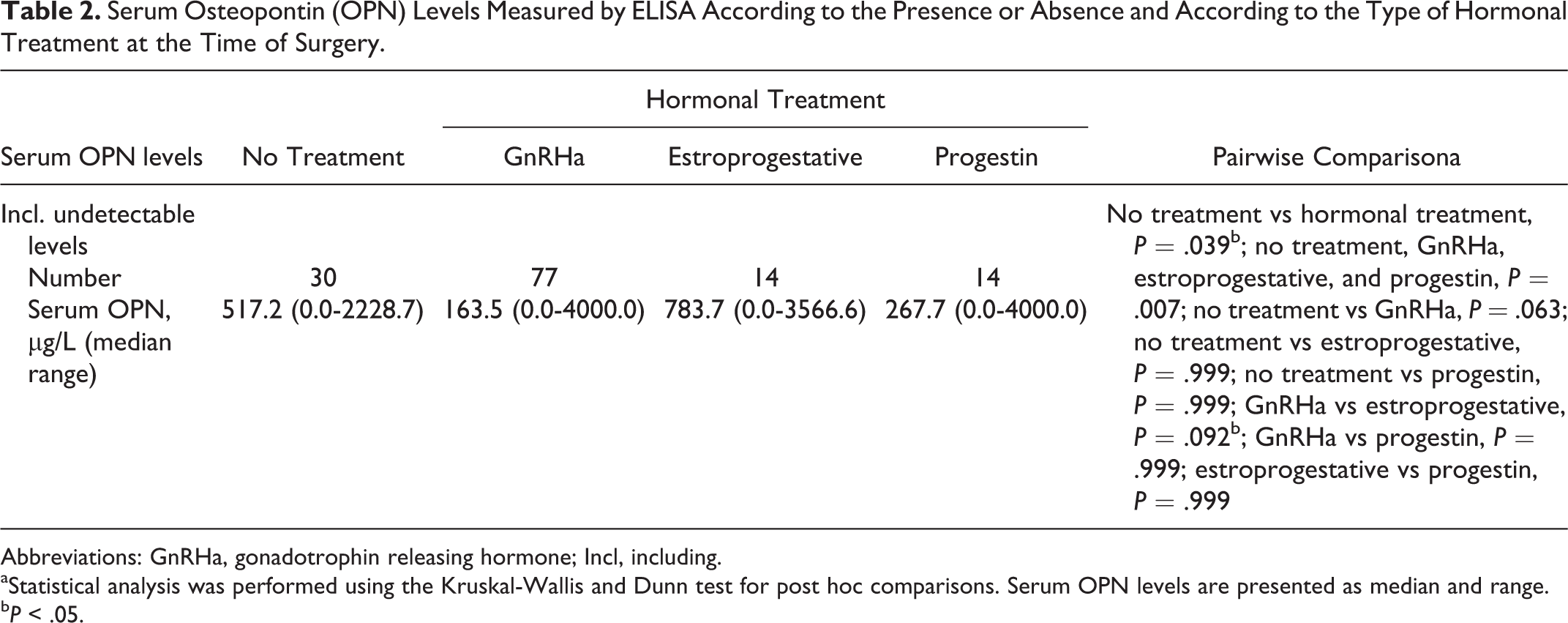
Abbreviations: GnRHa, gonadotrophin releasing hormone; Incl, including.
aStatistical analysis was performed using the Kruskal-Wallis and Dunn test for post hoc comparisons. Serum OPN levels are presented as median and range.
b P < .05.
As shown in Figure 2, OPN levels were lower in women with adenomyosis compared to controls (188.1 [0.0-4000.0] µg/L vs 507.3 [0.0-4000.0] µg/L); the difference did, however, not reach the level of significance (P = .066). We analyzed OPN levels according to the type of adenomyosis encountered. The difference according to the type of adenomyosis was significant (No-AM: 507.3 [0.0-4000.0] µg/L; DIF-AM: 309.6 [0.0-4000.0] µg/L; FOC-AM: n = 127.3 [0.0-4000.0] µg/L; P = .005). A post hoc pairwise comparison showed a significant difference between women with No-AM vs FOC-AM (P = .017) and DIF-AM vs FOC-AM (P = .019). There was no significant difference between No-AM versus DIF-AM (P = .99).
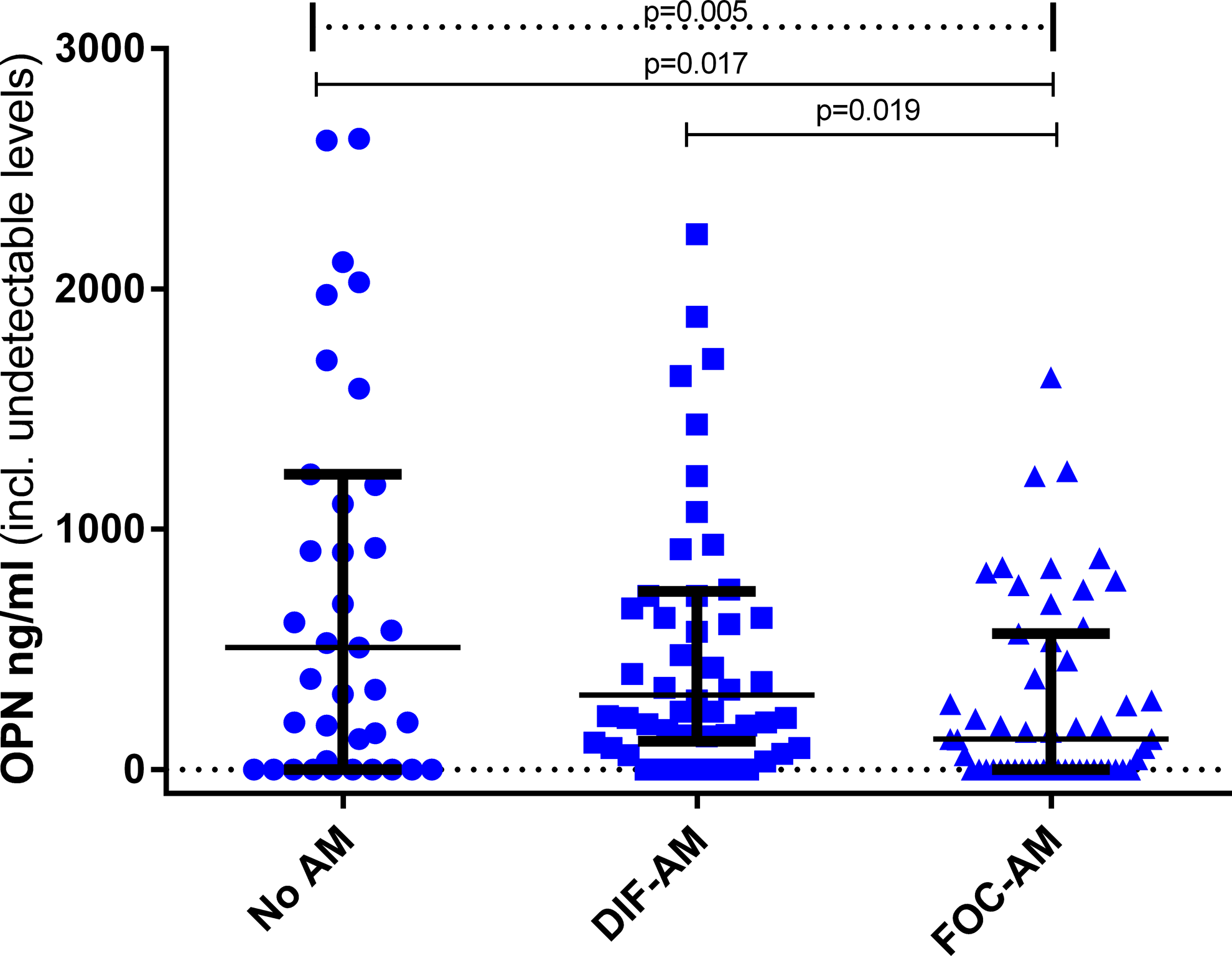
Serum osteopontin (OPN) levels measured by enzyme-linked immunosorbent assay (ELISA) in patients without adenomyosis (No-AM), with diffuse adenomyosis (DIF-AM), or focal adenomyosis (FOC-AM). Serum OPN levels differed significantly according to the type of adenomyosis (No-AM: n = 39; DIF-AM: n = 52; FOC-AM: n = 57; P = .005; dotted line). Post hoc pairwise comparison showed a significant difference between women with No-AM versus FOC-AM (adjusted P = .017; continuous line), DIF-AM versus FOC-AM (adjusted P = .019; continuous line). There was no significant difference between No-AM versus DIF-AM (adjusted P = .999). Statistical analysis was performed using Kruskal-Wallis test and Dunn test for post hoc multiple comparisons. Serum OPN levels are presented as median and interquartile range. Only significant (P < .05) comparisons are depicted.
Serum OPN levels did not differ according to the presence or the absence of endometriosis (215.4 [0.0-4000.0] µg/L vs 264.0 [0.0-4000.0] µg/L; P = .343). The difference in OPN levels according to the type of endometriosis was significant (P = .014; no endometriosis: 180.0 [0.0-4000.0] µg/L; SUP: 410.0 [0.0-2229.0] µg/L; OMA: 348.6 [0.0-1884.5] µg/L; DIE: 128.0 [0.0-4000.0] µg/L). Post hoc pairwise comparison showed a significant difference between women with SUP and DIE (adjusted P = .001) only; the difference between no endometriosis and DIE and the difference between no endometriosis and SUP were not statistically significant (adjusted P = .999 and P = .574, respectively; Figure 3).
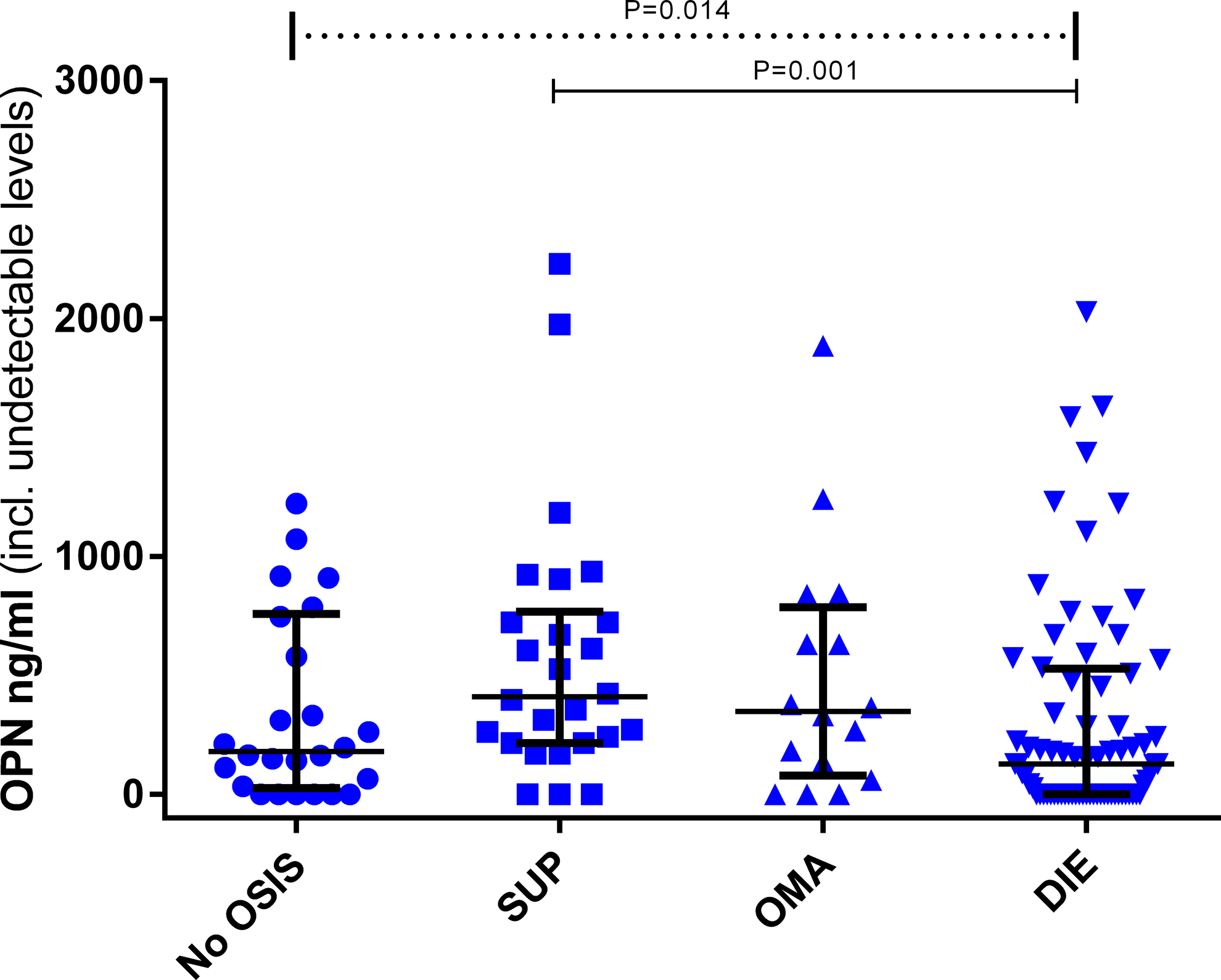
Serum osteopontin (OPN) levels measured by enzyme-linked immunosorbent assay (ELISA) in patients with no endometriosis and superficial peritoneal endometriosis (SUP), endometriomas (OMA), or deep infiltrating endometriosis (DIE). Serum OPN levels differed significantly according to the type of endometriosis (no endometriosis: n = 26; SUP: n = 26; OMA: n = 16; DIE: n = 80; P = .014; dotted line). Post hoc pairwise comparison showed a significant difference between women with SUP and DIE (adjusted P = .001; continuous line) only. Statistical analysis was performed using Kruskal-Wallis test and Dunn test for post hoc multiple comparisons. Serum OPN levels are presented as median and interquartile range. Only significant comparisons (P < .05) are depicted.
Serum OPN levels differed significantly according to the r-AFS stage (P = .001; no endometriosis median, 332.3 [0.0-4000.0] µg/L; stage 1: 903.8 [0.0-3666.6] µg/L; stage 2: 215.4 [0.0-4000.0] µg/L; stage 3: 332.3 [0.0-4000.0] µg/L; stage 4: 125.9 [0.0-1884.5] µg/L) (Figure 4). In the post hoc pairwise comparison, the difference was significant between stage 1 versus stage 2 (adjusted P = .040) and stage 1 versus 4 (adjusted P = .001).
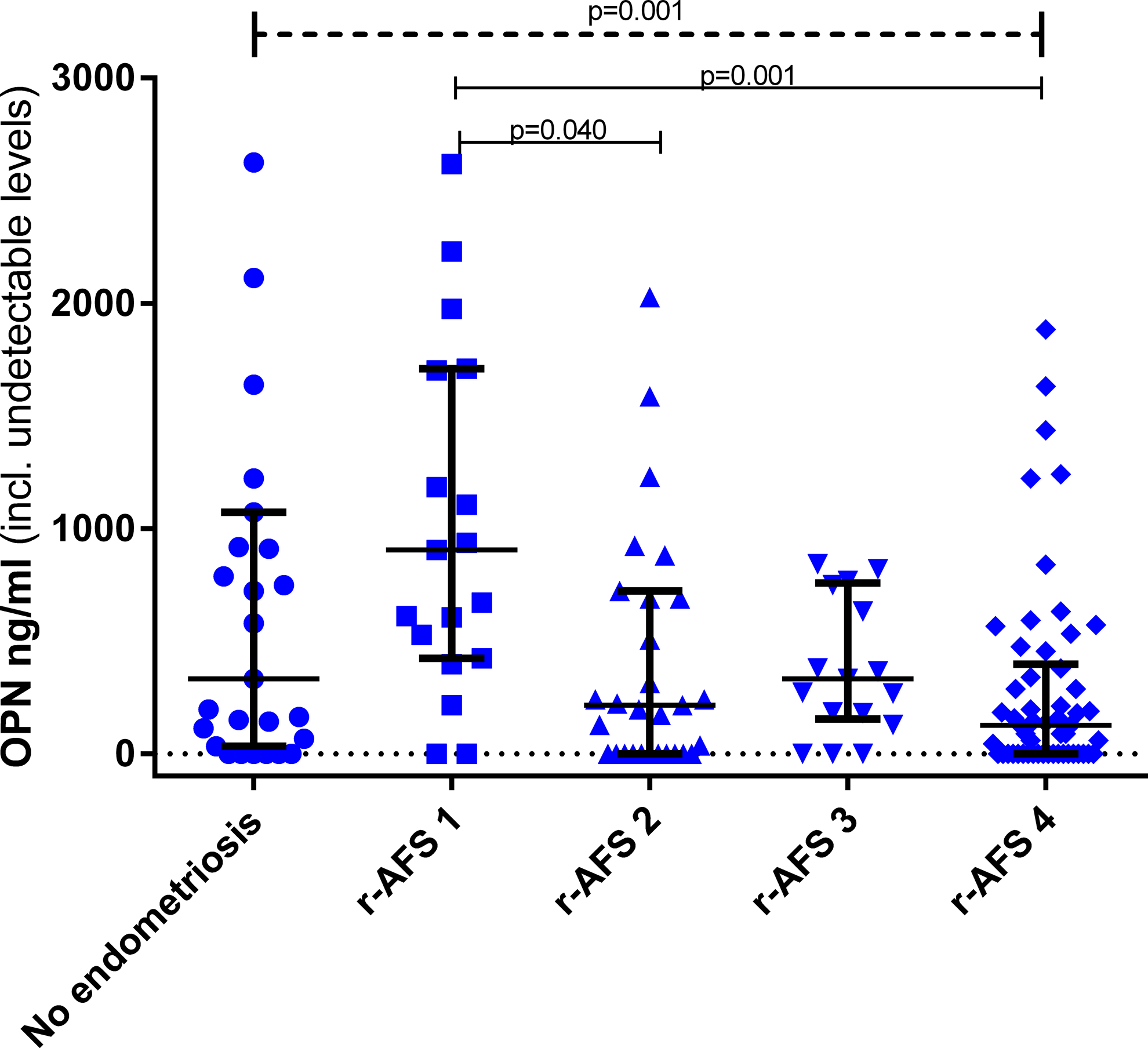
Serum osteopontin (OPN) levels measured by enzyme-linked immunosorbent assay (ELISA) in patients with no endometriosis and women with endometriosis according to revised American Fertility Society (r-AFS) stage. Serum OPN levels differed significantly according to r-AFS stage (no endometriosis: n = 26; stage 1: n = 19; stage 2: n = 31; stage 3: n = 17; stage 4: n = 54; P = .001; dotted line). Post hoc pairwise comparison showed a significant difference between stage 1 versus 2 (adjusted P = .040; continuous line) and in stage 1 versus 4 (adjusted P = .001; continuous line). Statistical analysis was performed using Kruskal-Wallis test and Dunn test for post hoc comparisons. Serum OPN levels are presented as median and interquartile range. Only significant comparisons (P < .05) are depicted.
Since FOC-AM, FD-AM, and DIE are associated in a great majority of cases (Table 1), we subdivided patients into 3 new categories: group A, no endometriosis and either No-AM or DIF-AM; group B, superficial endometriosis and either No-AM or DIF-AM; and group C, both DIE and FOC-AM. As shown in Figure 5, the difference in OPN levels was highly significant when comparing those 3 groups—group A: 179.5 (0.0-3106) µg/L; group B: 608.5 (0.0-2618.0) µg/L; group C: 59.0 (0.0-4000.0) µg/L (P = .001). Post hoc pairwise comparison found a significant difference in the comparison between groups B and C (P = .001) only.
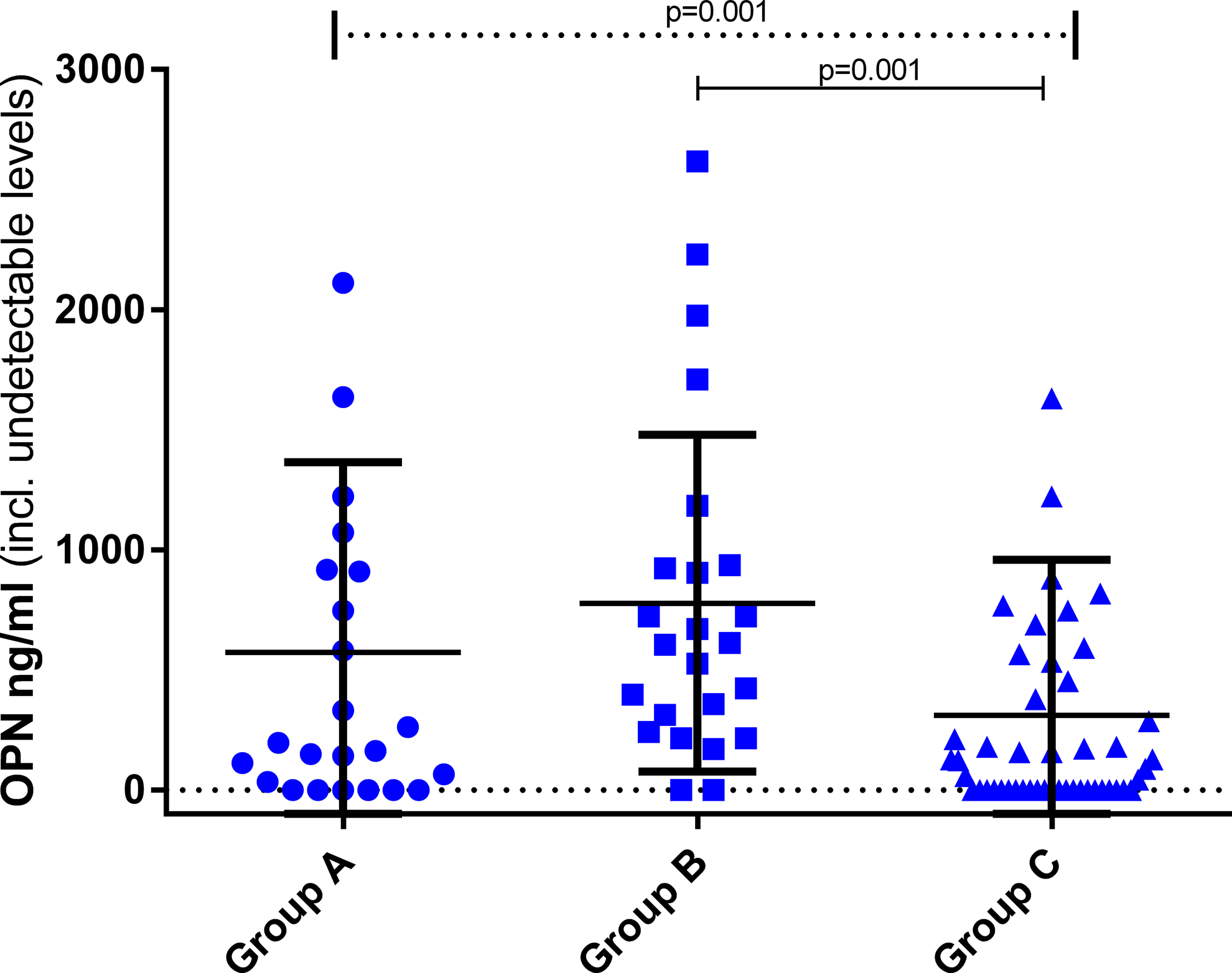
Serum osteopontin (OPN) levels measured by enzyme-linked immunosorbent assay (ELISA) in patients with no endometriosis and either no or diffuse adenomyosis (group A), superficial peritoneal endometriosis and either no or diffuse adenomyosis (group B), and focal adenomyosis and deep infiltrating endometriosis (group C). Serum OPN levels differed significantly according to the group (group A: n = 24; group B: n = 24; group C: n = 49; P = .001; dotted line). Post hoc pairwise comparison showed significant differences between groups B and C (adjusted P = .001; continuous line) only. Statistical analysis was performed using the Kruskal-Wallis and Dunn test for post hoc comparisons. Serum OPN levels are presented as median and interquartile range. Only significant comparisons (P < .05) are depicted.
A standard simultaneous multiple regression analysis was used to determine whether adenomyosis is associated with OPN levels after controlling for the presence of endometriosis and the type of hormonal treatment used. The β coefficient was at the limit of significance for the type of adenomyosis (β = −0.182; P = .05) but not for the type of endometriosis (β = −0.121; P = .217) or for the type of hormonal treatment used (β = −0.058; P = .528).
Clinical Correlations With Serum OPN Levels
Clinical, imagery, and surgical correlations with serum OPN (ie, detectable OPN levels) are reported as follows. None of the following correlation was significant: age (r = −0.112; P = .405), BMI (r = −0.115; P = .398), dysmenorrhea (r = 0.049; P = .716), deep dyspareunia (r = 0.1; P = .47), NCCPP (r = 0.135; P = .318), lower urinary tract symptoms (r = 0.072; P = .593), total number of endometriosis lesions (r = −0.079; P = .587), junctional zone thickness measured by MRI (r = −0.015; P = .912), total r-AFS score (r = −0.257; P = .058), and gastrointestinal symptoms (r = 0.139; P = .304).
Discussion
We here show for the first time that serum levels of OPN, a multifunctional cytokine implicated in acute and chronic inflammation as well as in cell migration and metastatic dissemination, are significantly decreased in women with focal forms of adenomyosis compared to women with no adenomyosis. When analyzing OPN levels according to the type of endometriosis, OPN levels were significantly decreased in women with DIE compared to women with SUP and in r-AFS stages II and IV of endometriosis compared to r-AFS stage I endometriosis. Our results also show a decrease in OPN levels in women with DIE + FOC-AM compared to women with SUP.
Literature on serum biomarkers of adenomyosis is scarce with the exception of Ca-125 and vascular endothelial growth factor. 33,34 Xiaoyu et al found 9 upregulated and 5 downregulated serum proteins in adenomyosis compared to endometriosis using isobaric tags of relative and absolute quantification and Western blot confirmation. 35 Upregulated proteins were implicated in molecular functions such as coagulation, complement activation, immune response, and regulation of proteolysis, whereas downregulated proteins played a role in apoptosis, inflammation, and hypoxia.
In adenomyosis, a single study reports decreased OPN staining and mRNA expression in the eutopic endometrium during the WOI and serum levels have never been studied. 25
Yang et al have recently shown that the migration of endometrial epithelial cells of women with endometriosis could be stimulated by the estrogen/OPN/MMP-9 pathway. 16 In transcriptome studies, OPN was upregulated in the eutopic endometrium during the WOI in women with normal menstrual cycles. 36 -38 Several studies have evaluated OPN expression in eutopic endometrium and ectopic lesions in women with endometriosis. 17,19 -21
In the eutopic endometrium, studies on OPN expression give conflicting results. Hapangama et al reported a weak endometrial staining in the proliferative phase and an increase during the secretory phase, with the highest immunostaining during the WOI in normal cycling women. 39 In women with endometriosis, the OPN immunoreactivity is greater than in controls during the luteal phase. Cho et al reported higher OPN mRNA expression in women with endometriosis but find no difference between the proliferative and secretory phases of the cycle. 17 Casals et al found no difference in OPN immunostaining during the WOI in women with minimal to mild endometriosis compared to controls, whereas Wei et al found a decreased immunostaining during the late luteal phase in women with r-AFS stages I to IV of endometriosis. 19,21
Only a few studies evaluate OPN expression in ectopic endometriosis implants. 18,20 D’Amico et al found a 3.8-fold increased OPN mRNA expression in ectopic implants compared to the eutopic endometrium from endometriosis-free controls. 18 In controls, OPN is localized in epithelial cells of the functional layer with no expression in stromal cells. In ectopic implants, OPN is localized in the cytoplasm of glandular epithelial cells and in several stromal macrophages. These results are in line with those reported by Odagiri et al in women with ovarian endometriosis cysts. 20
The strength of this study lies in the novelty of the topic and in the methodological design. Osteopontin levels are used as a diagnosis and prognostic biomarker in a wide variety of neoplastic and inflammatory diseases. Serum OPN levels have never been studied in adenomyosis, and only 2 previous studies report increased plasma OPN levels in women with endometriosis. 17,18 In both studies, mean plasma OPN levels were 2.5 and 1.6 times higher, respectively, in women with surgically and histologically confirmed endometriosis compared to control women with a benign gynecological condition. Osteopontin levels were not different in minimal to mild disease (r-AFS stages I-II) compared to moderate to severe disease (r-AFS stages II-III). Endometriosis and adenomyosis are heterogeneous diseases with different subtypes that can be differentially associated with biomarkers. The inclusion of women with a preoperative pelvic MRI and the complete surgical exploration of the pelvic cavity allows a precise division of patients according to subcategories of adenomyosis and endometriosis. Most studies on endometriosis fail to report the concomitant presence of adenomyosis. Furthermore, the study of biomarkers of inflammation according to the precise phenotypes of adenomyosis and endometriosis evaluated by surgery and histological confirmation for endometriosis and by MRI for adenomyosis has never been done before. Recent data from the Cochin cross-sectional study show no association between diffuse adenomyosis and endometriosis, whereas the association between focal adenomyosis and endometriosis, in particular DIE, is strong. These findings lead us to the subclassification of adenomyosis and endometriosis used in the present study. 40
Our study may have several shortcomings and/or biases in spite of all precautions taken. (1) The first limitation stems from the technical approach since we used a manual ELISA kit to measure OPN levels. Enzyme-linked immunosorbent assay kits are easily available and not too costly; they however have a complex protocol and low detection sensitivity. In our study, OPN levels were undetectable in 28.4% of the cases. The rate of undetectable OPN levels is a result per se, but the statistical handling of such results is controversial. We here report the result including undetectable levels. 41 The low detection sensitivity limits the use of OPN as a clinical biomarker in endometriosis and adenomyosis, and the results remain interesting in understanding the pathogenesis of the disease. Another limitation related to the use of ELISA methods for OPN determination comes from the fact that different studies on OPN as a serum/plasma biomarker of disease use different ELISA kits and that the range of normal OPN levels in the general healthy population is not determined. Osteopontin levels reported by different studies vary considerably. For instance, Bramwell et al reported mean OPN levels of 28.4 µg/L (median 26.3 µg/L, range 11.8-109 µg/L) in healthy women from the hospital staff aged 20 to 60 years using the human OPN enzyme immunoassay (ADI-900-142, Enzo Life Sciences, Ann Arbor, Michigan). 42 Cho et al reported mean ± standard error of the mean levels of 165.84 ± 19 µg/L in their control population of premenopausal women operated for benign gynecological indication excluding endometriosis using another ELISA kit (Immunobiological Laboratories, Gunma, Japan). (2) We conducted our study in a well-characterized prospective cohort of women with a preoperative MRI operated for benign gynecological indications. Imagery was performed only if required for preoperative investigation of gynecological symptoms and/or for characterization and mapping of endometriosis lesions and diagnosis of associated adenomyosis. This limitation explains the fact that women without adenomyosis and endometriosis (9 women without adenomyosis and endometriosis, 26 women without endometriosis, and 39 women without adenomyosis) are underrepresented in our study and that we do not have a sufficient disease-free control group. The small size of our control group and the wide distribution of OPN levels probably explain that our results couldn’t show a significant difference between women without endometriosis and different endometriosis subtypes and r-AFS stages. (3) Our department is a referral center for endometriosis. There is, therefore, an overrepresentation of severe cases of endometriosis (DIE) and of women with a history of endometriosis surgery. This overrepresentation of severe cases with lower OPN levels could explain why, contrary to what has been reported previously by Cho et al and D’Amico et al, we did not find different OPN levels between women with endometriosis and endometriosis-free women. 17,18 (4) A majority of women both with and without adenomyosis were under hormonal treatment at the time of surgery (68.4% and 84.0%, respectively). Most women with FOC-AM, which is associated with DIE in 86.0% of the cases, were under GnRH agonist treatment at the time of surgery (76.4%). GnRH agonist treatment was less frequent in women with No-AM (39.5%) or DIF-AM (44.4%). Our results show significantly different OPN levels according to the type of hormonal treatment used (P = .032). Since OPN levels are decreased in women with severe forms of endometriosis and FOC-AM who are for the great majority under GnRH agonist treatment, it is not possible to assess the influence of GnRH agonists on OPN levels in the present study. There are no published studies on the influence of hormonal treatments on serum OPN levels. Bautista et al found comparable OPN levels in healthy premenopausal and postmenopausal women. Although expression of OPN in hormone-sensitive tissue such as the endometrium has been shown to fluctuate throughout the menstrual cycle, in premenopausal women, serum OPN levels did not fluctuate according to the menstrual cycle phase. 43 In this line, Cho et al reported comparable serum OPN levels in the proliferative and secretory phases of the cycle evaluated by endometrial biopsy. 17 Overall, current data suggest the absence of influence of hormones implicated in the hypothalamo–pituitary feedback loop on serum OPN levels.
Endometriosis is an inflammatory disease. Proinflammatory cytokines such as interleukin (IL) 6, IL-1β, tumor necrosis factor (TNF)-α, and IL-17F are involved in macrophage activation, inflammation, cyclooxygenase 2 expression, angiogenesis, and cellular proliferation. 41 Many proinflammatory and anti-inflammatory cytokines and growth factors and other markers implicated in cell adhesion, apoptosis, and immunity have been studied in serum and peritoneal fluid as biomarkers of endometriosis. 41,44 Most proinflammatory cytokines have been shown to be overexpressed in endometriosis, leading to an inflammatory response favoring progression of the disease. Some inflammatory cytokines have been shown to be differentially expressed according to the severity of the disease, some being over expressed in early or superficial stages and others in severe forms of the disease. Interleukin 19, IL-20, and anti-inflammatory cytokines from the IL-10 family have been shown to decrease in women with OMAs, suggesting immunosuppressive effects favorable to the development of endometriosis. 41
Previous studies have focused on the role of OPN in inflammatory cells and have reported that OPN induces the expression of proinflammatory cytokines (IL-1β, IL-6, IL-8, interferon γ, and TNF-α) in inflammatory cells, such as macrophages, dendritic cells, and T cells. 45,46 Our study shows a decrease in OPN levels in focal adenomyosis, suggesting that OPN might not be implicated in the pathogenesis of this adenomyosis subtype. We also found higher OPN levels in women with SUP compared to women with DIE and focal adenomyosis. Our study failed to show a difference between these 2 phenotypes and controls without endometriosis/focal adenomyosis probably due to the small size of the control group and due to the wide distribution of OPN values. It could be speculated that OPN could be involved in the production of proinflammatory cytokines by epithelial cells and/or macrophages in the immune response during the development of early-stage peritoneal endometriosis and could potentially play a role in the regulation of peritoneal lesion spread. Our resultants lead to speculate about the implication of OPN in the peritoneal form of endometriosis and not in the deep infiltrating form of the disease. Further studies with a more important disease-free control group should be conducted to establish whether OPN levels are increased in women with superficial peritoneal forms of endometriosis compared to disease-free controls.
In conclusion, our results show decreased OPN levels in focal forms of adenomyosis. When associating phenotypes of adenomyosis and endometriosis, OPN levels were decreased in associated DIE and FOC-AM compared to SUP. Further studies should establish whether there is an association between different adenomyosis phenotypes, DIE, and clinical symptoms. Future research is necessary to determine whether focal and diffuse adenomyosis are different entities at the molecular level.
Footnotes
Acknowledgments
The authors thank Dr Anne-Elodie Millischer, the senior radiologist who performed and interpreted all pelvic MRIs in this study. The authors thank staff members from our surgical unit for their expert assistance with data collection. The authors also thankfully acknowledge Nathalie Girma for unabatedly managing the patient database.
Declaration of Conflicting Interests
The author(s) declared no potential conflicts of interest with respect to the research, authorship, and/or publication of this article.
Funding
The author(s) received no financial support for the research, authorship, and/or publication of this article.