
Abstract
Background:
Vascular endothelial growth factor (VEGF) plays a pivotal role in angiogenesis and is overexpressed in many kinds of malignant tumors. Soluble VEGF receptor 1 (sVEGFR-1/ soluble fms-like tyrosine kinase 1 [sFlt-1]) plays a role as an inhibitor of VEGF, and an antitumor effect has been shown in several studies using sFlt-1. Recently, in addition to its antiangiogenic effect, it was reported that sFlt-1 has direct cytotoxicity.
Materials and Methods:
Transfection of sFlt-1 plasmid DNA was performed in the BeWo choriocarcinoma cell line. Overexpression of sFlt-1 in BeWo cells was confirmed by ELISA. In order to evaluate cell proliferation, cell counting and BrdU uptake assay were performed. Cytotoxicity was tested by LDH assay. TdT-Mediated dUTP Nick end Labeling (TUNEL) staining and quantitative analysis of caspase-cleaved keratin 18 (ccK18) level were done to evaluate cell apoptosis.
Results:
The cell number was significantly less, and the ratio of cytotoxicity was significantly higher in sFlt-1 group compared to the control group. TUNEL staining and ccK18 level suggested nonapoptotic cell death.
Conclusion:
Soluble Flt-1 showed a cytotoxic effect on BeWo cells. Our results suggest that sFLT-1 could be therapeutic for malignant tumors.
Introduction
Remarkable progress has been achieved in therapy for malignant tumors in the past few decades. However, especially in advanced malignancies, it is still difficult to obtain a complete remission, even in cases where the tumor may show a temporary response. Therefore, the development of new therapies is important.
The growth of solid tumors is critically associated with angiogenesis. Vascular endothelial growth factor (VEGF), one of the angiogenic mediators, plays an important role in the progression of tumor neovascularization. 1
It has been reported that VEGF is overexpressed in many kinds of malignant tumors. 2 –7 Many studies have demonstrated a positive correlation between VEGF and tumor growth. A close association between VEGF and tumor angiogenesis has been also reported. 5,8-11
Based on its potential role in promoting tumor angiogenesis, VEGF has been an attractive target for therapeutic intervention in malignant tumors. Actually, tumor suppression has been shown in several studies by inhibiting VEGF or its receptor using neutralizing antibodies to VEGF 9,10,12 –14 or administration of adenovirus-mediated soluble VEGF receptor 1 (soluble fms-like tyrosine kinase 1 [sFlt1]) complementary DNA (cDNA) 15 –18 in a regional or systemic way. In addition to these studies, combined effect of adenovirus vector-mediated sFlt-1 administration and current chemotherapy has been reported in human tongue cancer. 19
Soluble Flt-1 is known as an endogenously expressed selective inhibitor of VEGF. It is an alternatively spliced version of Flt-1. 20 Soluble Flt-1 binds to VEGF with the same affinity and specificity to Flt-1 (full length-type receptor) and forms a receptor–ligand complex.
Several reports have been published on the antitumor effect of sFlt-1 due to its antiangiogenic effect. In addition to these reports, our group reported a direct cytotoxic effect of sFlt-1 on some kinds of human cancer cell lines. 21
We focused on choriocarcinoma which has similar characteristics to placenta. There is a report that VEGF promotes cell proliferation, and an anti-VEGF monoclonal antibody diminishes this effect on BeWo choriocarcinoma cell line. 3 However, there has been no study using sFlt-1 to investigate its antitumor effect.
In the present study, we aimed to elucidate the antitumor effect and cytotoxicity of sFlt-1, using the BeWo choriocarcinoma cell line.
Materials and Methods
Cell Culture
BeWo, a human choriocarcinoma cell line, was obtained from Riken Cell Bank (Ibaraki, Japan). BeWo cells were cultured in Dulbecco Modified Eagle Medium/Ham F-12 culture medium (Nacalai tesque, Japan). The medium was supplemented with 10% fetal bovine serum,
Lentiviral Plasmid
The lentiviral vector plasmid enhanced green fluorescent protein (pLV-EGFP) was prepared as described previously. 22,23 The pLV-hsFlt-1 were prepared by replacing EGFP cDNA with hsFlt-1 cDNA.
Cell Conditioning and Transfection
Cells were seeded in 6- and 12-well plates at densities of 8 × 10⁴ and 4 × 10⁴ cells in 2 and 1 mL of culture medium per well, respectively. The cells were transfected with pLV-hsFlt-1 or pLV-EGFP, using Lipofectamine LTX (Invitrogen, Carlsbad, California) at 24 hours postseeding. The pLV-EGFP was used as a control estimating the effect of transfection. The amounts of plasmid DNA and Lipofectamine reagents used were optimized according to the manufacturers’ protocol.
Cell Counting
Ninety-six hours after transfection, cells were detached by trypsinization, and the total number of cells in each well was counted with a hemocytometer. Experiments were repeated 6 times.
Cell Proliferation Assay
The rates of DNA synthesis in BeWo cells were determined by measuring bromodeoxyuridine (BrdU) incorporation with a cell proliferation enzyme-linked immunosorbent assay (ELISA), BrdU kit (Roche Diagnostics GmbH, Mannheim, Germany). Cells were seeded in 96-well plates at densities of 2 × 10³ cells in 100-µL culture medium per well. The cells were transfected with pLV-hsFlt-1 or pLV-EGFP, using Lipofectamine LTX at 24 hours postseeding. Ninety-six hours after transfection, the assay was performed according to the manufacturers’ protocol. Absorbance values were measured at 370 nm (reference wavelength: 492 nm) using a microplate reader (SH-9000Lab; CORONA ELECTRIC Co Ltd, Ibaraki, Japan). Experiments were performed on 9 samples and repeated 3 times.
Lactic Acid Dehydrogenase Cytotoxicity Assay
Cells were seeded in 6-well plates and transfected 24 hours after seeding. Cell culture supernatants in each well were collected 96 hours after transfection, and any particulate was removed by centrifugation. As maximum lactic acid dehydrogenase (LDH) release control, 200 μL of 10% Triton X-100 was added to a well 30 minutes before collecting supernatant. As spontaneous control, cells in one well were neither transfected nor any reagent was added before collecting supernatant. “Percent cytotoxicity” was calculated with Cyto Tox 96 Non-Radioactive Cytotoxicity Assay (Promega Corporation, Wisconsin), according to the manufacturers’ protocol. Experiments were repeated 3 times.
Soluble Flt-1 ELISA
Cell culture supernatants in each well were collected 96 hours after transfection, and particulate was removed by centrifugation. The supernatants after centrifugation were used as samples. The assay was performed with Human sVEGFR1/Flt-1 Immunoassay kit (R&D systems, Minneapolis, Minnesota), according to the manufacturers’ protocol. Absorbance values were measured at 450 nm (reference wavelength: 540 and 570 nm) using a microplate reader (Multiskan FC; Thermo Fisher Scientific, Inc, Massachusetts). Experiments were performed on 7 samples and repeated 3 times.
Placental Growth Factor and VEGF ELISA
Cell culture supernatants were collected by the same method as sFlt-1 ELISA. Free placental growth factor (PlGF) and free VEGF were measured. The assay was performed with Human PlGF Quantikine ELISA Kit (R&D systems) and Human VEGF Quantikine ELISA Kit (R&D systems), respectively, according to the manufacturers’ protocol. Absorbance values were measured as in the sFlt-1 ELISA. Experiments were performed on 4 samples and repeated 3 times, respectively.
TdT-Mediated dUTP Nick end Labeling Staining
Cells were seeded in appropriately prepared 4 glass slides (Lab-Tek IIChamber Slide w / Cover CC2 Glass Slide Sterile; Thermo Fisher Scientific). PLV-sFlt-1 was transfected in 1 slide 24 hours after cell seeding. As positive control, 25 µmol/L of etoposide was added to another slide 48 hours before staining to induce cell apoptosis. The other 2 slides were used for negative control and spontaneous control. Negative control did not have added terminal deoxynucleotidyl transferase (TdT). Spontaneous control was nontransfected cells with added TdT. TdT was also added to the positive control and experimental sFlt-1 slides. Staining was performed with the DeadEnd Fluorometric TdT-Mediated dUTP Nick end Labeling (TUNEL) System (Promega, Madison, Wisconsin), according to the manufacturers’ protocol. Experiments were repeated 3times.
Quantitative Analysis of Cell Apoptosis
During apoptosis, protein keratin 18 (K18) is cleaved by caspases at a unique aspartate, exposing an epitope that is recognized by the M30 antibody. The quantitative analysis of cell apoptosis was performed with The M30 Apoptosense ELISA (VLVbio, Nacka, Sweden) kit, quantifying the levels of soluble caspase-cleaved K18 (ccK18) fragment. Cell culture supernatants were collected using the same method as in the sFlt-1 ELISA. The assay was performed according to the manufacturers’ protocol. Absorbance values were measured the same method as in the sFlt-1 ELISA. Experiments were performed on 4 samples and repeated 5 times.
Statistical Analysis
All statistical analysis was performed with SPSS software. Student t test was used to analyze cell counting and LDH assays. Analysis of variance (ANOVA) was used for M30 ELISA. Mann-Whitney U test was used for other ELISA experiments and the BrdU assay. A P value <.05 was considered significant.
Results
sFlt-1 Expression Level in Cell Culture Medium
The transfection efficiency was estimated by calculating the percentage of cells with detectable EGFP among the live cells. Detectable EGFP was observed in about 40% of cells. The concentration of sFlt-1 in the culture media of each group was measured by ELISA (Figure 1). In the pLV-hsFlt-1 group, sFlt-1 showed a significantly higher level compared to the pLV-EGFP group (P < .01). There was no significant difference between pLV-EGFP and nontransfected groups (P = .46).
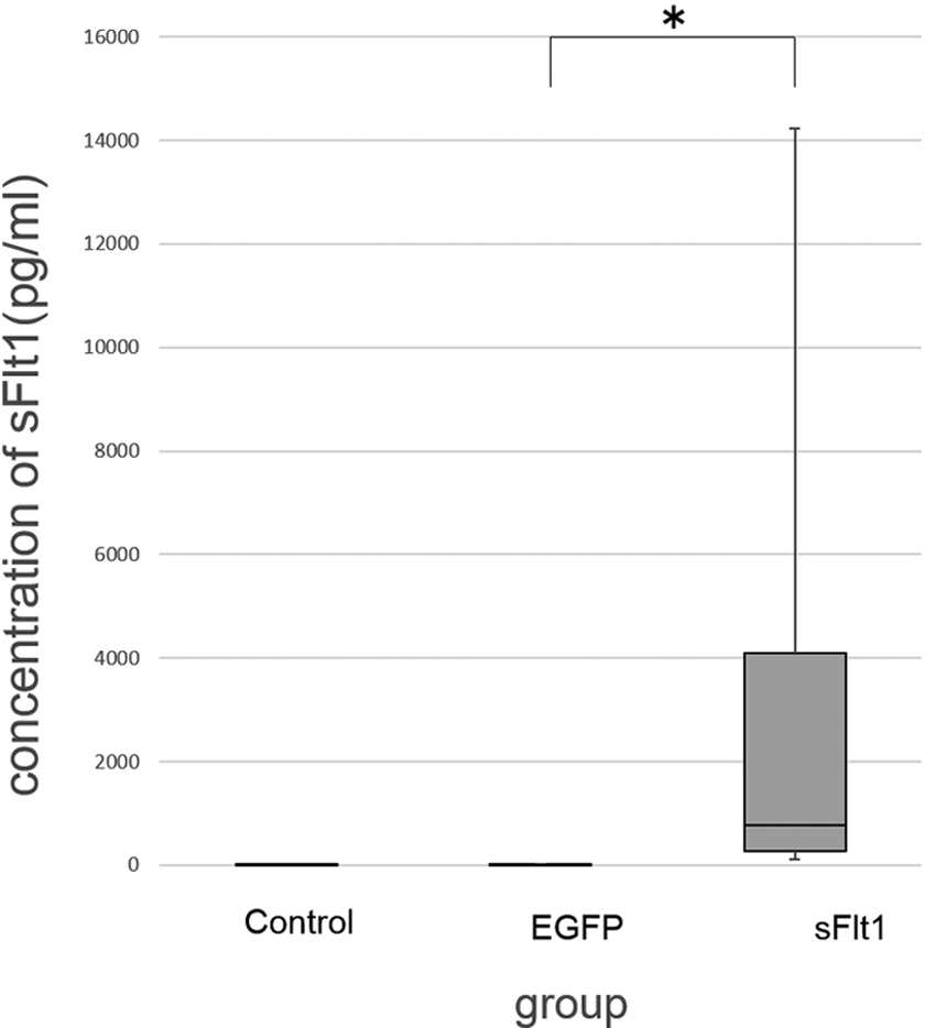
The concentration of soluble fms-like tyrosine kinase 1 (sFlt-1) in the culture media was measured by enzyme-linked immunosorbent assay (ELISA). pLV-hsFlt-1 transfected cells showed a significantly higher level of sFlt-1 compared to the lentiviral vector plasmid enhanced green fluorescent protein (pLV-EGFP) group (P <.01). Overexpression of sFlt-1 was successfully achieved. There was no significant difference between the pLV-EGFP and control groups (*P = .458). The data represent median, interquartile range (IQR), maximum, and minimum values from 3 separate experiments.
Expression Level of PlGF and VEGF in Cell Culture Medium
The concentration of PlGF (Figure 2) and VEGF (Figure 3) in the culture media of each group was measured by ELISA. There was no significant difference in PlGF levels between pLV-EGFP and pLV-hsFlt-1 groups (P = .197). Furthermore, VEGF levels between pLV-EGFP and pLV-hsFlt-1 groups showed no significant difference (P = .200), although there was a tendency for pLV-hsFlt-1 group to have a lower level of VEGF.
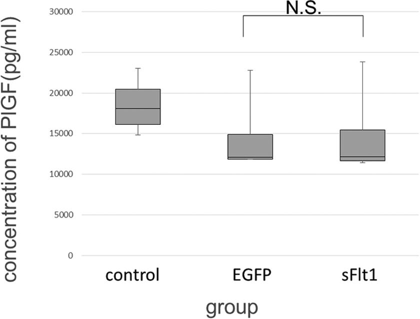
The concentration of platelet growth factor (PlGF) in the culture media of each group measured by enzyme-linked immunosorbent assay (ELISA). There was no significant difference in PlGF levels between lentiviral vector plasmid enhanced green fluorescent protein (pLV-EGFP) and pLV-hsFlt-1 groups (P = .197). The data represent median, interquartile range (IQR), maximum, and minimum values from 3 separate experiments.
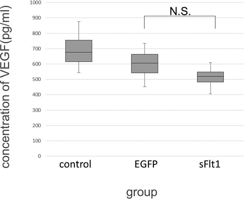
The concentration of vascular endothelial growth factor (VEGF) in each group was measured by enzyme-linked immunosorbent assay (ELISA). VEGF levels between lentiviral vector plasmid enhanced green fluorescent protein (pLV-EGFP) and pLV-hsFlt-1 groups showed no significant difference (P = .200). The data represent median, interquartile range (IQR), maximum, and minimum values from 3 separate experiments.
Effect of sFlt-1-Overexpression in Cell Proliferation
Ninety-six hours after transfection, the number of the cells in each group was counted. The cell number was significantly less in the pLV-hsFlt-1 group than in the pLV-EGFP group (P = .031; Figure 4). The cell number in the pLV-EGFP group was also less than in the nontransfected group because of the cytotoxicity caused by transfection itself.
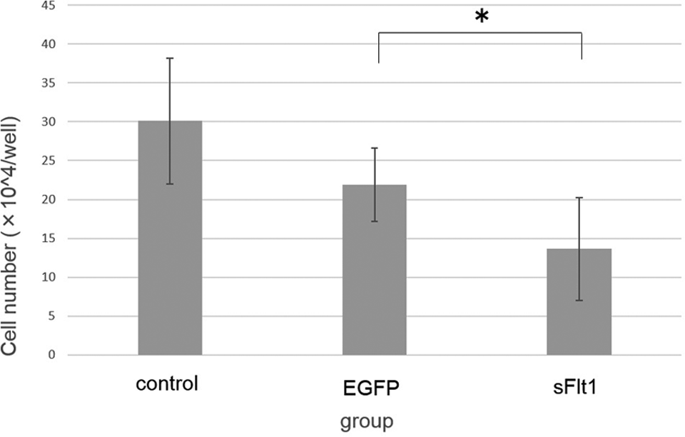
The cell number of each group at 96 hours after DNA transfection. The cell number was significantly lower in the pLV-hsFlt-1 group than in the lentiviral vector plasmid enhanced green fluorescent protein (pLV-EGFP) group (*P = .031). The data represent mean ± standard deviation (SD) from 3 separate experiments.
In order to demonstrate the effect of sFlt-1 on cell proliferation, BrdU incorporation was measured (Figure 5). The absorbance (370-492 nm) of pLV-EGFP group was significantly lower than in the nontransfected and pLV-hsFlt-1 groups (P = .005). There was no significant difference in absorbance between nontransfected and pLV-hsFlt-1 groups.
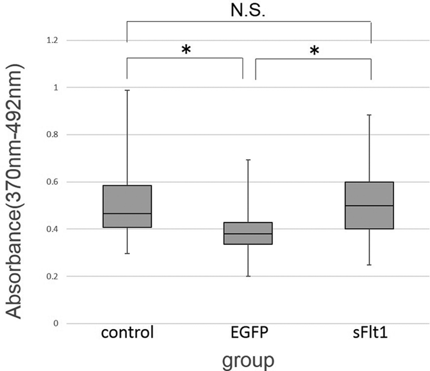
Bromodeoxyuridine (BrdU) incorporation in each group. There was no significant difference in the absorbance (370-492 nm) between nontransfected and pLV-hsFlt-1 groups. The absorbance of lentiviral vector plasmid enhanced green fluorescent protein (pLV-EGFP) group was significantly lower than the other groups. The data represent median, interquartile range (IQR), maximum, and minimum values from 3 separate experiments.
Effect of sFlt-1 Overexpression on Cytotoxicity
When cells are lysed, LDH is released around the cells. Thus, a higher LDH level in medium indicates higher cytotoxicity. The result of the LDH cytotoxicity assay revealed significant difference (P = .036) in the ratio of cytotoxicity between pLV-EGFP transfected and pLV-sFlt-1 transfected cells (Figure 6). This result indicates that sFlt-1 induces cytotoxicity in BeWo cells.
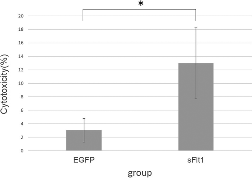
The result of lactate dehydrogenase (LDH) cytotoxicity assay. The “% cytotoxicity” in the pLV-sFlt-1 group was significantly higher than in the lentiviral vector plasmid enhanced green fluorescent protein (pLV-EGFP) group (P = .036). The data represent mean ± standard deviation (SD) from 3 separate experiments.
Staining of Apoptotic Cells in Each Group
Apoptotic cells were stained with TUNEL. In the positive control group with addition of 25 μmol/L of etoposide, approximately 40% of the cells were stained. In the pLV-sFlt-1 group, the cells were stained less than the positive control group. In the spontaneous group, the rate of stained cells was similar to the pLV-sFlt-1 group (Figure 7).
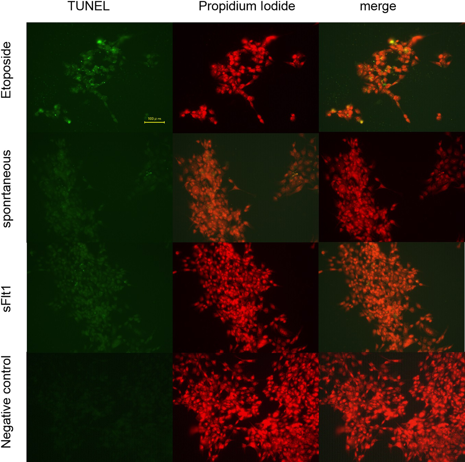
TdT-Mediated dUTP Nick end Labeling (TUNEL) staining of each group. Apoptotic cells were stained with TUNEL. In the positive control group (25 μmol/L Etoposide), approximately 40% of the cells were stained. In the pLV-sFlt-1 group, the cells stained were less than in the positive control. The rate of stained cells in the spontaneous group was similar to the pLV-sFlt-1 group.
Quantitative Analysis of Cell Apoptosis
Quantitative analysis of cell apoptosis was performed to evaluate the apoptotic effect of sFlt-1 in BeWo cells (Figure 8). There was a tendency for the pLV-hsFlt-1 group to show a higher level of ccK18 than the other groups, and the ccK18 levels between the nontransfected and pLV-hsFlt-1 groups showed a significant difference (P = .01). However, there was no significant difference between pLV-EGFP and pLV-hsFlt-1 groups.
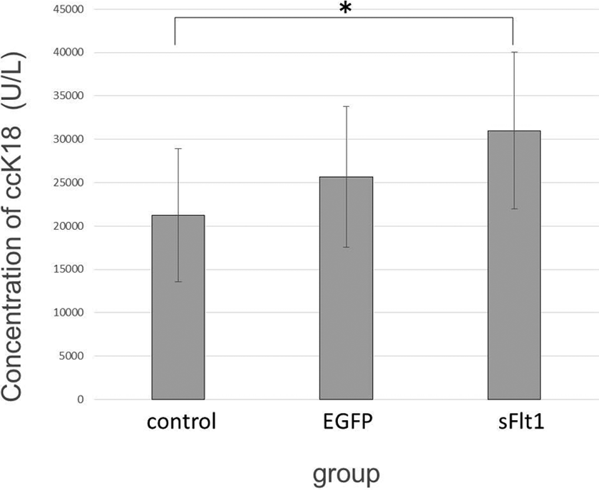
The concentration of caspase-cleaved K18 (ccK18) in each group is shown. The ccK18 levels between control and pLV-hsFlt-1 groups showed a significant difference. However, there was no significant difference between ccK18 levels of lentiviral vector plasmid enhanced green fluorescent protein (pLV-EGFP) and pLV-hsFlt-1 groups. The data represent mean ± standard deviation from 3 separate experiments.
Discussion
In this study, we showed cytotoxicity induced by sFlt-1 in BeWo cells. The results of quantitative analysis of ccK18 level and TUNEL staining did not indicate upregulation of apoptosis in BeWo cells.
Angiogenesis has a pivotal role in the growth of solid tumors. The VEGF is a major angiogenic mediator and is overexpressed in many kinds of human cancer cells. 3 –7 The VEGF receptor has 3 isoforms. These receptors are receptor tyrosine kinases having 7 immunoglobulin-like extracellular domains, 1 transmembrane region, and an intracellular kinase domain. Vascular endothelial growth factor receptor (VEGFR)-1 and VEGFR-2 are associated with angiogenesis, in contrast to VEGFR-3 which is associated with lymphangiogenesis. The VEGFR-1 is also called Flt-1 based on their structural similarity. The Flt-1 binds strongly to VEGF compared to VEGFR-2, but its kinase activity is about one order of magnitude weaker than that of VEGFR-2. These results indicate that VEGFR-2 is the major transducer of angiogenic signals, and Flt-1 plays a regulatory role. Soluble Flt-1 is an alternatively spliced isoform of Flt-1. Soluble Flt-1 carries only the ligand-binding region consisting of 6 immunoglobulin-like domains and has high affinity with VEGF, the same as full-length Flt-1. 20
The function of sFlt-1 has not been totally elucidated. The VEGFR-1 knockout mouse is embryonically lethal caused by overgrowth of vascular endothelial cells. 24 However, mice that lack the tyrosine kinase domain of Flt-1 are basically healthy. 20 This indicates that the role of Flt-1 in early embryogenesis is derived from its soluble form. Therefore, sFlt-1 can be considered to have other functions which are different from those of full-length Flt-1.
Soluble Flt-1 is also strongly associated with preeclampsia. Our group previously made a mouse model of preeclampsia by placenta-specific overexpression of sFlt-1 using lentiviral vectors. Placentae and fetuses from preeclampsia mice model were significantly smaller than those of the control group. 22 Immunostaining for CD31 (platelet/endothelial cell adhesion molecule 1) revealed suppression of vascular bed development in the placenta of the model mouse and also indicated sparse construction of the placenta. Based on this result, we focused on the possibility that sFlt-1 was cytotoxic by itself.
Several reports have been published on antitumor effect of sFlt-1 due to its antiangiogenic effect. Actually, tumor suppression has already been shown in several studies using neutralizing antibodies to VEGF and regional or systemic administration of adenovirus-mediated sFlt-1 cDNA. Our group previously reported that sFlt-1 induces cytotoxicity in ovarian and colorectal cancer cells. 21 However, to the best of our knowledge, no study has been reported on antitumor effect of sFlt-1 in BeWo cells. In this study, we showed an antitumor effect in BeWo cells. It may be difficult to distinguish the direct effects of sFLT-1 on the tumor cells from its antiangiogenic effects. Therefore, in this study, we treated cell lines with sFLT-1 in vitro in which the angiogenesis effect is negligible. It is reasonable to think that cell death is derived from direct cytotoxicity of sFlt-1 and not from the effect of neutralizing VEGF because no evidence has shown that VEGF directly promotes cell proliferation except in vascular endothelial cells. This idea is also supported by the observation that VEGF and PlGF concentration in culture supernatants was not significantly different between pLV-EGFP and pLV-hsFlt-1 groups (Figures 2 and 3).
The mechanism of the cell death induced by sFlt-1 has been controversial. In this study, the results did not indicate upregulation of apoptotic effects in BeWo cells. However, several reports have shown that sFlt-1 induces apoptosis in other cell lines. 15,16 There was also a tendency for pLV-hsFlt-1 group in this study to show higher level of ccK18 than other groups, although there was no significant difference between pLV-EGFP and pLV-hsFlt-1 groups (Figure 8). Our findings do not deny the apoptosis observed by other researchers. However, our study clarifies that nonapoptotic effects might also play an important role in tumor cell damage.
In clinical conditions, choriocarcinoma is commonly treated with multiagent regimens because the tumor cells are likely to develop drug resistance if single-agent therapy is administered. However, the therapeutic outcome becomes much poor when there is metastasis except for the lung or when primary chemotherapy is not effective. Therefore, it is important to establish additional therapies leading to cure of current therapy-resistant cases. Soluble Flt-1 might have this potential ability.
Soluble Flt-1 has another advantage in being used as a therapeutic agent. Side effect of sFlt-1 has been well documented and investigated with regard to preeclampsia. The representative adverse effects induced by sFlt-1 are high blood pressure and proteinuria, and other severe side effects are rarely observed. Unlike VEGF antibodies, sFlt-1 is a physiological substance naturally existing in the human body. Furthermore, sFlt-1 is metabolized within 48 hours in vivo. 25 Therefore, using sFlt-1 as an antitumor agent may have fewer complications than conventional chemotherapy.
Conclusion
Soluble Flt-1 has a cytotoxic effect on BeWo cells and could be a therapeutic candidate for malignant tumors.
Footnotes
Acknowledgments
We thank Dr Stephen J. Gadd, who worked in the Department of Pathology, University of Adelaide, for critical reading of the manuscript.
Declaration of Conflicting Interests
The author(s) declared no potential conflicts of interest with respect to the research, authorship, and/or publication of this article.
Funding
The author(s) disclosed receipt of the following financial support for the research, authorship, and/or publication of this article: This work was supported by The Osaka Medical Research Foundation for Intractable Diseases.