
Abstract
Cannabis sativa L. extracts were characterized by ultra performance liquid chromatography-mass spectrometry (UPLC-MS) using tetrahydrocannabinol (THC), cannabidiol (CBD), and tetrahydrocannabinolic acid (THCA) as marker compounds. The inhibitory effects of various extracts were determined on the survival and progression of highly metastatic breast cancer cells. A higher amount of CBD was found in the dichloromethane extract, and this was found to be effective in inhibiting breast cancer cell growth in vitro and in angiogenesis. Collectively, it may be concluded that CBD, THC, and THCA in the African variety of C. sativa can be used as marker compounds in UPLC-MS analysis. The ability of the plant to inhibit breast cancer cell survival and progression may affirm the traditional use of the drug as an anticancer agent.
Cannabis sativa L. (marijuana) has been used both medicinally and recreationally for many centuries in China, India, Southeast Asia, South Africa, and South America. 1,2 However, the discovery of the chemical structures of their unique active components as cannabinoids (CBDs) in early 1970s caused new insights into cannabis research. 2 To date, approximately 108 cannabinoids have been discovered in C. sativa. Cannabidiol (CBD), Δ9-tetrahydrocannabinol (THC), and tetrahydrocannabinolic acid (THCA) are considered as the most relevant because of their high biological potency and abundance in plant preparations. 3,4 However, the secondary metabolite content of the plants may vary due to geographical differences. 5 Therefore, it is always a good idea to validate the plant using standard lead molecules for exploring regional variations. CBDs, THC, and THCA, as well as other phytochemicals such as flavonoids and terpenes, have demonstrated a variety of possible therapeutic uses. 3 However, the flavonoids and terpenes act synergistically with CBD and THC for their biological activity. 6 CBDs are the most important non-psychoactive component of C. sativa; these have been associated with the largest number of pharmacological effects and THC is the most psychoactive cannabinoid, while THCA, which is a precursor of THC, also showed neuroprotective, antioxidant, and anti-inflammatory effects. 7
Presently, South Africa is one of the world’s largest producers and exporters of high-quality cannabis and day by day is becoming the main African regional hub for cannabis cultivation and trading. 5 The South African government and medical research community are still waiting for the scientific validation of the medicinal claims of the plant prior to approving legalization of medical marijuana in the country. 8 -10 Therefore, scientific validation and chemical characterization of the African variety of C. sativa may lead to policy change and subsequent approval for the cultivation and production of medicinal cannabis in South Africa.
In the present study, the African variety of C. sativa was analyzed and characterized by ultra performance liquid chromatography-mass spectrometry (UPLC-MS) using THC, CBD, and THCA as marker compounds. The inhibitory effects of the cannabis extracts on the survival and progression of highly metastatic breast cancer cells (MDA-231) were also scientifically undertaken.
Three extracts were generated from authenticated C. sativa plants, ie, n-hexane, dichloromethane (DCM), and methanol extracts. The highest percentage yield was found for the n-hexane extract (10.5%, w/w, raw material), whereas the percentage yields from the DCM and methanol extracts were 3.8% and 6.7%, respectively. A reverse phase UPLC-MS analytical chromatographic method was developed for the reasonable separation of all targeted marker compounds in the DCM and methanol extracts.
The n-hexane extract is known to extract non-polar compounds, including oils, which can block the UPLC column and hence was not used for this analysis.
The peaks which correspond to the molecular formula and exact masses of the three compounds in the DCM extract in negative ESI mode are shown in Figure 1(a). The peaks with retention times (minutes) of 11.31, m/z 359.2227; 13.18, m/z 357.2064; and 13.48, m/z 357.2063 confirm the presence of CBD/THC in peak 1, and THCA in peaks 2 and 3, with iFit confirmation >97% . In positive ESI mode only THC and CBD were detected at a retention time of 12.61, m/z 315.2325 (Figure 1(b)).
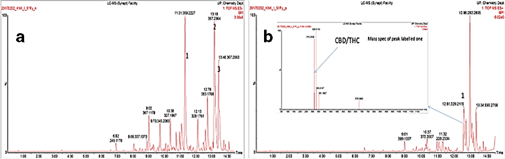
UPLC chromatogram of the DCM extract: (a) ESI negative mode; (b) ESI positive mode.
In the UPLC chromatogram of the methanol extract, one peak with a specific retention time of 5.99 minutes (m/z = 357.1193) showed the presence of THCA, with an iFit of 99.6%. Another peak, with a retention time of 13.68 minutes (m/z = 359.2227), confirmed the presence of THC/CBD in the methanol extract.
3-(4,5-Dimethylthiazol-2-yl)-−2,5-diphenyltetrazolium bromide (MTT) assay was performed to determine the cytotoxicity to MDA-231 cells of the DCM extract after 24 and 48 hours of exposure to the extract. The extract did not show any major effect toward the MDA cancer cell line (5 × 10⁴ cell densities) after 24 hours of exposure, although a concentration-dependent reduction of cell survival was observed (10-100 µg/mL). An IC50 value of 76.6 ± 15.2 µg/mL was determined. After 48 hours of exposure, the DCM extract remarkably inhibited the breast cancer cell growth in a concentration-dependent manner and an IC50 value of 27.8 ± 5.0 µg/mL was determined (positive control, doxorubicin, IC50 at 24 hours 50.3 ± 6.1 and at 48 hours 19.7 ± 4.7 µg/mL).
The DCM extract of C. sativa also inhibited blood vessel formation at day 11 of vascular endothelial growth factor (VEGF) and L-arginine-induced chick chorioallantoic membrane (CAM) formation (Figure 2). The angiogenic index was significantly (* P<0.05) increased by VEGF as compared with the vehicle control. CAM and DCM extract of C. sativa at a concentration of 40 µg significantly inhibited the angiogenic index (** P<0.01). The effect of cannabis DCM extract at 40 µg was equal to that of the standard angiogenic inhibitor tinzaparin (20 µg) (Figure 2(b)).
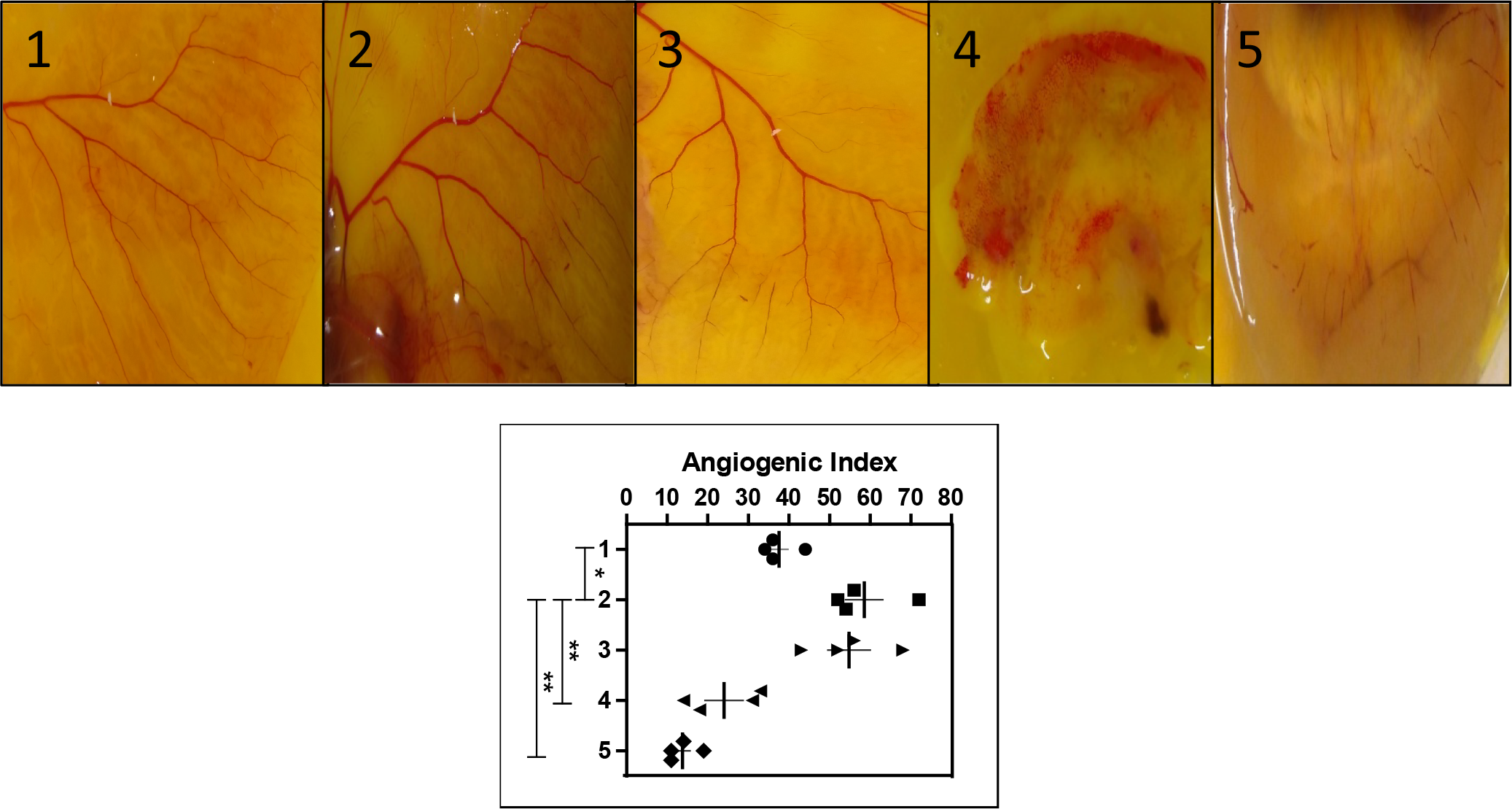
(a) Representative figures of CAM assay (n = 4). 1: VC; 2: VEGF + L Arg.; 3: DCM extract (20 µg); 4: DCM extract (40 µg); 5: tinzaparin (20 µg). (
The identification of active lead molecules is an important requirement for quality control and dose determination of plant-related drugs. 11 LC and MS have been applied widely for the quality control of traditional medicine. 12 The present study aimed to characterize the African “Dagga” variety of C. sativa using UPLC-MS analysis and to confirm the inhibitory effect of these standardized C. sativa extracts on in vitro breast cancer cells (MDA-231) and angiogenesis in experimental CAM assay models.
UPLC-MS analysis was performed using electro spray ionization; data recorded included retention times and iFit% confirmations in negative and positive modes. Both the DCM and methanol extracts showed the presence of the three marker compounds (CBD, THC, and THCA), although CBD and THC have the same accurate mass and hence are non-distinguishable through MS analysis. In the DCM extract, two similar peaks appeared at two different retention times with the same accurate mass as THCA, which may confirm the presence of a similar THCA isotope. The percentage of the marker compounds was higher in the DCM extract than the methanol extract. The DCM extract therefore was selected for further studies on breast cancer cell survival and progression assay.
Breast tumors are one of the most common human neoplasms and one of the leading causes of death among women. 13 Although the antiproliferative effect of cannabinoids on different tumor cells has been extensively confirmed both in vitro and in vivo, 14 a recent report indicates that THC may enhance breast cancer cell growth under certain circumstances. 15 The African “Dagga” variety of C. sativa inhibited the most metastatic breast cancer MDA-231 cell growth in a concentration-dependent manner with an IC50 of 27.8 ± 5.0 µg/mL after 48 hours of exposure.
Cancer cells need ample blood supply to enable metastatic processes and cell progression and, therefore, angiogenesis is the key regulator for extreme metastatic cancers like breast cancer. 16 To check the efficacy of C. sativa on vasculature formation, a CAM angiogenesis model was used. Angiogenesis was induced by VEGF and L-arginine (L-Arg). VEGF, along with L-Arg, significantly increased the angiogenic index (P < 0.05) as compared with the vehicle control. The DCM extract of C. sativa at a concentration of 40 µg was able to inhibit the blood vessels formation significantly at day 11 of CAM formation. The effect of cannabis was also found comparable to that of the standard anti-angiogenic drug tinzaparin (20 µg) (P < 0.01).
Tumor cells always establish angiogenesis by secreting various angiogenic factors like VEGF, MMPs, NO, and IL-8. 17 Inhibition of vasculature rather than inhibiting the tumor cells directly is considered as the most reliable approach to antitumor research these days. 18 In vitro and in vivo experiments showed that cannabinoids directly inhibit vascular endothelial cell migration and survival, as well as decrease the expression of proangiogenic factors like VEGF and MMPs. 19
Experimental
Collection, Authentication and Extraction
Cannabis sativa L. aerial parts were collected in Lesotho with a permit (number POS 064/2016/1017) for handling cannabis in the laboratory. The plant was authenticated by “Geo Potts Herbarium” at the University of the Free State, Bloemfontein, South Africa. The plant sample was extracted sequentially with n-hexane, DCM, and methanol (MeOH) by 1 hour of maceration, followed by filtration and evaporation in a rotary vacuum evaporator.
UPLC-MS Analysis
Compound separation and detection was performed using a Waters UPLC system with a Waters Synapt G2 QTOF instrument. Only the DCM and MeOH extracts were analyzed through UPLC-MS. The DCM extract was dissolved in 80% acetone (ACN) and 20% water, while the methanol extract was dissolved in 80% MeOH and 20% water. Approximately 1 mg of each extract was analyzed.
The analysis was accomplished using an Acquity UPLC BEH C18 1.7dµm (2.1 × 100 mm column). Gradient mobile phases were used: A, water: 0.1% formic acid; and B, ACN + 0.1% HCO2H. The elution rate was 0.400 mL/min. Gradient elution was used for the separation of the extracts. Elution started with 97% A and 3% B; this remained linear until 14 minutes, then from 14 to 16 minutes, elution was kept constant with 0% A and 100% B; a linear gradient of 97% A and 3% B was then used to reach completion after 20 minutes. Both negative and positive modes were used in the MS analysis. Data were accumulated using Software Masslynx V4.1.
Cytotoxicity in MDA-231 Breast Cancer Cells
The strongly metastatic breast cancer cell line MDA-MB-231 was grown in Dulbecco’s modified Eagle’s medium (ATCC® 30-2002™) supplemented with 10% fetal bovine serum (Hyclone, Thermo Fisher Scientific Inc, USA). The cells were maintained at 37°C in an atmosphere containing 5% CO2. Cell growth was measured under a light microscope and 70% to 80% confluence of the cells was used in all experiments. MDA-231 breast cancer cells at cell densities of 5 × 10⁴ were seeded in 96/48 well tissue culture plates and the cells were allowed to adhere for 24 hours at 37°C in the CO2 incubator. On the next day, the cells were exposed to different concentrations of extracts (0–100 µg/mL) and doxorubicin (0–100 µg/mL) for 24 and 48 hours, respectively. A cell viability assay was then performed using MTT assay. 20 Data were analyzed by plotting % cell survival vs concentration, and IC50 values were calculated in the plot of % inhibition vs concentration using Graph Pad Prism software version 5.
VEGF-Induced Angiogenesis Assay in Chick Chorioallantoic Membrane
Twenty fertile chicken eggs were obtained from a local farmer in Bloemfontein, South Africa, and incubated (“Prolab” Model PL-0001) at 37ᵒC ± 1ᵒC. Sixty percent humidity was maintained by keeping a tray full of sterile distilled water in circulating air condition. On day 8, approximately a 1 cm2 hole was prepared on the surface of the egg shells in aseptic conditions and 1 cm2 (diameter) Whatman filter paper (Sigma-Aldrich, USA) shocked with VEGF (0.2 nM/CAM)/L-Arg(10 µg/CAM)/extract (20–40 µg) as per grouping, inserted and sealed with adhesive tape. Tinzaparin, at 20 µg/CAM, was used as a positive angiogenesis inhibitor in the experiment. All the eggs (n = 4) were returned to the incubator aseptically until day 11 of CAM development. The developed CAM was opened on day 11 in Petri dishes and the blood vessels formed were observed, recorded, and analyzed under a microscope to represent the data. 21 The intact blood capillaries extended from the main blood vessels were counted microscopically and analyzed to calculate the angiogenic index as the average number of blood capillaries formed in each CAM. 22
Statistical Analysis
Statistical significance (p) was calculated in all experiments using Graph Pad Prism version 4.03 and Graph Pad Instat software (Graph Pad Software Inc., San Diego, CA, USA) and analyzed with one-way ANOVA followed by Dunnett’s post hoc test of significance where *P < 0.05 and **P < 0.01 were considered to be significant and highly significant, respectively. 23
Footnotes
Acknowledgement
Authors are thankful to NRF and DST-IKS Based Technology, South Africa (Research Development and Innovation on Medical Cannabis Project Funding agreement Number: DST/CON 0162/2015), for financial support and University of the Free State for institutional support. Dr Asis Bala sincerely acknowledges the University of the Free State, Bloemfontein, South Africa for a Post-Doctoral Research Fellowship.
Declaration of Conflicting Interests
The author(s) declared no potential conflicts of interest with respect to the research, authorship, and/or publication of this article.
Funding
The author(s) disclosed receipt of the following financial support for the research, authorship, and/or publication of this article: This work was supported by NRF and DST-IKS Based Technology, South Africa (Research Development and Innovation on Medical Cannabis Project Funding agreement Number: DST/CON 0162/2015).