
Abstract
Objective
This study aimed to evaluate the phenolic and flavonoid composition of the hydro-ethanolic extract of Psidium guajava L. leaves harvested from the El Oued region (southeastern Algeria) and to investigate its various biological activities.
Methods
Dried P. guajava leaves were defatted with petroleum ether and then extracted with ethanol by cold maceration using 50% aqueous. Total phenolic content (TPC) and total flavonoid content (TFC) were quantified using standard spectrophotometric methods, phenolic compounds were identified using high-performance liquid chromatography coupled with diode-array detection (HPLC-DAD). The extract's antioxidant activity was determined by DPPH radical scavenging assay. In vitro assays were also conducted to evaluate anti-hemolytic, antibacterial (against Pseudomonas aeruginosa, Escherichia coli, Staphylococcus aureus, and Bacillus subtilis), anti-inflammatory (based on bovine serum albumin [BSA] denaturation), and α-amylase inhibitory activities.
Results
The results that the extract contained high levels of total phenolics and flavonoids, HPLC-DAD identified major compounds such as gallic acid and quercetin. The extract demonstrated potent antioxidant activity (IC50 = 17.24 ± 0.01 µg/mL), which was less effective than that of standard ascorbic acid. However, it showed superior α-amylase inhibitory activity compared to acarbose (IC50 = 168.32 ± 1.12 vs 299.26 ± 17.81 µg/mL). Additionally, the extract exhibited moderate anti-inflammatory activity (IC50 = 459.14 ± 27.49 µg/ml) compared to aspirin and moderate anti-hemolytic activity compared to vitamin E, antibacterial activity was more pronounced against Gram-positive strains.
Conclusion
The findings highlight the therapeutic potential of P. guajava leaves as a natural source of bioactive compounds with antioxidant, anti-inflammatory, antibacterial, and antihyperglycemic properties.
Keywords
Introduction
Medicinal plants have constituted a cornerstone of human healthcare since antiquity, valued for their therapeutic efficacy, accessibility, and relatively low side effects. 1 In recent decades, scientific research has shown renewed interest in investigating the bioactive constituents of these plants and their associated biological activities. 2 These natural resources represent a vast reservoir of secondary metabolites, such as alkaloids, terpenes, phenolics, flavonoids, and essential oils, 3 many of which exhibit a broad spectrum of pharmacological properties, including antidiabetic, anti-inflammatory, anticancer, antioxidant, and antimicrobial effects. 4 This highlights the necessity of systematic scientific studies to elucidate their chemical profiles and harness their potential for developing safe and effective pharmaceuticals. 5
Psidium guajava L. (guava), a medicinal species belonging to the Myrtaceae family, thrives in tropical and subtropical climates. Native to areas such as Mexico, Central America, and northern areas of South America, this plant is morphologically characterized by its oppositely arranged leaves and notably short petioles.6,7 Guava leaves have long been integrated into traditional medicinal practices throughout Latin America and the Caribbean, where they have been widely used to treat gastrointestinal disorders such as diarrhea, dysentery, and gastroenteritis and to alleviate abdominal discomfort and indigestion.7,8 Notably, the leaves contain higher bioactive compounds compared to other plant parts, which underscores their value in pharmaceutical, nutritional, and cosmetic applications. 9
The health-promoting properties of P. guajava leaves are primarily attributed to their richness in phenolic compounds, including gallic, ferulic, and caffeic acids, as well as flavonoids such as quercetin, kaempferol, myricetin, rutin, cinnamic acid, and epicatechin. 9 These phytoconstituents display significant antioxidant activity by counteracting oxidative damage in biological systems, potentially preventing chronic diseases such as diabetes, cancer, and cardiovascular disease. 10 Traditional uses also include treatments for cough, hypercholesterolemia, and weight management, while recent studies have confirmed their hypoglycemic, cardioprotective, antimicrobial, and antispasmodic properties.7–11
The antioxidant potential of phenolic compounds is intrinsically linked to their structural features, particularly the number and position of hydroxyl (-OH) groups on aromatic rings. These molecules act as hydrogen or electron donors to neutralize reactive oxygen species (ROS), such as superoxide and hydroxyl radicals), thereby stabilizing their structures and reducing oxidative stress, a major factor in the pathgenesis of cancer and hypertension.10–12 The chemical profile of plant-derived extracts can vary considerably depending on factors such as species origin, extraction method (eg, hydro-ethanolic extraction), temperature, and solvent concentration. 13
Hydro-ethanolic extraction (ethanol-water mixture) is widely recognized as one of the most effective techniques for isolating polyphenols and flavonoids given its ability to solubilize both polar and semi-polar constituents. 14 Several studies have demonstrated that hydro-ethanolic extraction of P. guajava leaves yields higher phenolic content and stronger antioxidant activity compared to aqueous, ethanolic, and methanolic extracts. In particular, a 50% hydro-ethanolic solvent ratio has been identified as the most efficient for enhancing antioxidant properties in P. guajava leaf extracts. 15
Analytical investigations employing high-performance liquid chromatography coupled with diode-array detection (HPLC-DAD) have revealed considerable variation in the polyphenolic profile of ethanolic, 16 hydroethanolic,14–17 methanolic, 18 and aqueous, 9 extracts of P. guajava leaves, reinforcing their promise as a source for innovative therapeutic agents. 9 Current research in increasingly focused on comprehensive analysis of secondary metabolites in P. guajava leaves with the aim of elucidating their mechanisms of action, optimizing extraction protocols, and evaluating therapeutic efficacy while minimizing adverse effects. 19 These studies highlight the pivotal role of polyphenols in mitigating inflammation and oxidative damage, thereby supporting the development of natural, sustainable plant-based pharmaceuticals. 16
The present study aims to investigate the biological potential of P. guajava cultivated in the El Oued region, southeastern Algeria, through comprehensive laboratory analyses including: (1) determination of total phenolic and flavonoid contents, (2) HPLC-DAD characterization of polyphenolic constituents in hydro-ethanolic leaf extracts, and (3) in vitro evaluation of antioxidant, anti-hemolytic, antibacterial, anti-inflammatory, and antidiabetic activities. The overarching objective is to assess this plant as a source of novel therapeutic solutions for modern health challenges, particularly those linked to oxidative stress, metabolic disorders, and infectious diseases.
Materials and Methods
Chemicals and Reagents
This study employed various analytical-grade chemicals and reagents, used without further purification. The material includes ascorbic acid (AA), 2,2-diphenyl-1-picrylhydrazyl (DPPH•), absolute ethanol, ethanol 96%, Folin–Ciocalteu reagent, sodium carbonate (Na2CO3), aluminum chloride (AlCl3), methanol, ethylene diamine tetra acetic acid (EDTA), phosphate-buffered saline (PBS), quercetin (Q), petroleum ether, gallic acid (GA), formic acid, phosphate buffer, sulfuric acid (H2SO4), double-distilled water (DD), ammonium molybdate, sucrose solution, hydrogen peroxide (H2O2), acetonitrile, vitamin E (tocopherol), gentamicin, bovine serum albumin (BSA), distilled water, sodium phosphate, dimethyl sulfoxide (DMSO), aspirin, α-amylase, hydrochloric acid (HCl), sodium chloride (NaCl), acarbose, iodine solution, and starch.
Collection and Preparation of Psidium guajava Leaves
Leaves of P. guajava were collected in November 2024 from a farm in the El Bayadha region, located south of Oued Souf, Algeria. The leaves were thoroughly rinsed with distilled water to remove surface impurities and air-dried in the shade at ambient temperatures (20-25 °C) for two weeks. The dried leaves were finely milled into powder and stored in airtight plastic containers at room temperature. 20
Preparation of Hydro-Ethanolic Extract
A total of 20 g of powdered plant material was accurately weighed to initiate the extraction process with a hydro-ethanolic solvent. The sample was first defatted by immersion in petroleum ether under magnetic stirring for 24 h at ambient temperature. The suspension was then filtered using Whatman No. 1 filter paper . The filtrate was discarded, while the solid residue was retained for further use. The defatted plant residue was subsequently macerated in 200 mL of an ethanol–water mixture (1:1, v/v) and subjected to constant agitation at room temperature for an additional 24 h. Filtration was performed again using Whatman No. 1 filter paper. This extraction procedure was performed twice using fresh solvent each time.
All resulting filtrates were combined and concentrated using a rotary evaporator. The concentrated extract was subsequently dried in a hot-air oven at 45 °C overnight. Finally, the dried material was preserved in a refrigerator maintained at 4 °C until further use.
The employed extraction method produced a yield of 29.258% relative to the dry leaf powder weight, calculated as follows:
HPLC Analysis of Phenolic Compounds
The detection and characterization of phytochemical constituents were carried out using HPLC-DAD on an Agilent 1200 series system outfitted with a Kinetex Evo C18 analytical column. Initially, standard compounds were first injected to determine their retention times under the specified chromatographic settings. Subsequently, a 20 µL aliquot of the plant extract was analyzed under the same chromatographic conditions, including a mobile phase flow rate set at 1 mL/min, a detection wavelength fixed at 324 nm, and a column temperature maintained at 30 °C. The chromatographic peaks were compared with those of the reference standards, enabling both qualitative and quantitative assessment of phenolic constituents present in the sample, as presented in Table 1. 21
Experimental Conditions for the Separation of Phenolic Compounds in HPLC-DAD Analysis.
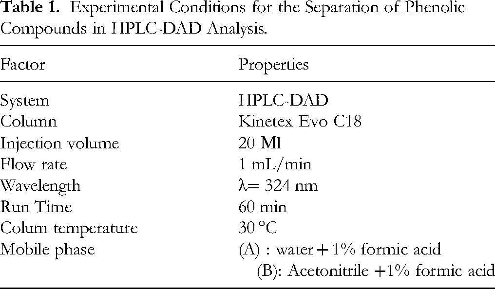
The HPLC method was validated, demonstrating excellent linearity (R2 > 0.999) over the tested concentration range, high repeatability (RSD < 5%), and recovery rates of 80–90%, confirming analytical accuracy and reliability of the method.
Determination of Total Phenolic Content (TPC)
The TPC was quantified employing the Folin–Ciocalteu colorimetric assay with slight modifications based on the procedure reported by Chihi et al 22 Briefly, 1 mL of each appropriately diluted extract was combined with 0.5 mL of Folin–Ciocalteu reagent, previously diluted 1:10 with distilled water. After allowing the mixture to react for 5 min, 2 mL of sodium carbonate solution (7.5%, w/v) was added. The mixture was then incubated in the dark at room temperature for 30 min. Absorbance was subsequently measured at 765 nm using a UV-Vis spectrophotometer. A calibration curve was established using gallic acid in the concentration range of 0.006 to 0.05 mg/mL. The phenolic content of the extracts was quantified as milligrams of gallic acid equivalents per gram of extract (mg GAE/g extract).
Determination of Total Flavonoid Content (TFC)
The TFC was assessed following a modified version of the aluminum chloride colorimetric method according to the procedure reported by Oliveira et al 23 In brief, 0.5 mL of each diluted plant extract was combined with an equal volume (0.5 mL) of 2% (w/v) aluminum chloride solution prepared in absolute ethanol. The reaction mixture was allowed to stand in the dark for 45 min at ambient temperature. The absorbance was measured at 415 nm using a spectrophotometer. Quercetin served as the standard reference compound over a concentration range of 0.005–0.035 mg/mL. Results are presented as milligrams of quercetin equivalents (mg QE/g extract).
Antioxidant Activity
DPPH Assay
The antioxidant capacity of the hydro-ethanolic extract was evaluated using the DPPH (2,2-diphenyl-1-picrylhydrazyl) radical scavenging assay following the method described by Aqil et al 24 with minor modification. A stock solution of DPPH was freshly prepared by dissolving 4 mg of the violet crystalline compound in 50 mL of methanol, ensuring complete dissolution to obtain a uniform deep-purple solution. Ascorbic acid served as the positive control.
Serial dilutions were prepared for both the reference standard (2-20 µg/mL) and the plant extract (5-35 µg/mL) using methanol as the solvent. For each tested concentration, 0.5 mL of solution was placed in test tubes, then combined with 1 mL of the DPPH reagent. The tubes were gently shaken and incubated in the dark at room temperature for 30 min; absorbance was then recorded at 517 nanometers using a UV-Visible spectrophotometer.
The percentage of radical scavenging activity was determined by employing the following equation:
Where:
The antioxidant potential was determined by calculating the IC50 value, which refers to the required extract concentration (µg/mL) to neutralize 50% of DPPH free radicals.
Total Antioxidant Contents (TAC)
The total antioxidant capacity (TAC) was determined using the phosphomolybdenum method, which is based on the reduction of molybdenum(VI) ions (Mo6+) to molybdenum(V) ions (Mo5+) by the antioxidants present in the extract, leading to the formation of a green- colored phosphomolybdenum complex under acidic conditions. 25 For the assay, 0.2 mL of the diluted extract was mixed with 2 mL of the reagent solution, which contained a mixture of 0.6 M sulfuric acid, 28 mM sodium phosphate, and 4 mM ammonium molybdate prepared in 100 mL of distilled water. The mixtures were incubated in a water bath at 95 °C for 90 min, then cooled to room temperature before measuring the absorbance at 695 nm using a spectrophotometer. Gallic acid, at concentrations ranging from 0.01 to 0.14 mg/mL, was employed to generate a standard calibration curve. The antioxidant capacity of the extracts was expressed based on the linear regression equation correlating gallic acid concentrations with their corresponding absorbance values. 26
Anti-Hemolytic Activity Ex Vivo
The anti-hemolytic activity of P. guajava extract was evaluated using a previously established spectrophotometric method,27,28 with minor modifications. The assay was performed in a 96-well microplate format. Whole blood samples were obtained from healthy volunteers and collected into ethylenediaminetetraacetic acid (EDTA)-containing tubes. The samples were centrifuged at 2000 rpm for 10 min to isolate erythrocytes. The supernatant was discarded, and the erythrocyte pellets were washed three times with PBS, followed by centrifugation under the same conditions. The final pellet was thoroughly suspended using PBS to obtain a 10% erythrocyte suspension.
Plant extracts were prepared in PBS at final concentrations ranging from 10 to 40 mg/mL. In each well of a microplate, 20 µL of a red blood cell suspension was mixed with 40 µL of the extract solution. Hemolysis was then induced by adding 40 µL of hydrogen peroxide (H2O2) prepared in PBS. The plates underwent incubation at 37 °C for 3 h. To terminate the reaction, 80 µL of PBS and 30 µL of a 20% sucrose solution were added to each well. Finally, the degree of hemolysis was assessed by measuring the absorbance at 540 nm using a microplate reader.
A positive control was prepared by treating the erythrocyte suspension with H2O2 alone to represent 100% hemolysis, while PBS alone served as the negative control. α-Tocopherol (vitamin E), prepared at the same concentration range (10-40 mg/mL), was used as a reference antioxidant and subjected to identical experimental conditions. All experiments were performed in triplicate, and the anti-hemolytic activity was determined as a percentage using the following formula:
Evaluation of the Antimicrobial Activity of the Hydro-Ethanolic Extract of P. guajava
To evaluate the antimicrobial therapeutic potential of the hydro-ethanolic extract of P. guajava leaves, three main analytical approaches were employed: the agar well diffusion assay, the broth microdilution technique to determine the minimum inhibitory concentration (MIC), and the determination of the minimum bactericidal concentration (MBC) using the spot inoculation technique. The evaluation focused on four standard bacterial strains, including two Gram-negative bacteria (Pseudomonas aeruginosa ATCC 27853 and Escherichia coli ATCC 25922) and two Gram-positive bacteria (Staphylococcus aureus ATCC 25923 and Bacillus subtilis ATCC 25973). All experiments were performed under standardized conditions, with each assay conducted in triplicate to ensure the accuracy and reliability of the results, following the recommended protocols established by reference organizations in pharmaceutical microbiology.
Agar Diffusion Method (Well Method)
Petri dishes containing Mueller–Hinton agar (for bacterial testing) were aseptically seeded with microbial suspensions standardized to 106 CFU/mL, derived from freshly cultured bacterial or yeast isolates. The inoculation was carried out evenly across the agar surface using sterile cotton swabs. After seeding, the plates were left to dry briefly to allow proper absorption before further processing. Wells were created at the center of each plate using the upper tip of a sterile Pasteur pipette. Each well was then filled with approximately 50 µL of the extract dissolved in water at concentrations of 60, 30, 15, and 5 mg/mL. Gentamicin (30 µg/disc) served as the positive control.
The plates were incubated at 37 °C for 24 h for bacterial cultures and 48 h for yeasts. The antimicrobial activity was assessed by observing the formation of clear inhibition zones around the wells. The diameters of these zones were measured in millimeters to evaluate the efficacy of the extract.29,30 (see Figure 1)
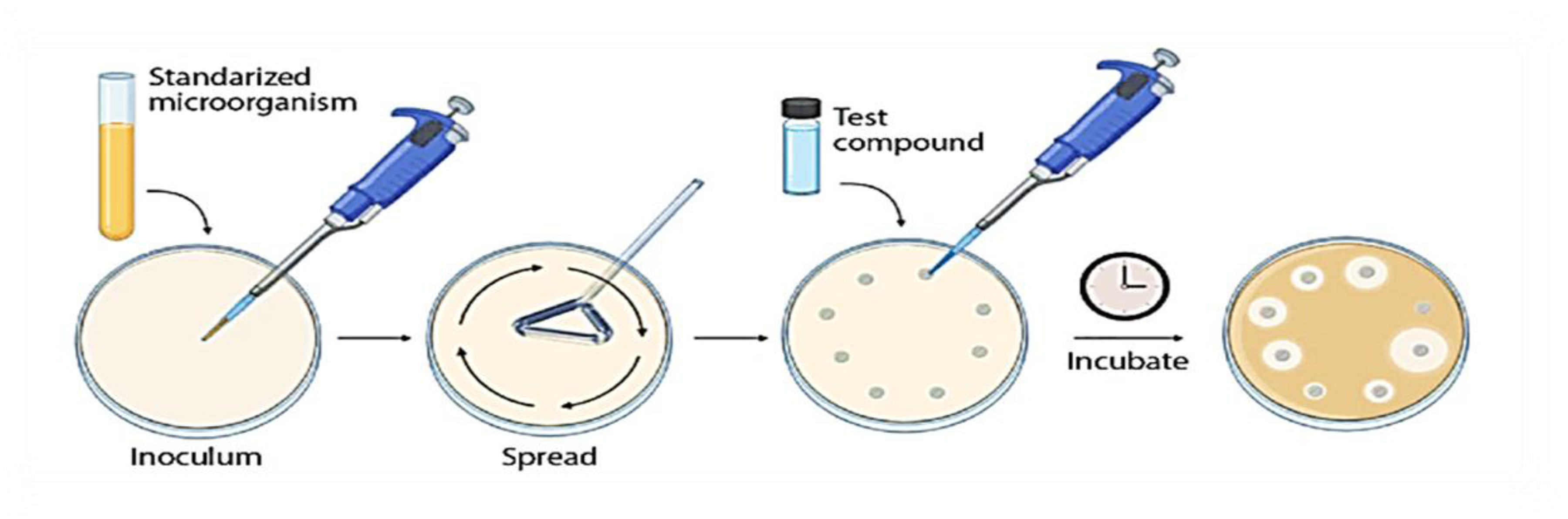
Diagram Illustrating the Well Diffusion Method on Agar Medium. 31
Broth Microdilution Method for MIC and MBC Determination
The antimicrobial efficacy of P. guajava extract was evaluated using the broth microdilution method in 96-well microplates, following the guidelines of the Clinical and Laboratory Standards Institute (CLSI). 32 test Bacterial suspensions were initially prepared by culturing the test strains on Mueller–Hinton agar (MHA), followed by inoculation into cation-adjusted Mueller–Hinton broth (MHB) until visible turbidity was achieved. The suspensions were then standardized to a turbidity matching the 0.5 McFarland standard, corresponding to approximately 1.5 × 10⁸ CFU/mL using the BioMérieux DensiCHEK Plus device (VITEK 2 Systems).
A stock solution of the extract was prepared at a concentration of 80 mg/mL by dissolving the dry plant extract in dimethyl sulfoxide (DMSO). Serial dilutions were made to obtain final concentrations between 80 and 2.5 mg/mL. In the microplate assay, 100 μL of each extract dilution was added to the wells, followed by 50 μL of the standardized bacterial suspension. Each assay included a growth control (without extract or antibiotic) and a sterility control (broth only). Plates were incubated at 37 °C for 24 h.
The MIC was determined as the lowest concentration of the extract that completely inhibited visible bacterial growth. 33 To determine the MBC, 10 μL from each well showing no visible growth was subcultured on MHA plates free of the extract and incubated under the same conditions. The MBC was defined as the minimum concentration at which bacterial growth was completely inhibited on the agar surface. All experiments were performed in triplicate to ensure reproducibility of the results. 34
Anti-Inflammatory Activity In Vitro
The anti-inflammatory potential of the hydro-ethanolic extract was evaluated based on its ability to inhibit heat-induced denaturation of BSA following a previously established protocol with minor modifications.35,36
Various concentrations of the extract and the standard anti-inflammatory agent aspirin were prepared in distilled water. For each treatment, 50 µL of the test extract was combined with 450 µL of a 0.5% (w/v) aqueous BSA solution. The mixtures were incubated at 37 °C for 20 min, followed by heat treatment at 57 °C for 5 min to induce protein denaturation. A control sample was prepared by replacing the extract with double-distilled water (DD). After cooling at room temperature, the absorbance of each sample was measured at 255 nm using a UV–Visible spectrophotometer. Aspirin (acetylsalicylic acid) served as the positive control. All experiments were carried out in triplicate to ensure the reproducibility and reliability of the results. The percentage inhibition of protein denaturation was calculated using the following equation:
Vt: Absorbance of the test sample; Vc: Absorbance of the control. The IC50 value, which corresponds to the concentration of the extract required to inhibit 50% of protein denaturation, was determined from the dose–response curve.
Evaluation of The Inhibitory Activity of P. guajava Extract Against α-amylase Enzyme
The inhibitory potential of the hydro-ethanolic extract of P. guajava leaves against α-amylase activity was assessed using a modified spectrophotometric method. 37 In this assay, the reaction mixture was formed by mixing 50 µL of the extract at various concentrations with 50 µL of the enzyme solution (0.5 mg/mL in 0.02 mM sodium phosphate buffer (pH 6.9) containing 0.006 M NaCl), followed by incubation at 37 °C for 10 min. Subsequently, 50 µL of a 1% starch solution (prepared in 2 mM phosphate buffer) was added to initiate the enzymatic reaction, and the mixture was then incubated for an additional 30 min at the same temperature. To terminate the reaction, 30 µL of 1 M HCl was added, followed by 70 µL of iodine reagent in each tube. The absorbance was recorded at 580 nm using a UV–Visible spectrophotometer.
The experiment included various controls: a blank sample (extract replaced by phosphate buffer), acarbose as a standard inhibitor, an enzyme-free control, and a starch-only control. All measurements were carried out in triplicate to ensure result accuracy. The percentage of inhibition was calculated using the following formula:
The IC50 value was determined through regression analysis.
Statistical Analysis
Most experiments were repeated three times (n = 3), and results were recorded as mean ± standard deviation. All statistical data and IC50 values were calculated using origin pro 2018 and Microsoft Excel 2016 software.
Results
Determination of Total Flavonoid and Phenolic Contents
The total phenolic and flavonoid contents of the P. guajava extract were quantified as 268.30 ± 0.66 mg of gallic acid equivalents per gram of extract (mg GAE/g) and 16.96 ± 0.10 mg quercetin equivalents per gram of extract (mg QE/g), respectively. These values were determined using calibration curves constructed using gallic acid and quercetin as standard references. The detailed results are presented in Table 2.
Phenolic and Flavonoid Contents of P. guajava Extract (n = 3).

HPLC Analysis of Phenolic Compounds
HPLC analysis of the hydro-ethanolic extract from P. guajava leaves collected in the Oued Souf region identified eleven distinct phenolic compounds. Identification was achieved by matching retention times with authentic standards (Table 3), while the chromatographic profile (Figure 2) demonstrated significant variability in both the composition and concentration of these phytochemicals. The detected phenolics included gallic acid, chlorogenic acid, caffeic acid, p-coumaric acid, sinapic acid, verbascoside, coumarin, isorhamnetin-3-O-rutinoside (narcissin), quercetin, apigenin, and isorhamnetin.
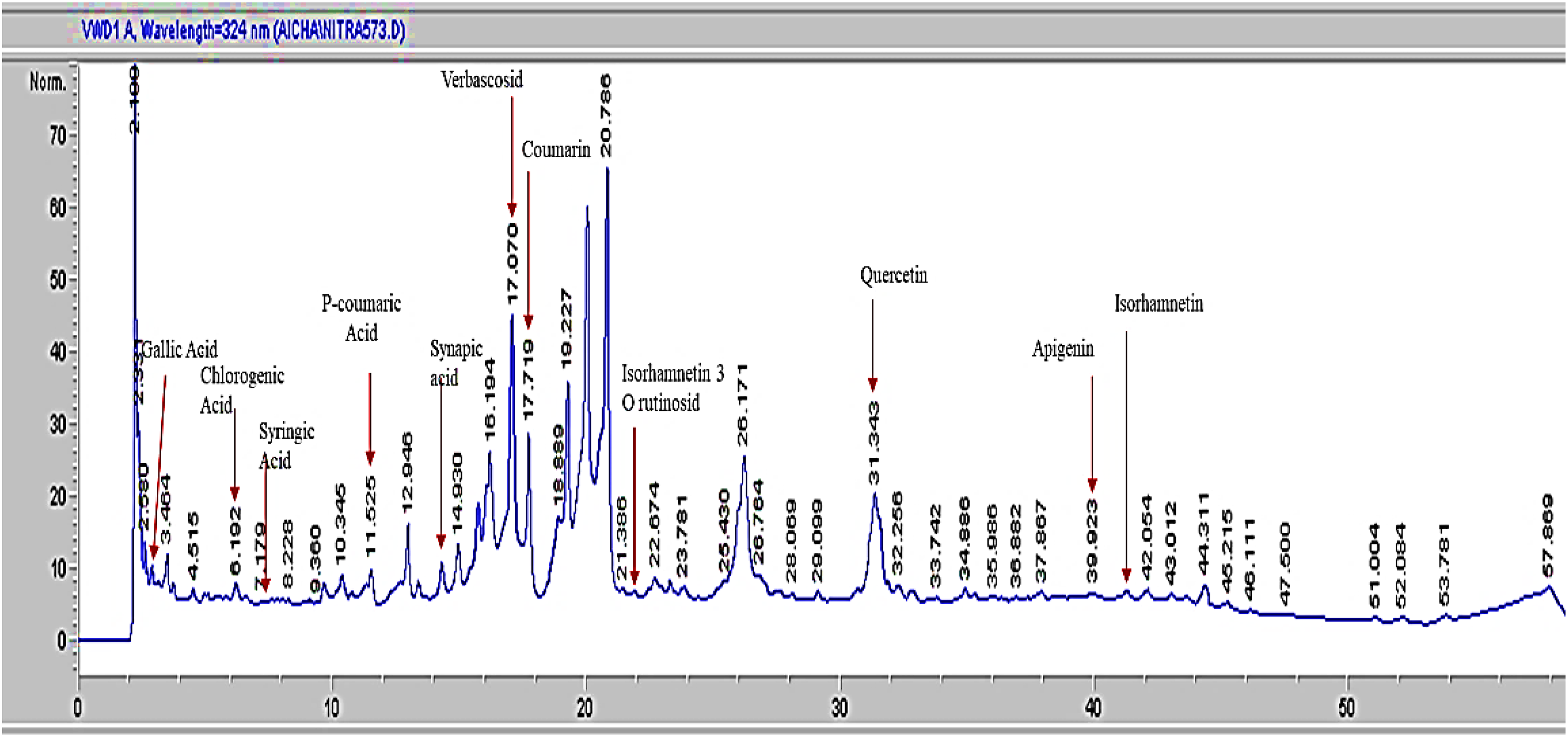
HPLC Chromatogram of Phenolic Compounds in Hydro-Ethanol Extract of P. guajava.
Concentrations of Phenolic Compounds in the Hydro-Ethanolic Extract and Their Retention Time as Determined by HPLC-DAD (n = 3).

Antioxidant Activity
In Vitro
Table 4 summarizes the antioxidant properties of hydro-ethanolic extract, revealing significant antioxidant capacity. The TAC reached 491.95 ± 2.45 mg GAE/g extract, as determined by the phosphomolybdenum method. DPPH radical scavenging assays demonstrated potent activity, with the extract showing an IC50 of 17.24 ± 0.01 μg/mL - comparable to ascorbic acid (reference standard). These results indicate that lower IC50 values correlate with enhanced antioxidant efficacy, confirming the extract's strong free radical neutralization potential.
Total Antioxidant Capacity (TAC) and IC50 Values for DPPH Test of P. guajava Extract (n = 3).
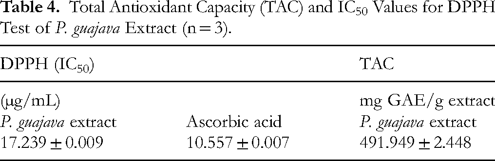
IC50: Half Maximal Inhibitory Concentration.
Anti-Hemolytic Activity Ex Vivo
The protective capacity of the P. guajava extract against erythrocyte hemolysis induced by H2O2 was assessed to determine its anti-hemolytic activity. The extract demonstrated a moderate antioxidant effect, with hemolysis inhibition increasing proportionally to the extract concentration, as illustrated in Figure 3. At a concentration of 40 mg/mL, the extract achieved an inhibition percentage of 43.00 ± 5.06%. In contrast, the reference antioxidant α-Tocopherol (Vit E) exhibited a significantly higher inhibition rate of 82.95 ± 2.03% at the same concentration (Table 5).

Anti-Hemolytic Inhibitory Activity of P. guajava Leaf Hydro-Ethanolic Extract Compared with Reference Standard.
Percentage Inhibition of the Anti-Hemolytic Activity of P. guajava Extract and Standard Compound. Data are Expressed as Mean ± SEM (n = 3).

Antimicrobial Activity
Table 6 (Figure 4) presents the antibacterial activity of the hydro-ethanolic extract of P. guajava as assessed by the diffusion method. The extract demonstrated antibacterial effects against three bacterial strains—P. aeruginosa, S. aureus, and B. subtilis—exhibiting clear inhibition zone diameters of 8 mm, 10 mm, and 13 mm, respectively, at a concentration of 60 mg/mL. No inhibitory effect was detected at lower concentrations. Furthermore, the extract showed no antibacterial effect against E. coli. These findings were compared with the positive control gentamicin, which produced inhibition zones of 31, 30, 27, and 22 mm against E. coli, P. aeruginosa, S. aureus, and B. subtilis, respectively.
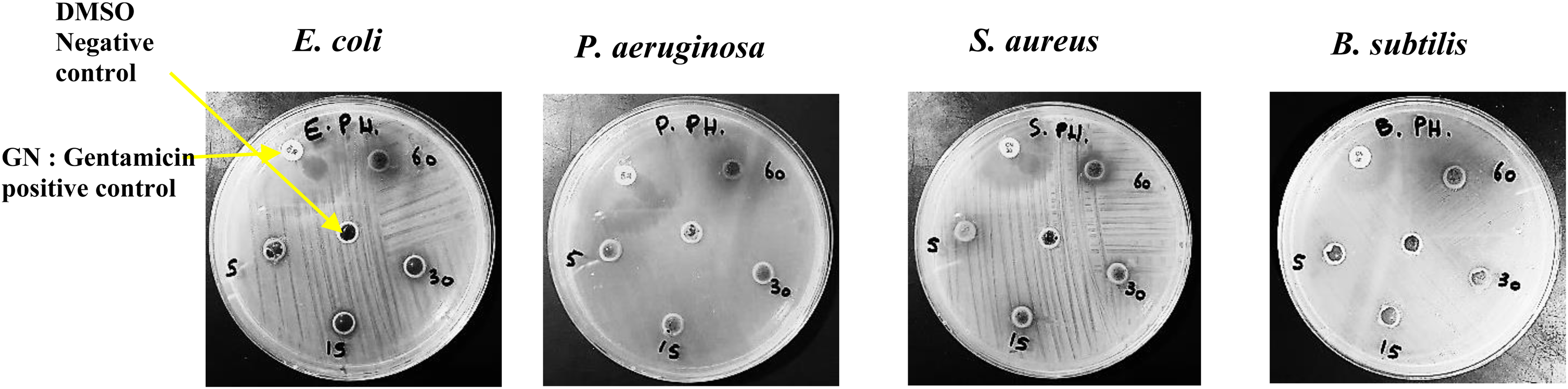
Antibacterial Assay by the Wells Diffusion Method for Hydro-Ethanolic Extract on Agar Plate.
Results of Antibacterial Activity of P. guajava Leaf Extract by Well Method (mm).

NI = No Inhibition, CN = Gentamicin (CN) 30ug Discs.
Using the broth microdilution method, the minimum concentrations required to inhibit growth (MIC) of the P. guajava extract were determined as 80, 40, 40, and 80 mg/mL against E. coli, P. aeruginosa, S. aureus, and B. subtilis, respectively. The corresponding MBC were 80, 80, 40, and 80 mg/mL, respectively. Among the tested strains, S. aureus was the most susceptible, with both MIC and MBC values of 40 mg/mL. An MBC/MIC ratio of 1 indicates the extract's complete bactericidal activity, as observed for S. aureus. For P. aeruginosa, an MBC/MIC ratio of 2 was recorded, suggesting the extract's significant bactericidal efficacy against this strain. These data are detailed in Table 7 (Figure 5).

Photo of Broth Microdilution Method Used for Antimicrobial Activity of Hydro-Ethanolic Extract Against Bacteria Strains.
MIC and MBC of P. Guajava Extract Against Different Bacteria and MBC/MIC Ratio.

The experimental results for both tests are shown in Figure 4 and Figure 5, respectively.
In Vitro Denaturation of Bovine Serum Albumin (BSA)
The anti-inflammatory potential of the hydro-ethanolic extract was evaluated based on its ability to inhibit the heat-induced denaturation of BSA. At a concentration of 1000 µg/mL, the extract exhibited significant inhibitory activity (67.70 ± 0.66%). In comparison, the reference compound aspirin showed a superior inhibitory effect (94.13 ± 2.20%) at a lower concentration (500 µg/mL), which represented the maximum observed effect. The calculated IC50 values were 459.14 ± 27.49 µg/mL for the extract and 7.86 ± 0.15 µg/mL for aspirin, highlighting the markedly higher potency of aspirin. A dose-dependent increase in inhibition was observed for both the extract and the standard compound, as presented in Table 8. These findings indicate that the hydro-ethanolic extract exhibits moderate inhibitory activity against BSA denaturation compared to aspirin.
In Vitro Inflammatory Effect of P. guajava Extract and IC50 Value in Comparison to the Standard. Data are Presented as the Mean Value ± SEM (n = 3).

In Vitro α-amylase Inhibitory Activity
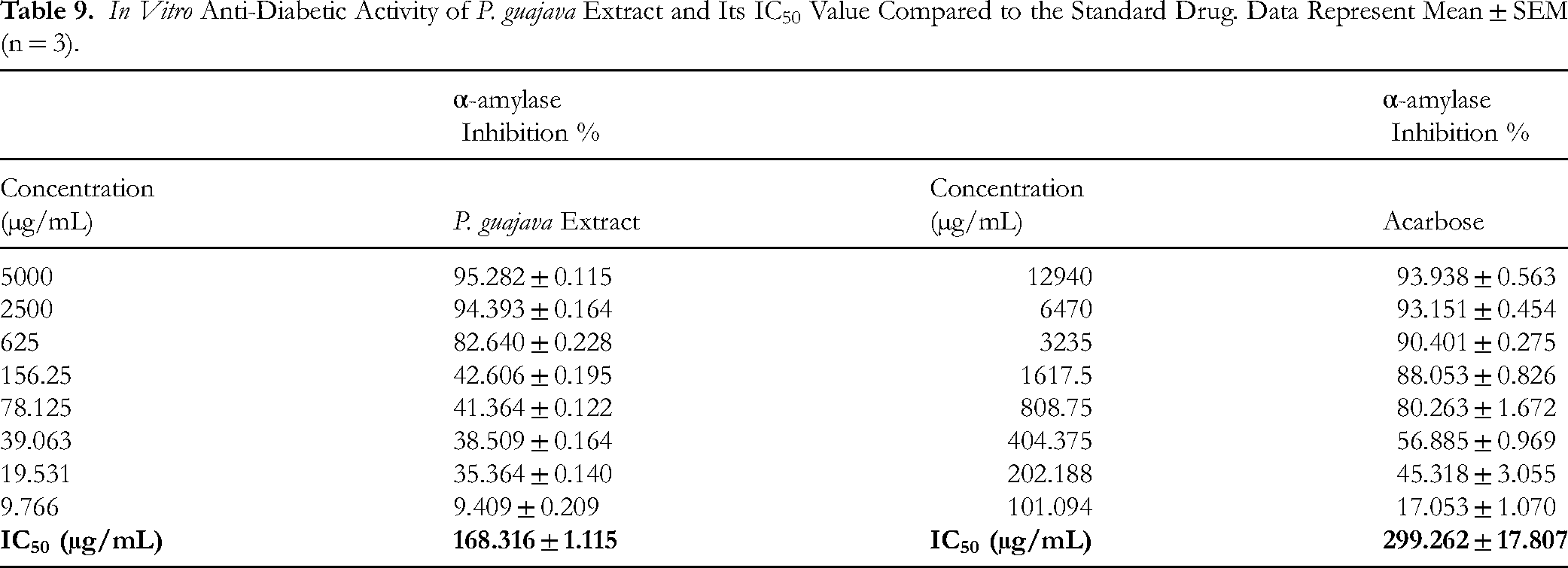
The hydro-ethanolic extract of P. guajava exhibited significant α-amylase inhibitory potential, with an IC50 value of 168.32 ± 1.12 µg/mL—markedly lower than that of the standard reference compound, acarbose (299.26 ± 17.81 µg/mL). The extract demonstrated a potent, dose-dependent inhibition of α-amylase activity, performing comparably to, and in some instances surpassing, the efficacy of acarbose. These findings clearly indicate that P. guajava extract contains strong α-amylase inhibitory constituents. Complete data are presented in Table 9.
In Vitro Anti-Diabetic Activity of P. guajava Extract and Its IC50 Value Compared to the Standard Drug. Data Represent Mean ± SEM (n = 3).
Discussion
P. guajava a species of the Myrtaceae family, is widely cultivated in tropical and subtropical regions due to its adaptability and high nutritional value. In traditional medicine, P. guajava leaves have long been used to manage a wide range of health conditions, such as gastrointestinal disturbances (notably diarrhea), respiratory tract infections, diabetes, cardiovascular disorders, and cancer. These therapeutic uses are largely attributed to its diverse phytochemical profile, particularly its abundance of phenolic compounds and flavonoids, which are recognized for their antioxidant, anti-inflammatory, and hypoglycemic activities.7–38
In the present study, the extraction yield obtained using 50% hydro-ethanolic solvent infusion was approximately 29.258%, which is considered satisfactory. A comparable study by Park et al 39 using the same solvent system reported a yield of 25.8 ± 2.54%. Hydro-ethanol (50%) is widely regarded as one of the most effective solvents for phytochemical extraction, offering an optimal balance between polarity and solubility, and generally outperforming absolute ethanol or water alone. Park et al further demonstrated that ethanol 30% achieved the highest yield (35.8%), while the yield decreased significantly with ethanol 70% (17.1%), suggesting that the intermediate solvent concentration (50%) offers excellent quantitative and qualitative extraction advantages. Other reports also indicate that medium hydro-ethanol concentrations improve total phenol recovery and antioxidant activities compared to other solvents. 15
The antioxidant capacity of P. guajava leaves is closely associated with their capacity to scavenge ROS and mitigate oxidative stress attributable to their phenolic and flavonoid constituents. 40 In this study, HPLC-DAD analysis revealed the presence of various phenolic acids—including gallic, sinapic, chlorogenic, p-coumaric, and caffeic acids—as well as major flavonoids such as quercetin and apigenin in the hydro-ethanolic extract of P. guajava leaves. These results are consistent with previous findings confirming the presence of these bioactive molecules in P. guajava and underline their essential role in the plant's biological activity.9–16,41
Previous studies (Nursanty et al; 42 Huynh et al, 19 ) have identified a broad range of phenolic compounds—including gallic acid, chlorogenic acid, isorhamnetin, and catechin derivatives, quinic acid, epigallocatechin, and flavonoids such as quercetin, apigenin-7-β-D-glucuronopyranoside and naringenin-4'-O-glucopyranoside—in P. guajava leaf extracts. Variability in TPC and TFC across studies can be explained by differences in extraction and analytical protocols, 43 may reflect the influence of specific environmental characteristics in the Oued Souf region, where the desert climate likely stimulates the biosynthesis of secondary metabolites related to environmental stress. 44 Our study used a 50% hydro-ethanolic extract and HPLC analysis, while others relied on traditional spectroscopic methods, 45 or advanced techniques like LC-MS, 42 or UPLC-QTOF-MS/MS. 19
Leaf maturation stages, harvest season, drying, and storage significantly influence chemical composition, as confirmed by multiple seasonal studies. 46 However, the lack of mass spectrometry (MS) in our study limits precise compound identification, necessitating the application of advanced analytical techniques such as LC-MS in future research.
Regarding bioactivity, quantitative estimates in this study revealed high levels of total phenolic (268.304 ± 0.659 mg GAE/g) and flavonoids (16.959 ± 0.099 mg QE/g), reflecting notable antioxidant capacity. The extract exhibited strong DPPH radical scavenging activity (IC50 = 17.239 ± 0.009 µg/mL) and a TAC of 491.949 ± 2.448 mg GAE/g.
The extract also demonstrated 43% anti-hemolytic activity (43% inhibition at 47 mg/mL compared to 82% for vitamin E), likely due to the ability of flavonoids and polyphenols to stabilize erythrocyte membranes and reduce lipid peroxidation. 48
In terms of antimicrobial properties, the extract showed selective efficacy against Gram-positive bacteria such as S. aureus and B. subtilis, while being less effective against Gram-negative bacteria like E. coli. This is attributed to the resistance of the latter due to their lipopolysaccharide-rich outer membrane. 49
The extract also displayed moderate anti-inflammatory activity, as evidenced by its inhibition of BSA denaturation. This effect may be attributed to compounds such as gallic acid, caffeic acid, chlorogenic acid, p-coumaric acid, sinapic acid, quercetin, and apigenin, known for their anti-inflammatory properties. 50 However, while this assay is preliminarily acceptable for assessing anti-inflammatory activity, it is insufficient alone to explain the mechanism of action, necessitating more advanced models (eg, COX-2 or TNF-α inhibition). 51
Finally, the extract showed significant α-amylase inhibitory activity (IC50 = 168.3 ± 1.12 µg/mL), surpassing the pharmaceutical standard acarbose (IC50 = 299.2 µg/mL), This suggests potential antidiabetic properties, although the current data are limited to in vitro assays, Further studies are needed to confirm in vivo efficacy and safety, identify the active compounds, and explore clinical applications.¹¹ Dose-dependent α-amylase inhibition by ethanolic and methanolic P. guajava extracts has also been reported in previous studies.11,48 The hydro-ethanolic extract of P. guajava leaves shows promising properties as a rich natural source of bioactive compounds, particularly phenolics and flavonoids, with significant antioxidant, antimicrobial (especially against Gram-positive), anti-inflammatory, and α-amylase inhibitory activities. While these findings highlight the therapeutic potential of P. guajava, further research is required to isolate specific active molecules, elucidate mechanisms, and validate pharmacological effects through advanced experimental models and clinical studies.
The novelty of this study lies in its comprehensive in vitro evaluation of multiple bioactivities of P. guajava leaf extract obtained in a specific arid region (Oued Souf, Algeria), highlighting the potential influence of environmental stress on the biosynthesis of secondary metabolites. In addition, the identification of key phenolic compounds via HPLC-DAD strengthens the chemical basis of the observed activities.
However, this study has several limitations. All bioassays were conducted in vitro, and no in vivo or clinical validation was performed. and the plant material was from a single geographic location, which may not capture chemotypic diversity. In addition, although the activities observed are promising, the exact mechanisms of action remain to be elucidated.
Overall, the findings support further research on P. guajava as a source of bioactive phytochemicals. Future studies should focus on compound isolation, mechanistic studies using molecular targets (eg, COX-2, TNF-α), and validation by in vivo and clinical models to assess safety, efficacy, and therapeutic relevance
Conclusion
The findings of this study indicate that the hydro-ethanolic extract of P. guajava leaves exhibits notable in vitro biological activities, including antioxidant, antibacterial, anti-inflammatory, and anti-diabetic effects. These effects may be associated with the extract's high content of phenolic and flavonoid compounds, as revealed by HPLC-DAD analysis. However, these results are based solely on in vitro experiments, and the plant material was collected from a single geographic location, which may limit the generalizability of the findings.
While further in vivo and clinical investigations are necessary to confirm these preliminary observations, the present study suggests that P. guajava leaf extract warrants further exploration as a potential source of bioactive compounds for future applications.
Abbreviations
To enhance accuracy and readability, all abbreviations have been systematically reviewed and defined as follows
Total Phenolic Content
Total Flavonoid Content
High-Performance Liquid Chromatography coupled with Diode-Array Detection.
Total Antioxidant Contents
Reactive Oxygen Species
Phosphate-Buffered Saline
Bovine Serum Albumin
Minimum Inhibitory Concentration
minimum bactericidal concentration
Milligrams of Gallic Acid Equivalents per Gram of sample extract
Milligrams of Rutin Equivalents per Gram of sample extract
Hydrolysable Polyphenols
Condensed Tannins
An inducible enzyme involved in prostaglandin synthesis, playing a key role in inflammation and pain.
A pro-inflammatory cytokine that regulates immune responses and is implicated in various inflammatory and autoimmune diseases.
Dimethyl Sulfoxide.
Footnotes
Acknowledgments
We would like to thank the editor and the anonymous reviewers for their thoughtful and accurate comments on this manuscript, as well as everyone who helped us complete and proofread this manuscript.
Ethical Approval
This study is not applicable to both human and/or animal studies that require ethical approval.
Author Contributions
All authors contributed to the study.
Funding
The authors declare that no funding was received for this study.
Competing Interests
The authors declare that they have no known competing financial interests or personal relationships that could have appeared to influence the work reported in this paper.
Data Availability
The data that supports the findings of this study are available within the article.