
Abstract
Objectives
Nekemias grossedentata is a traditional herb known for its high flavonoid content, particularly dihydromyricetin (DMY), which exhibits a significant antioxidant and favorable safety profile, making it a promising candidate for mitigating skin aging. This study aims to develop a simplified and scalable protocol for obtaining N. grossedentata extract (NGE) with high-purity DMY, and to evaluate the antioxidant and anti-glycation activities of the extract.
Methods
A series of assays were conducted to evaluate the antioxidant activity of NGE, including 2,2-diphenyl-1-picrylhydrazyl (DPPH) and 2,2′-azino-bis (3-ethylbenzothiazoline-6-sulfonic acid (ABTS). Additionally, intracellular reactive oxygen species (ROS) level and the activities of antioxidant enzymes (superoxide dismutase, SOD; glutathione peroxidase, GSH-Px) were analyzed in H2O2-induced HaCaT cells. Anti-glycation activity was assessed by monitoring advanced glycation end-product (AGE) formation in Human primary dermal fibroblasts (HDFs)-methylglyoxal (MGO) systems.
Results
A rapid extraction method yielded NGE with 90.4% DMY purity. NGE exhibited potent radical scavenging activity (DPPH IC50 = 6.89 ± 0.51 μg/mL; ABTS IC50 = 4.37 ± 0.84 μg/mL). In cellular models, NGE significantly reduced H2O2-induced ROS and restored SOD and GSH-Px activities. Furthermore, NGE showed significant antiglycation activity (55.04%).
Conclusion
Through the synergistic integration of direct antioxidant interventions, enhancement of antioxidant enzyme activity, and anti-glycation mechanisms, NGE exhibits multi-target anti-aging properties. These findings underscore its potential as a natural ingredient in cosmetic and dermatological formulations.
Introduction
Aging is associated with a spectrum of degenerative conditions, and the skin, as the body’s largest organ, is particularly susceptible to endogenous damage induced by biochemical processes such as oxidation and glycation. The accumulation of advanced glycation end products (AGEs), formed non-enzymatically via the Maillard reaction between reducing sugars and free amino groups in proteins, lipids, and nucleic acids, contributes significantly to skin aging.1,2 AGEs promote oxidative stress, inflammatory reactions, and protein cross-linking, resulting in dullness, wrinkles, and a loss of skin elasticity.3,4 Research indicates that when AGEs interact with receptors for advanced glycation end products (RAGEs), they activate the reduced nicotinamide adenine dinucleotide phosphate-oxidase system (NADPH-oxidase), which in turn generates intracellular reactive oxygen species (ROS). 5 An imbalance between ROS and the cellular antioxidant defense system, which includes enzymes like catalase (CAT), glutathione peroxidase (GSH-Px), and superoxide dismutase (SOD), leads to oxidative stress. Cellular damage occurs when ROS levels exceed the antioxidant capacity, hastening the aging process of the skin and causing inflammation and hyperpigmentation. 6 Therefore, treatments targeting both oxidative and glycation processes have the potential to prevent skin aging and preserve skin health.
In recent years, natural plant-derived active metabolites isolated from various plants have garnered increased interest as promising alternatives due to their rich content of bioactive compounds and low incidence of side effects. 7 Flavonoids, as the most abundant functional compounds distributed in higher plants, exhibit a variety of bioactive properties, including natural antioxidant, antiviral, anti-inflammatory, anti-diabetic, anti-cancer, and anti-aging effects. 8 Vine Tea, derived from the young stems and leaves of Nekemias grossedentata (Hand.-Mazz.) J.Wen & Z.L.Nie (syn. Ampelopsis grossedentata) is a traditional beverage and herbal medicine also known as musty tea, white tea, sweet tea, or longevity vine. For centuries, it has been extensively utilized as a health-promoting beverage among the Zhuang, Tujia, and Yao ethnic groups.9,10 N. grossedentata contains a diverse array of bioactive constituents, including flavonoids, phenols, steroids, terpenoids, polysaccharides, and other volatile components. Among these, flavonoids represent the most abundant class, comprising up to 43%, 11 with dihydromyricetin (DMY) being the predominant flavonoid at concentrations of up to 38% (w/w). 12 DMY exhibits a wide range of pharmacological activities, including antibacterial, anticancer, anti-inflammatory, antioxidant, hepatoprotective, cardioprotective, antidiabetic, and antihypertensive effects.13-16 Furthermore, recent studies have demonstrated the beneficial effects of DMY on mitigating skin aging, such as improving age-related methylation patterns induced by UV, 17 inhibiting oxidative stress and inflammation caused by UVA, 18 alleviating skin damage resulting from UVB, 19 exhibiting anti-acne effects, 20 and inhibiting melanin production. 21
Although progress has been made in characterizing the anti-glycation properties of plant polyphenol extracts from sources like coffee, berries and grape byproducts,22-24 there hasn’t been a systematic evaluation of the specific application of N. grossedentata leaf extract, a source that is distinguished by its exceptionally high DMY content, to skin-relevant anti-glycation endpoints. Furthermore, the majority of current anti-glycation research uses acellular protein-sugar model systems (such as BSA-glucose or BSA-fructose), 25 which might not accurately capture the biological intricacy of glycation in real dermal cells. It is crucial to assess to both antioxidant and anti-glycation in an integrated, cell-based framework because of the interaction between AGEs buildup and oxidative stress in the skin. In order to fill these gaps,the current study developed a simplified, scalable ethanol-based extraction and crystallization procedure that produces an N. grossedentata extract (NGE) with more than 90% DMY purity in a single step. Then employed a series of chemical and cell-based assays to comprehensively evaluate the antioxidant and anti-glycation of NGE. This integrated method offers a foundational investigation supporting the potential application of NGE in the development of cosmetic products aimed at mitigating skin aging.
Materials and Methods
Chemicals and Reagents
Nekemias grossedentata was purchased from Jiulongyuntengcha Professional Cooperative in Zhetu Township, Guangnan County. 2, 2-diphenyl-1-picrylhydrazyl (DPPH) was purchased from TCI (Shanghai) Chemical Industry Development Co., Ltd. (Shanghai, China). ABTS was purchased from Dalian Meilun Biotechnology Co., Ltd. (Dalian, China). Assay kits for the determination of cellular glutathione peroxidase (GSH-Px), superoxide dismutase (SOD), reactive oxygen species (ROS), bicinchoninic acid (BCA), and 2,7-Dichlorodihydrofluorescein diacetate (DCFH-DA) were purchased from Beyotime Institute of Biotechnology (Shanghai, China). Dihydromyricetin (DMY) (CAS Number 27200-12-0, purity ≥ 98%) standard was purchased from Yunnan Xili Biotechnology Co., Ltd. (Kunming, China). MGO, MTT and aminoguanidine were purchased from Sigma Aldrich Trading Co (Shanghai, China).
Extraction Preparation of NGE
1.5 kg of powdered N. grossedentata leaves were extracted with 95% ethanol using a solid-to-solvent ratio of 1:12 (w/v) at 5°C for 72 hours. The resulting supernatants were filtered and combined. The combined filtrate was concentrated under reduced pressure to remove ethanol and then subjected to crystallization at room temperature for 12 hours. The resulting precipitate was collected and freeze-dried. The final product, known as N. grossedentata extract (NGE), was stored at 5°C until further use.
Determination of Total Flavonoid Content
A UV spectrophotometer (UV-1800, Shimadzu, Kyoto, Japan) was used to measure the total flavonoid content of the N. grossedentata extract in accordance with the method modified from Zhang et al. 26 Briefly, 0.15 mL of the stock solution was mixed with 3 mL of 5% aluminum chloride (diluted to 10 mL with 95% ethanol) for total flavonoid quantification, and the absorbance was recorded at 311 nm. Using the series concentration C (μg/mL) of the dihydromyricetin standard solution as the abscissa and the absorbance A as the ordinate, a standard curve was plotted (A=0.0775C-0.0459, R2=0.9994, linear range 2.52-17.64 μg/mL).
Determination of Dihydromyricetin Content
The purity of dihydromyricetin (DMY) was assessed using high-performance liquid chromatography (HPLC) on a Symmetry C18 column (3.5 × 150 mm, Waters, USA). The mobile phase consisted of methanol (A) and 0.1% aqueous phosphoric acid (B) at a ratio of 55:45 (v/v). The flow rate was maintained at 1.0 mL/min with a column temperature of 25 °C. The injection volume was 10 μL, and the detection wavelength was set at 291 nm27.
To establish the calibration curve, DMY was accurately weighed and dissolved in ethanol in a 25 mL volumetric flask to prepare a stock solution (0.8567 mg/mL). Standard working solutions were obtained by serial dilution with ethanol. The calibration curve was constructed by plotting the peak area A against the mass concentration of the sample C (mg/mL), yielding the linear regression equation (A=14792C-14.871, R2=0.9999, linear range 0.08567-0.8567 mg/mL). Accurately weighed NGE was dissolved in ethanol at 0.20 mg/mL in ethanol. The injection volume was set at 10 μL, and the absorbance at 291 nm was recorded.
Antioxidant Capacity (DPPH·, ABTS·+)
The DPPH and ABTS free radical scavenging capacity of NGE was assessed according to a modified method. 27 Briefly, 200 µL of a range of sample solutions (3.125–1,000 μg/mL) were combined with a 0.2 mM DPPH solution. Absolute ethanol was used to replace the sample solution as a blank and replace the DPPH solution as a control. After incubation in the dark at room temperature for 30 min, the absorbance was measured at 517 nm using a spectrophotometer (UV2550, SHIMADZU, Kyoto, Japan). Vitamin C (VC) acted as the positive control28,29 and all tests were conducted in triplicate.
For ABTS free radical scavenging capacity, 30 µL of serially diluted NGE (3.125 –100 μg/mL) was mixed with 300 µL of the ABTS working solution. After mixing well, the samples were incubated at room temperature for 6 min without exposure to light. The remaining experimental procedure is essentially identical to the DPPH assay, the only difference is the ABTS assay detects the absorbance value at 405 nm. The following formula was used to determine DPPH· and ABTS·+ radical scavenging activities:

The IC50 values were calculated by using GraphPad Prism 10.1.2 software.
Antioxidant Assays Based on Cell Models
Cell Viability
HaCaT cells (Catalog Number: BNCC339817, Beina Biotechnology Co., Ltd., Shangcheng, China) were cultivated for 24 hours in 96-well plates. After washing with PBS, fresh medium was added to the negative control. The sample groups were treated with media with varying amounts of NGE (313–5000 μg/mL). Following 24 hours of incubation, cell viability was assessed using the MTT assay,
30
with absorbance measured at 490 nm. The three highest concentrations that maintained cell viability above 90% were selected for subsequent experiments. The following formula was used to determine the cell viability:

Intracellular ROS Level Assay
A ROS Assay Kit (Beyotime Institute of Biotechnology, Shanghai, China) was used to quantify intracellular ROS levels. HaCaT cells were planted in 96-well black plates and 24-well plates and allowed to adhere for 24 hours. The cells were divided into negative control, model control, positive control (10 μg/mL VC), and NGE-treated groups (156, 313, and 625 μg/mL), each with three replicates. After 24 hours of treatment, all groups except the blank control were exposed to 250 μM H2O2 for 30 minutes. The cells were then washed with PBS and incubated with 10 μM dichlorofluorescein diacetate (DCFH-DA, a fluorescent probe) at 37 °C for 30 minutes. 31 Cell morphology was examined using fluorescent microscopy (Olympus BX43, Tokyo, Japan) and captured on camera using the microplate reader (excitation/emission: 488/525 nm). 29
GSH-Px and SOD Levels Assay
HaCaT cells were seeded in 6-well plates at a density of 2 × 10⁵ cells/well and allowed to adhere overnight. Subsequently, all groups except the negative control were exposed to 250 μM H2O2 for 30 minutes to induce oxidative stress. Following H2O2 exposure, cells were washed with PBS, lysed on ice using RIPA lysis buffer, and centrifuged at 12,000 × g for 10 min at 4 °C. The resulting supernatants were collected and stored at −80 °C until analysis. Superoxide dismutase (SOD) and glutathione peroxidase (GSH-Px) activities were quantified using commercially available assay kits (Nanjing Jiancheng Bioengineering Institute, Nanjing, China), according to the manufacturer’s instructions.32,33
Anti-Glycation Assay With HDFs-MGO Model
Cell Viability
Human primary dermal fibroblasts (HDFs) (Catalog Number: Fb230131, Guangdong Boxi Biotechnology Co., Ltd., Guangdong, China) were seeded in 96-well plates and cultured until 50-60% confluent. The sample groups treated with medium containing different concentrations of NGE (7.813- 125 μg/mL). The MTT assay was used to measure cell viability. 30 After 4h of incubation, the optical density (OD) was measured at 490 nm using a microplate reader in accordance with the kit’s instructions.
Immunofluorescence Detection of Protein-Bound CML
Based on previously reported methods, 34 a cellular glycation model was created by treating HDFs with methylglyoxal (MGO) to induce high N-carboxymethyl lysine (CML) expression. Cells were cultured in 24-well plates until 30–50% confluent and then exposed to MGO (21.6 μg/mL) alone or in combination with NGE (62.5 μg/mL) or aminoguanidine (184.7 μg/mL, positive control) for 48 h in a CO2 incubator at 37°C with 5% CO2. Cells were fixed and incubated overnight at 4°C with a primary antibody against carboxymethyl lysine (anti-carboxymethyl lysine antibody [CML26], Abcam, Catalog Number: ab125145, mouse monoclonal, dilution 1:200) in blocking buffer. After washing, cells were incubated with a fluorophore-conjugated secondary antibody and visualized using a fluorescence microscope (Olympus BX43, Tokyo, Japan) with excitation/emission at 370/440 nm.
Fluorescence intensity was quantified with Image-Pro Plus software, and the inhibition rate was calculated as follows:

Statistical Analysis
All data were statistically processed using GraphPad Prism 10.1.2. The test data were represented as mean ± standard deviation (SD), and statistical significance between groups was analyzed by one-way analysis of variance (ANOVA) and Tukey’s test. *p< 0.05 was accepted as significant difference.
Results
Yield of NGE and Content of Dihydromyricetin
The crystallization of DMY was conducted at room temperature (25°C). A single crystallization step yielded 24% (w/w) of the dried extract, designated as NGE (96.5% total flavonoids). To determine the purity of the obtained DMY in NGE, high-performance liquid chromatography (HPLC) analysis was conducted with a standard DMY. As shown in Figure 1A-B, the chromatogram displayed that the main peak in NGE is DMY. Furthermore, we estimated that the DMY content in NGE with a purity of 90.4%. Our approach provides a simplified and scalable protocol to obtain high-purity DMY with superior efficiency. In addition, the structure of DMY is shown in Figure 1C, and the UV absorption spectra of DMY and NGE are shown in Figure 1D. Dihydromyricetin determination in NGE by high-performance liquid chromatography (HPLC). (A) The HPLC profiles of DMY; (B) The HPLC profiles of NGE; (C) Chemical structure of DMY; (D) The UV spectrophotometer of DMY and NGE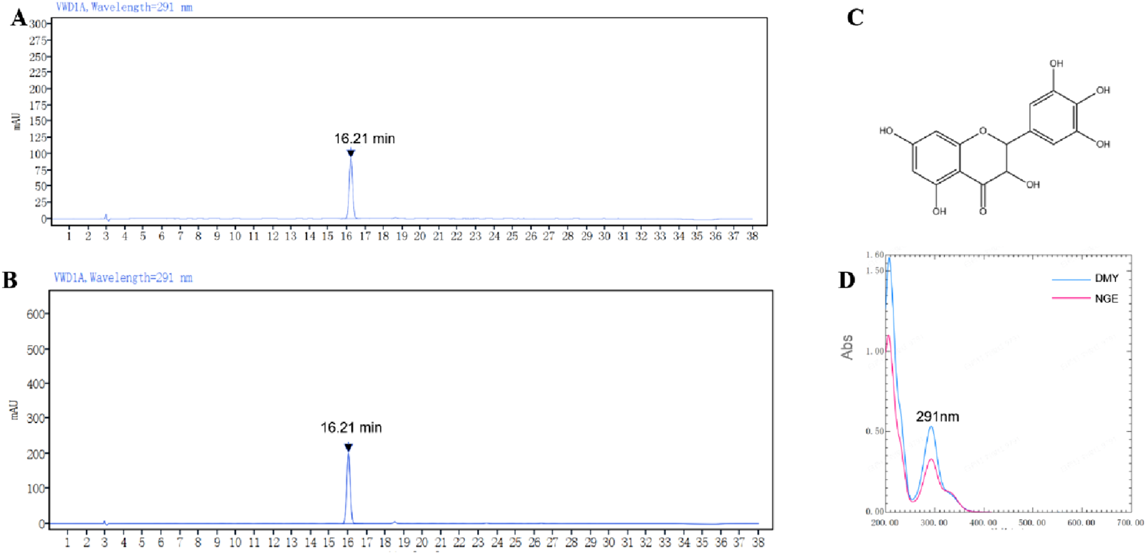
Antioxidant Activities of NGE by Chemical Assays
As illustrated in Figure 2, NGE exhibited dose-dependent antioxidant activity within the concentration range of 3.15–100 μg/mL. For DPPH and ABTS radical scavenging assays, NGE displayed potent activity with IC50 values of 6.89 and 4.72 µg/mL, respectively. Whereas the positive control, Vitamin C (VC), showed IC50 values of 11.73 and 10.56 µg/mL. NGE demonstrated markedly stronger radical scavenging activity compared to the classical antioxidant VC, which confirms the potential of NGE as a potent antioxidant, particularly at high concentrations. Antioxidant potency of NGE extract and VC. (A) DPPH scavenging activity; (B) ABTS scavenging activity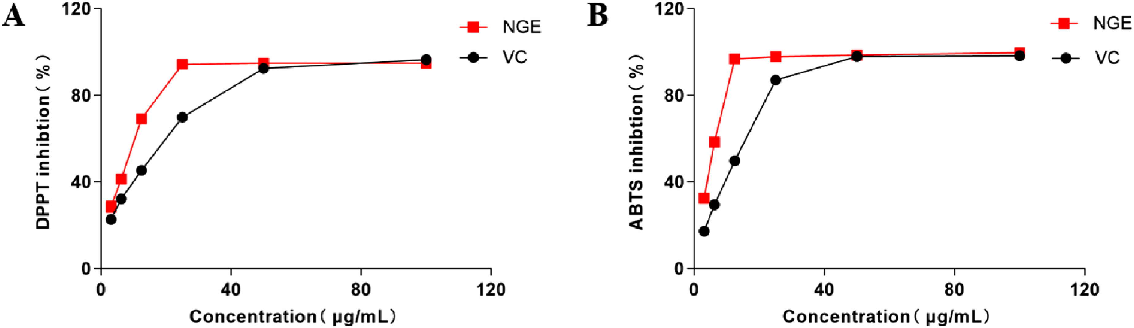
Antioxidant Activities of NGE Based on Cell Models
Cytotoxicity of NGE at Different Concentrations on HaCaT Cells
As shown in Figure 3A, the cytotoxicity of NGE on HaCaT cells was determined by using the MTT assay. The results showed that NGE concentrations less than 625 μg/mL did not display any obvious cell toxicity. Therefore, subsequent studies used concentrations of 625 μg/mL, 313 μg/mL, and 156 μg/mL to assess the biological effects of NGE against H2O2-induced keratinocyte damage. Effects of NGE on ROS and the intracellular antioxidant enzymes in H2O2-irradiated HaCaT cells. (A) Cytotoxicity of NGE on HaCaT cells; (B) Intracellular ROS level; (C) SOD activity; (D) GSH-Px activity; (E) Images of HaCaT by fluorescence microscope (100 x). All results were triplicates of the mean ± SD. *p < 0.05, ** p < 0.01, *** p < 0.001 compared with model group; ### p < 0.001 compared with control group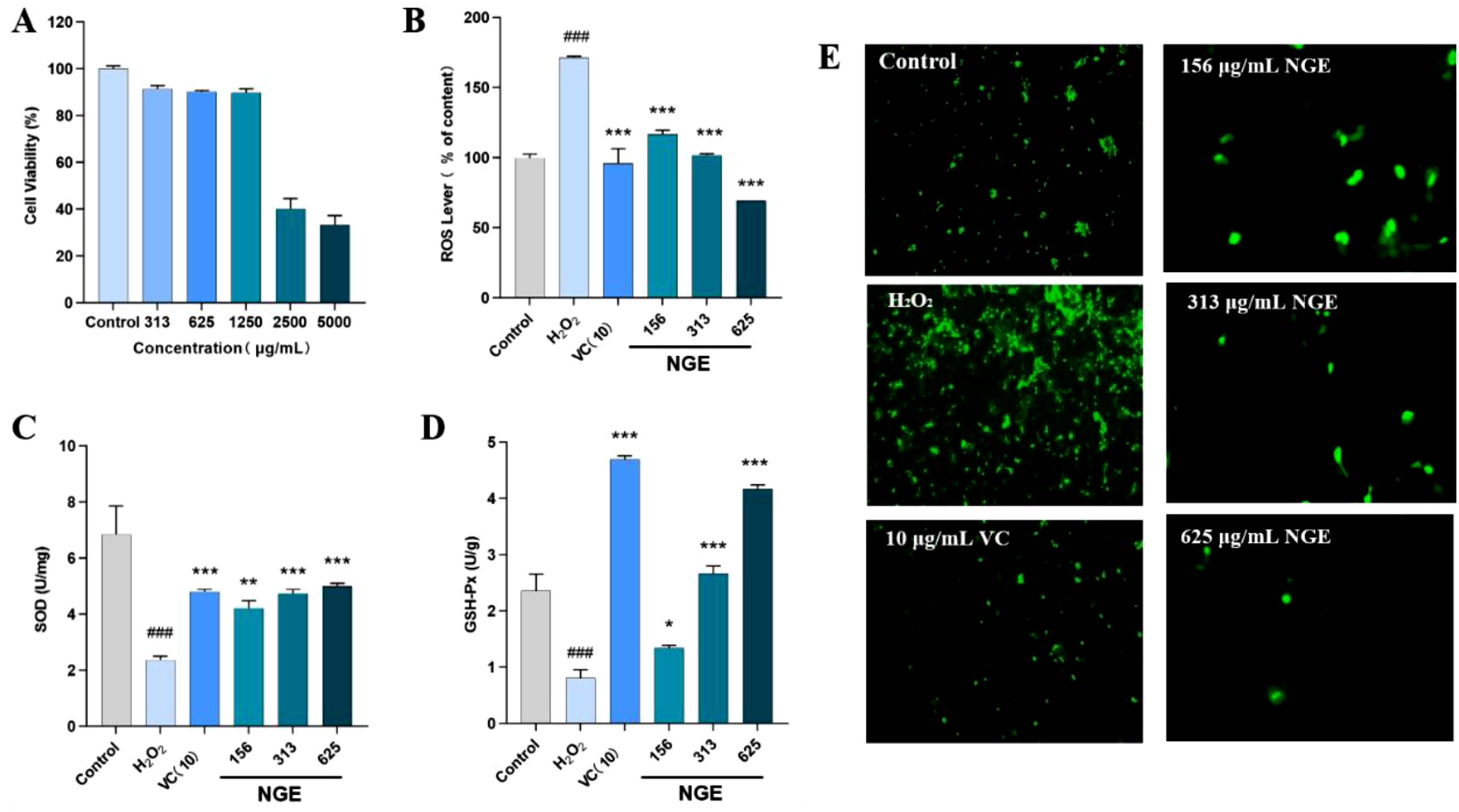
NGE Attenuates H2O2-Induced ROS Generation in HaCaT Cells
Reactive oxygen species (ROS), including hydroxyl radical (·OH), superoxide anion (O2·-), and hydrogen peroxide (H2O2), function as key signaling molecules but can trigger oxidative damage and cell death when overproduced. Therefore, intracellular ROS levels serve as a critical indicator of oxidative stress. In this study, we evaluated the protective effect of NGE against H2O2-induced ROS production by using the fluorescent probe DCFH-DA to monitor intracellular ROS levels. As depicted in Figure 3B and 3E, treatment with 250 μM of H2O2 induced a marked increase in ROS levels (see Supplemental Material). However, pretreatment with different concentrations of NGE could notably reduce this H2O2-induced ROS generation in a dose-dependent manner. These results demonstrate that NGE effectively protecting keratinocyte from H2O2-induced oxidative stress by inhibiting intracellular ROS generation.
Effects of NGE on SOD and GSH-Px Activities in H2O2-Treated HaCaT Cells
Overproduction of ROS resulted in oxidative stress, meanwhile, the intracellular antioxidant defense systems can be activated to protect cells against such oxidative damage. Intracellular enzymes, particularly SOD and GSH-Px, are vital for eliminating free radicals. In the present study, exposure to 250 μM H2O2 significantly decreased the activities of both SOD and GSH-Px in HaCaT cells. However, as shown in Figures 3C-D, retreatment with NGE dose-dependently restored these levels. The enzyme activities in the NGE-treated groups were significantly higher than those in the H2O2-only group. These results demonstrate that NGE exerts its protective effect by enhancing the cellular enzymatic antioxidant defense system.
In Vitro Anti-glycation Effect of NGE Extract
Cytotoxicity of NGE at Different Concentrations on HDF Cells
The cytotoxicity of NGE on HDFs was determined by using the MTT assay, as shown in Figure 4A. The results demonstrated that NGE at concentrations up to 62.5 μg/mL did not display any significant cytotoxicity. Therefore, a concentration of 62.5 μg/mL was selected for subsequent studies to evaluate the biological activity of NGE against MGO-induced fibroblast damage. Anti-glycation activity of NGE extract. (A) Cytotoxicity of NGE on HDF cells. (B) Effect of CML expression in MGO-induced HDFs by NGE extract. (C) Representative images of HDFs by fluorescence microscope, scale bar =200 μm. All results were triplicates of the mean ± SD. *** p < 0.001 compared with model group; ### p < 0.001 compared with control group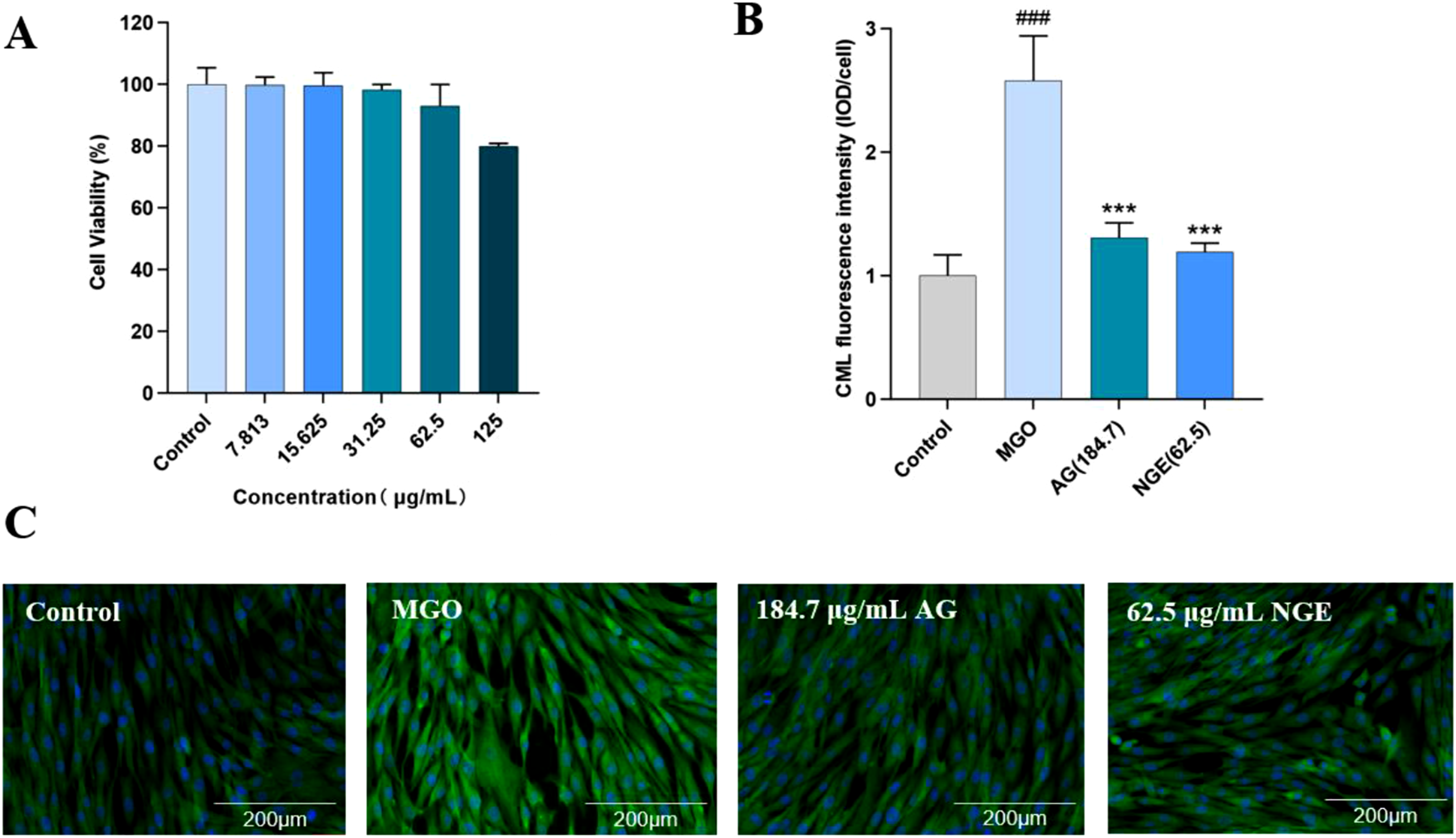
Effect of NGE on CML Levels in MGO-Induced HDFs
Methylglyoxal (MGO), a key precursor in AGE formation, reacts with proteins to generate carboxymethyl lysine (CML), a major non-fluorescent AGE in skin. In this study, HDFs were treated with MGO to induce glycation, and CML levels were assessed by immunofluorescence (IF). As shown in Figure 4C, strong CML-specific green fluorescence was detected in MGO-induced cells, whereas only a minimal signal was observed in the non-induced control (see Supplemental Material). Treatment with aminoguanidine (AG), a known AGE inhibitor, significantly reduced CML levels by 49.22%. Notably, at the non-cytotoxic concentration of 62.5 μg/mL (as indicated in Figure 4B), NGE treatment exhibited even stronger inhibition, reducing CML content by 55.04% compared to the model group (Figure 4C). These findings indicate that NGE possesses potent anti-glycation activity in a cellular context, outperforming the positive control at the tested concentration.
Discussion
Nekemias grossedentata (commonly known as vine tea or tengcha) is a rich source of bioactive compounds, including flavonoids, polysaccharides, and polyphenols. 35 This aligns with the increasing interest in natural antioxidants for cosmetic and pharmaceutical applications. This study presents a simplified ethanol-based extraction method that achieved a 24% yield of N. grossedentata extract (NGE), with the primary component, DMY, constituting up to 90.4% of the extract. Notably, this protocol requires only a single crystallization step, whereas a previously reported aqueous method demanded eight successive crystallizations to reach 98% DMY purity. 36 In comparison to conventional techniques such as heat reflux or ultrasonic extraction,37,38 this method provides practical benefits and demonstrates potential for industrial-scale application.
The antioxidant capacity of NGE was substantiated through a series of chemical assays, demonstrating potent radical scavenging activity against DPPH (IC50 = 6.89 ± 0.51 µg/mL) and ABTS (IC50 = 4.37 ± 0.84 µg/mL). DMY played a crucial role in scavenging DPPH·, ABTS·+ radicals in the flavonoid-rich extracts from vine tea, which is in agreement with previously reported results.27,39 Notably, the H2O2 treatment significantly increased intracellular ROS generation in keratinocytes; however, pretreatment with NGE significantly decreased ROS levels in a dose-dependent manner, confirming its strong antioxidant properties. These findings suggest that vine tea extracts could mitigate oxidative stress. And DMY plays a dominant role in vine tea’s antioxidant activities, indicating its potential as a natural antioxidant resource.
The mechanism underlying the antioxidant activity of flavonoids may involve direct interactions with reactive oxygen species (ROS) and/or the activation of cellular antioxidant systems. In this study, NGE significantly reduced H2O2-induced ROS production in HaCaT cells and restored the activities of key antioxidant enzymes, including SOD and GSH-Px, which are major antioxidant enzymes that play a crucial role in scavenging free radicals. 40 Data from other researchers also indicate the potent antioxidant effects of vine tea extracts and DMY, which are related to their influence on cellular enzyme systems. He et al reported that dihydromyricetin pretreatment exhibited a protective effect by inhibiting UVA-induced ROS generation, enhancing GSH-Px activity, 18 and decreasing the content of MDA in UVA ray-treated HaCaT cells. Additionally, other studies have shown that treatment of mice with the water extract of Ampelopsis grossedentata alleviates oxidative stress, reduces levels of pro-inflammatory cytokines, and increases the antioxidant enzyme activity of CAT. 41 Liao et al also demonstrated that DMY treatment significantly inhibited the marked increase in antioxidant enzyme levels (SOD, GPX, and CAT) induced by 2, 2′-azobis (2-amidinopropane) dihydrochloride (AAPH) during human erythrocyte hemolysis. 36
In addition to its antioxidant properties, NGE demonstrated significant anti-glycation activity. Protein glycation results in the formation of advanced glycation end products (AGEs), which accumulate in skin tissues and contribute to the loss of elasticity and the acceleration of aging. In this study, NGE notably inhibited AGE formation in MGO-induced models, indicating its potential in preventing glycation-related skin damage. This finding aligns with the result previously reported by Wang et al, 42 which suggests that DMY may act as a potential RAGE inhibitor, suppressing AGE-RAGE signaling activation and promoting skin cell viability. Beyond RAGE antagonism, DMY may also directly trap reactive carbonyl species, a well-recognized antiglycation mechanism of flavonoids. Its nucleophilic meta-polyhydroxylated A-ring can deplete important AGE precursors by forming adducts with MGO 43 and potentially capture upstream Amadori rearrangement products, 44 thereby depleting key AGE precursors. However, the C-4 carbonyl group may attenuate this trapping efficiency relative to flavanols 44 ; therefore, future mass spectrometry-based adduct analysis would be required to explicitly verify this process for DMY. 45
Several limitations of this study should be acknowledged. First, the cellular anti-glycation assay was conducted at a single concentration of NGE (62.5 μg/mL) in the HDFs-MGO model; a full dose–response evaluation would better characterize inhibitory potency. Second, we acknowledge that Trolox is widely recognized as a standard antioxidant reference owing to its well-defined radical scavenging mechanism.46,47 This study’s sole use of vitamin C as a positive control may restrict direct comparison with research that uses Trolox. Trolox and vitamin C should be used as parallel references in future research to improve cross-study comparability. Third, the exact mechanism by which NGE inhibits the formation of AGE was not directly elucidated; further potential mechanisms, including direct carbonyl (MGO) trapping by DMY via nucleophilic adduct formation, RAGE antagonism, or indirect suppression of ROS-mediated glycation acceleration, remain to be distinguished through dedicated biochemical and molecular studies. Finally, direct evidence of protein-level alterations, such as protein cross-linking and aggregation profiles analyzed by SDS-PAGE, 48 as well as oxidative modifications indicated by fluorescent markers including dityrosine, kynurenine and N'-formylkynurenine were not obtained. 49 These criteria would provide a more thorough assessment of the protective function of DMY at the protein structure level as well as glycation-induced protein damage. Future studies incorporating these complementary analyses will be necessary to fully elucidate the antiglycation mechanism and to establish a more integrated profile of DMY-mediated protection.
In summary, this study provides comprehensive evidence supporting the multifunctional potential of NGE in skin protection, encompassing both antioxidant and anti-glycation activities. Although variations in plant sourcing and processing may impact flavonoid composition, our findings establish a solid basis for the quality evaluation and further development of N. grossedentata as a promising natural ingredient in cosmeceutical and dermatological formulations.
Conclusion
In this study, we developed a simplified extraction method to obtain a flavonoid-rich extract from N. grossedentata (NGE), with dihydromyricetin (DMY) content reaching 90.4%. NGE exhibited potent antioxidant activity in both chemical and cellular models, demonstrating significant free radical scavenging capacity against DPPH and ABTS radicals. In H2O2-induced HaCaT cells, NGE effectively reduced intracellular ROS levels and enhanced the activity of key antioxidant enzymes, including GSH-Px and SOD. Furthermore, NGE displayed notable anti-glycation effects by inhibiting CML accumulation in MGO-induced HDF cellular models. Collectively, these findings provide a robust experimental foundation for the potential application of NGE in cosmetic formulations aimed at alleviating oxidative stress and glycation-related damage.
Supplemental Material
Supplemental Material - Antioxidant Activity and Anti-glycation Effects of Nekemias grossedentata Leaf Ethanol Extract
Supplemental Material for Antioxidant Activity and Anti-glycation Effects of Nekemias grossedentata Leaf Ethanol Extract by Junhong Liu, Zengshang Wang, Ting Ruan, Qianghua Quan, Xue Shao, Guoqing Li in Natural Product Communications
Footnotes
Consent to Participate
There are no human subjects in this article and informed consent is not applicable.
Funding
This work was supported by the Major Science and Technology Project of Yunnan Provincial Department of Science and Technology (Grant No. 202502AA310027).
Declaration of Conflicting Interests
The authors declared no potential conflicts of interest with respect to the research, authorship, and/or publication of this article.
Statement of Human and Animal Rights
This article does not contain any studies with human or animal subjects.
Supplemental Material
Supplemental material for this article is available online.
References
Supplementary Material
Please find the following supplemental material available below.
For Open Access articles published under a Creative Commons License, all supplemental material carries the same license as the article it is associated with.
For non-Open Access articles published, all supplemental material carries a non-exclusive license, and permission requests for re-use of supplemental material or any part of supplemental material shall be sent directly to the copyright owner as specified in the copyright notice associated with the article.