






Abstract
Objective
This study aimed to isolate and identify bioactive compounds from Salvia miltiorrhiza. and evaluate their anti-inflammatory effects.
Methods
Purification of the compounds was performed using conventional column chromatography and semi-preparative liquid chromatography. Subsequent structural elucidation was accomplished by spectroscopic methods, including NMR, HR-MS, UV, IR, and ECD. Anti-inflammatory activity of the compounds was evaluated in lipopolysaccharide (LPS)-stimulated murine BV-2 microglial cells and RAW264.7 cells.
Results
A new diterpenoid, miltiorrhizanene A (1), and a previously undescribed enantiomer of a known phenylpropanoid glycoside, miltiorrhizaglycoside A (2), were isolated from Salvia miltiorrhiza. Of these, compound
Conclusion
Two compounds, including a new diterpenoid with significant anti-inflammatory activity, were isolated from Salvia miltiorrhiza. These findings expand the chemical diversity of this medicinal plant and highlight miltiorrhizanene A (
Keywords
Introduction
Salvia miltiorrhiza, a member of the Lamiaceae family, is native to East Asia, with a primary distribution in China (particularly the northern and eastern provinces), as well as Vietnam, Japan, and Korea.
1
With a rich history spanning over 2000 years in Traditional Chinese Medicine (TCM), the dried root of this plant, known as “Danshen,” has been valued for its therapeutic properties. Traditionally, it has been used to promote blood circulation, alleviate cardiac discomfort, and reduce inflammation, as well as to treat skin infections and promote tissue regeneration.2-5 Clinically, Danshen can be administered either as a single herb or in combination with other medicinal substances.6,7 A notable example is Danhuang Ointment, a compound preparation of S. miltiorrhiza and Rheum palmatum (Dahuang), which has demonstrated significant efficacy in treating diabetic foot ulcers.
8
Phytochemical investigations have identified diverse secondary metabolites in S. miltiorrhiza, including diterpenoids, phenolic acids, flavonoids, and polysaccharides.9-11 Among these, diterpenoids and phenolic acids are particularly noteworthy for their broad spectrum of pharmacological properties, such as anti-inflammatory, antioxidant, antidepressant, antitumor, and cardioprotective effects.12-14 As part of our ongoing search for anti-inflammatory agents from traditional Chinese medicines, we investigated the chemical constituents of S. miltiorrhiza roots. This phytochemical study led to the isolation of two compounds: a new diterpenoid, miltiorrhizanene A (1), and a previously undescribed enantiomer of a known phenylpropanoid glycoside, miltiorrhizaglycoside A (2) (Figure 1). Their isolation, structural elucidation, and anti-inflammatory activities are reported herein. Structures of compounds 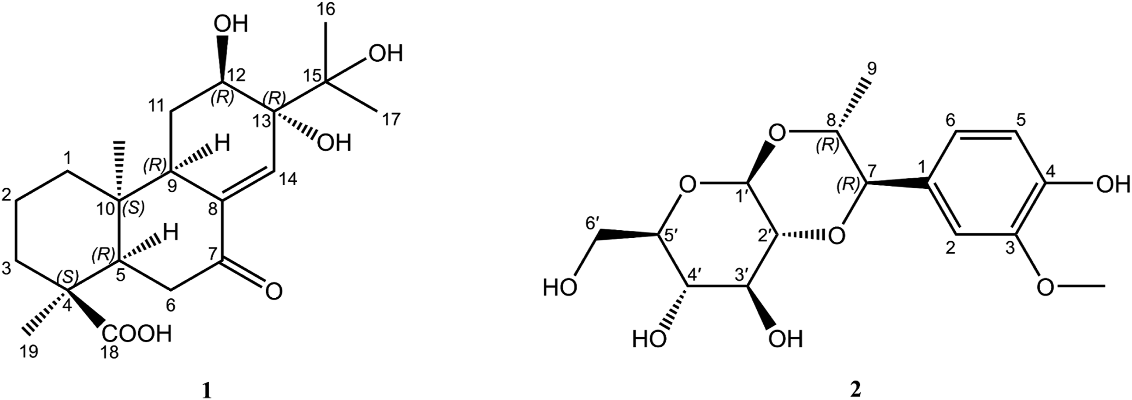
Materials and Methods
General
NMR spectroscopic data were collected using Bruker NEO 600 MHz (Bruker, Germany). Standard pulse sequences were employed for 1H, 13C, HMQC, HMBC, 1H−1H COSY, and ROESY experiments. Chemical shifts (δ) are reported in ppm relative to tetramethylsilane (TMS) or the residual solvent signals as internal standards. HR-ESI-MS data were obtained on a Thermo Fisher Q Exactive mass spectrometer (Thermo Fisher Scientific, USA). Optical rotations were measured at 25 °C on a Rudolph A21102 API/1W automatic polarimeter (Rudolph Research Analytical, USA). UV spectra were acquired using a UV-2700 spectrometer (Shimadzu, Japan) and a V-730 spectrometer (JASCO, Japan). IR spectra were recorded on an IR Tracer-100 spectrometer (Shimadzu, Japan) and an FT/IR-4600 spectrometer (JASCO, Japan). Column chromatography (CC) was performed with D-101 macroporous resin (Tianjin Haiguang Chemical Co., Ltd., China), silica gel (Qingdao Marine Chemistry Co. Ltd, China), Toyopearl HW-40F, and octadecylsilyl silica gel (ODS) (YMC Co. Ltd., Japan). Reversed-phase high-performance liquid chromatography (HPLC) was performed using a Shimadzu LC-20A series instrument (Shimadzu, Japan) equipped with a Neptune C18 column (5 μm, 250 × 10 mm; Phenomenex, USA).
Plant Material
The roots of S. miltiorrhiza were collected in October 2024 from Qingzhen Town, Guiyang City, Guizhou Province, P. R. China, at an altitude of 1200–1300 m above sea level (coordinates: N 26.648754°, E 106.299670°). The species identification was carried out by one of the authors (Xu Sun). A voucher specimen (No.DC202410-1) is deposited at the Guizhou Provincial Institute for Food and Drug Control, China.
Extraction and Isolation
The air-dried roots of S. miltiorrhiza (5 kg) were pulverized and subsequently extracted with 50% aqueous ethanol under reflux (3 × 2 h). The combined extract solutions were concentrated under reduced pressure to yield a brown residue. The residue was suspended in water and successively partitioned with petroleum ether, ethyl acetate, and n-butanol. The n-butanol fraction (95.8 g) was subjected to D101 macroporous resin, successively eluted by water and methanol. The methanol fraction (64.5 g) was subjected to ODS column chromatography and eluted with a stepwise gradient of MeOH–H2O (0:10 to 10:0, v/v), yielding six subfractions (Fr.1–Fr.6). Fr.1 (18.3 g) was chromatographed on a normal phase silica gel CC using a stepwise gradient of CH2Cl2–MeOH (9:1 to 5:5, v/v), yielding five fractions (Fr.1.1–Fr.1.5). Fr.1.3 (5.4 g) was separated to give three fractions (Fr.1.3.1–Fr.1.3.3) by using Toyopearl HW-40F CC with MeOH–H2O (5:5, v/v). Fr.1.3.3 (1.2 g) was further purified by ODS column chromatography (MeOH–H2O, 5:5 to 7:3, v/v) and then by preparative HPLC (MeOH–H2O, 7.6:2.4, v/v; flow rate 3 mL/min; detection at 220 nm) to yield compound
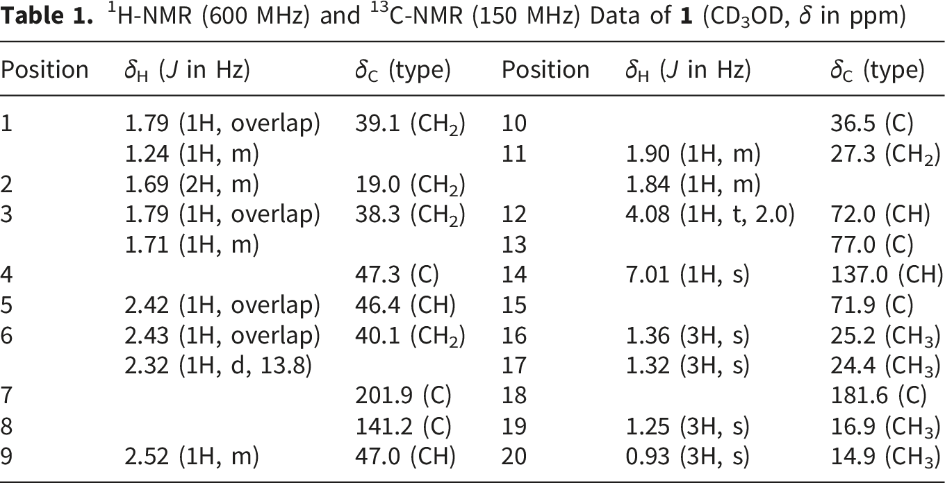
1H-NMR (600 MHz) and 13C-NMR (150 MHz) Data of
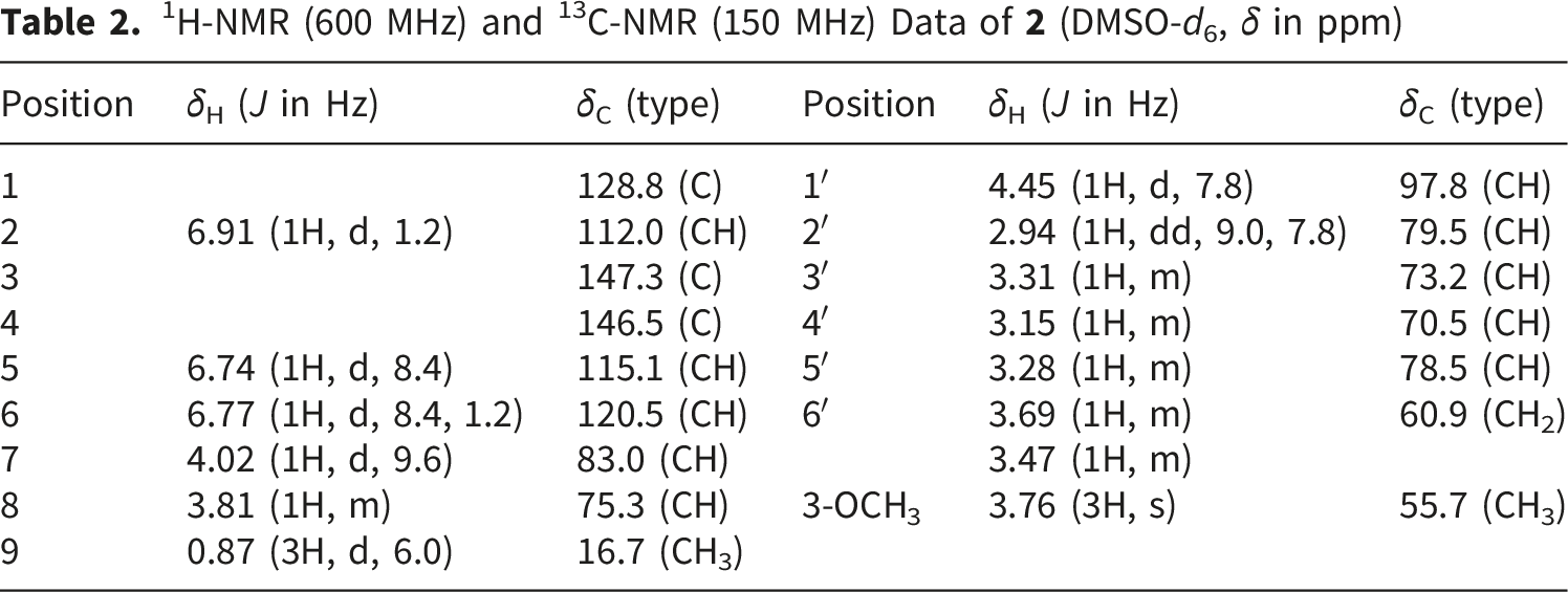
1H-NMR (600 MHz) and 13C-NMR (150 MHz) Data of
Acid Hydrolysis
Compound 2 (1.0 mg) was hydrolyzed with 2 M HCl (2.0 mL) at 95 °C for 3 h. The hydrolysate was allowed to cool to room temperature and then extracted with EtOAc (3 × 2 mL). The aqueous layer was evaporated to dryness under reduced pressure to remove residual HCl. The residue was dissolved in pyridine (0.4 mL), and L-cysteine methyl ester hydrochloride (1.0 mg) was added. The mixture was heated at 60 °C for 1 h, followed by addition of trimethylsilyl imidazole (0.15 mL). The reaction was kept at 60 °C for another 1 h. After completion, the reaction mixture was evaporated to dryness under a gentle stream of nitrogen. The resulting residue was dissolved in water (1.0 mL) and extracted with n-hexane (0.5 mL). The n-hexane layer was then analyzed by gas chromatography (GC). The absolute configuration of the glucose was confirmed by comparison of its retention time with those of authentic D-glucose and L-glucose standards.15,16
Cell Culture
Murine BV-2 microglia cells were kindly provided by the Cell Culture Center, Institute of Basic Medical Sciences, Chinese Academy of Medical Sciences (Beijing, China). Murine RAW264.7 cells (ZQ0098) were obtained from Zhong Qiao Xin Zhou Biotechnology Co., Ltd. (Shanghai, China). Both cell lines, BV-2 and RAW264.7, were originally derived from the China Center for Type Culture Collection (CCTCC), with catalog numbers GDC0311 and GDC0143, respectively. BV-2 cells were maintained in Dulbecco’s Modified Eagle Medium (DMEM) supplemented with 10% fetal bovine serum (FBS) and 1% penicillin-streptomycin, and incubated at 37°C in a humidified atmosphere containing 5% CO2. Cells in the logarithmic growth phase were used for subsequent experiments. RAW264.7 cells were cultured as described above for BV-2 cells.
CCK-8 Cell Viability Assay
BV-2 cells were seeded in a 96-well plate at a density of 1 × 10⁵ cells per well (in 100 μL of medium) and divided into blank, control, and compound-treated groups. After 24 h of incubation, the cells were treated with 100 μL of the corresponding test compound solutions. Subsequently, 10 μL of CCK-8 solution (abbkine scientific co.,ltd, China, Cat. ATYG01021) was added to each well, and the plate was incubated for another 1.5 h at 37°C. The optical density (OD) was then measured at 450 nm using a microplate reader. Cell viability of RAW264.7 cells was determined using the same method as described for BV-2 cells. The CCK-8 cell viability assay was performed according to the manufacturer’s instructions and as previously described. 17
Assay for Inhibition Against Nitric Oxide Production
The NO production analysis was performed using a previously reported method.17,18 BV-2 cells and RAW 264.7 macrophages were seeded in 96-well plates at densities of 1 × 10⁵ and 3 × 10⁵ cells per well (in 100 μL of medium), respectively, and incubated for 24 h. To induce inflammation, cells were pretreated with various concentrations of the test compound for 3 h, followed by stimulation with 250 ng/mL LPS for an additional 24 h; untreated cells served as the control. After the incubation period, the culture supernatants were collected, and NO levels were measured using an NO Assay Kit following the manufacturer’s instructions.
Statistical Analysis
All bioassays were performed in triplicate. Statistical analysis was conducted using SPSS software (version 20.0). Differences between groups were evaluated using Student's t-test or one-way analysis of variance (ANOVA) for multiple comparisons. A P-value of less than 0.05 was considered statistically significant.
Results
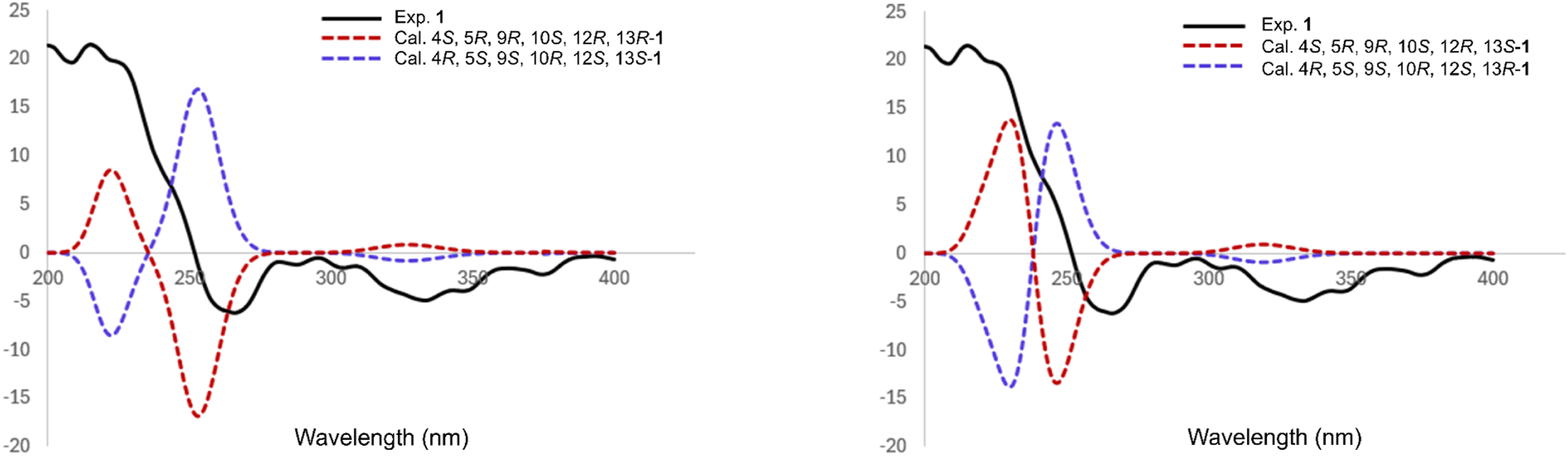
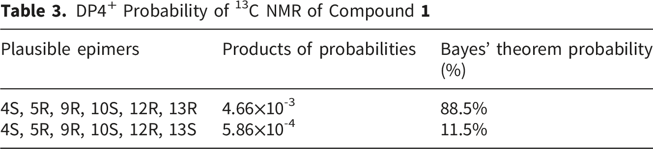
Compound Key HMBC, COSY, and NOESY correlations of compound Comparison of the experimental CD spectrum of DP4+ Probability of 13C NMR of Compound 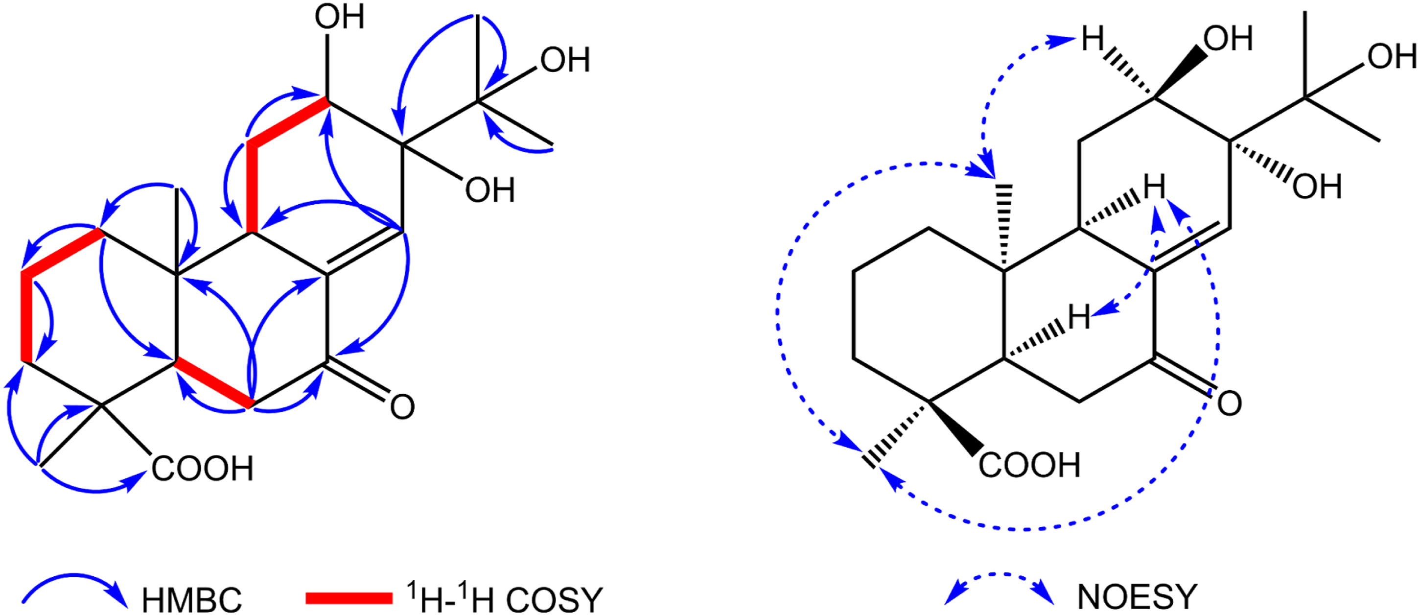
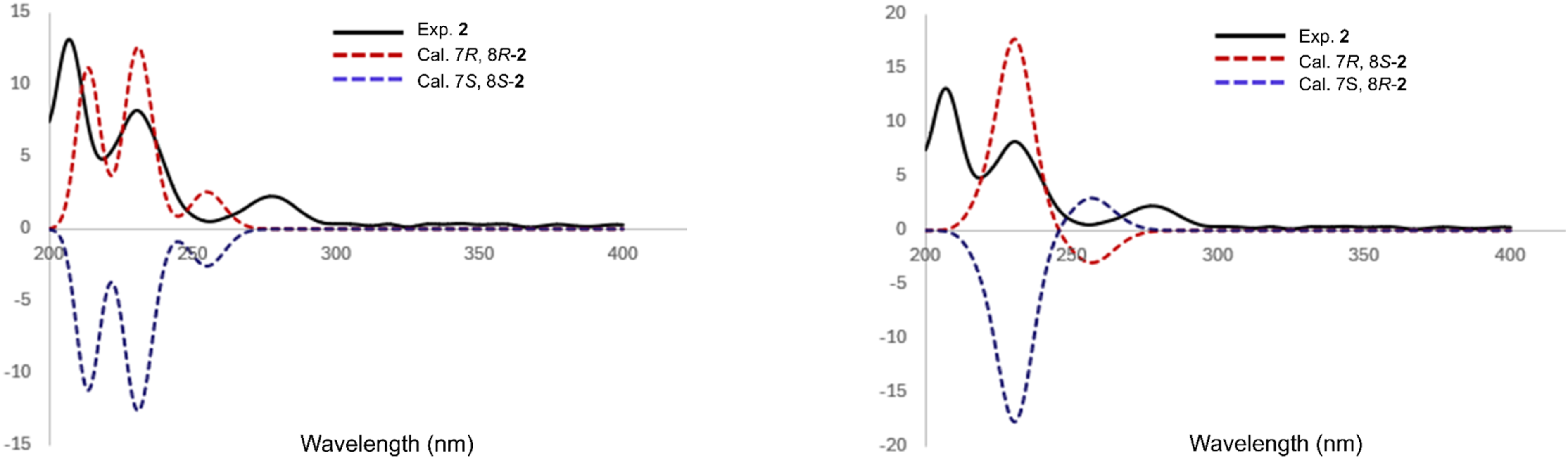
Compound Key HMBC and COSY correlations of compound Comparison of the experimental CD spectrum of 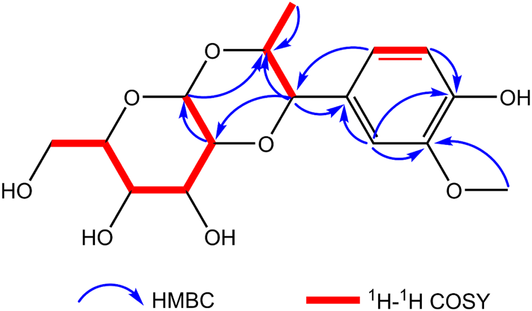
Inhibitory Activity of Compounds Against NO Production in LPS-Stimulated BV-2 and RAW 264.7 Cells

aThese data represent the average values of three repeated experiments (mean ± S.D.).
bPositive control.
Discussion
In the present study, two compounds were isolated from the roots of S. miltiorrhiza: a new diterpenoid, miltiorrhizanene A (
The anti-inflammatory effects of the isolated compounds were evaluated by measuring NO production in LPS-stimulated BV-2 microglial and RAW264.7 macrophage cells.
Novelty of
Despite these promising findings, several limitations of this study should be acknowledged. First, although
Looking forward, future studies should focus on the following aspects. To elucidate the molecular mechanism, the effects of compound 1 on iNOS and COX-2 expression at both the mRNA and protein levels should be investigated by RT-PCR and Western blot analysis. Additionally, given that many diterpenoids exert anti-inflammatory effects by suppressing the NF-κB and MAPK signaling pathways,26,27 it would be worthwhile to examine whether
Conclusion
In summary, two compounds were isolated from the roots of S. miltiorrhiza: miltiorrhizanene A (
Supplemental Material
Supplemental Material - A New Diterpenoid and a Previously Undescribed Enantiomer of a Known Phenylpropanoid Glycoside From Salvia Miltiorrhiza Bunge
Supplemental Material for A New Diterpenoid and a Previously Undescribed Enantiomer of a Known Phenylpropanoid Glycoside From Salvia Miltiorrhiza Bunge by Sheng Zhao, Lixin Wang, Qingqing Yu, Shan Zhao, Chuanjing Yang and Xu Sun in Natural Product Communications.
Footnotes
Acknowledgements
This study was financially supported by the Science and Technology Program of Guizhou Province, the Zunyi City Science and Technology Program, and the Guizhou Administration of Traditional Chinese Medicine.

Ethical Considerations
Ethical approval is not applicable to this article.
Consent to Participate
There were no human subjects included in this study, and informed consent was not available.
Author Contributions
Sheng Zhao, Lixin Wang, and Qingqing Yu performed all experiments; Sheng Zhao and Lixin Wang analyzed the data; Sheng Zhao and Shan Zhao wrote the manuscript. Chuanjing Yang and Xu Sun were the project leaders and designed the experiments.
Funding
The authors disclosed receipt of the following financial support for the research, authorship, and/or publication of this article: This work was supported by the Science and Technology Program of Guizhou Province (No. ZK [2022] general 553), the Zunyi City Science and Technology Program (No. HZ (2023) 397), and the Guizhou Administration of Traditional Chinese Medicine (No. QZYYZDXK(JS)-2023-11 and QZYY-2025-121).
Declaration of Conflicting Interests
The authors declared no potential conflicts of interest with respect to the research, authorship, and/or publication of this article.
Statement of Human and Animal Rights
This article does not contain any studies with human or animal subjects.
Supplemental Material
Supplemental material for this article is available online.
References
Supplementary Material
Please find the following supplemental material available below.
For Open Access articles published under a Creative Commons License, all supplemental material carries the same license as the article it is associated with.
For non-Open Access articles published, all supplemental material carries a non-exclusive license, and permission requests for re-use of supplemental material or any part of supplemental material shall be sent directly to the copyright owner as specified in the copyright notice associated with the article.
