






Abstract
Objective
Diabetes mellitus and its complications, specifically diabetic retinopathy (DR), require novel therapeutics with enhanced safety profiles. Guided by the traditional use of Taxus mairei, this study aimed to isolate bioactive constituents from its leaf extracts and investigate their dual potential in regulating metabolic enzymes and protecting retinal cells against hyperglycemic stress.
Methods
The bioactive n-butanol fraction of T. mairei leaves was subjected to phytochemical isolation and structural elucidation. The isolated compounds were evaluated for inhibitory activities against α-glucosidase and α-amylase, supported by molecular docking analysis to understand structure–activity relationships. Furthermore, the potential retinal-protective effects were assessed using ARPE-19 retinal pigment epithelial cells under high-glucose conditions, focusing on cell viability and vascular endothelial growth factor A (VEGF-A) secretion.
Results
Nine compounds were identified, including a novel phenolic ester, 3-hydroxy-2-(3-hydroxy-4-methoxyphenyl)propyl 2-(4-hydroxyphenyl)acetate (
Conclusions
This study highlights T. mairei as a source of bioactive compounds with complementary antidiabetic potential. The identified biflavonoids offer promise for postprandial glycemic control, whereas the novel phenolic ester showed a distinct protective activity profile against high-glucose-induced VEGF-A overproduction. These findings suggest that T. mairei constituents have the potential to be developed into multifunctional therapeutics for diabetes and its microvascular complications.
Introduction
Diabetes mellitus is a global metabolic disorder characterized by chronic hyperglycemia resulting from impaired insulin secretion, insulin resistance, or both. It is broadly classified into Type 1 diabetes mellitus (T1DM), involving autoimmune destruction of pancreatic β-cells, and Type 2 diabetes mellitus (T2DM), which is strongly associated with obesity, insulin resistance, and progressive β-cell dysfunction.1,2 Diabetes leads to serious long-term complications affecting both macrovascular systems (heart, brain, peripheral vasculature) and microvascular tissues (retina, kidney, nerves), which collectively contribute to disability and premature mortality. 3 Although pharmacologic agents such as metformin, thiazolidinediones, sulfonylureas, and α-glucosidase inhibitors are widely used, their clinical application is often limited by adverse effects including gastrointestinal discomfort, hypoglycemia, or undesired weight changes. Therefore, the discovery of safer, natural compounds with multifunctional antidiabetic properties remains an important therapeutic goal.
Postprandial hyperglycemia control is central to T2DM management, and digestive enzyme inhibitors targeting α-glucosidase and α-amylase play a crucial role in delaying carbohydrate absorption and reducing glucose excursions. At the same time, early diabetic retinopathy (DR), one of the most prevalent microvascular complications, is increasingly recognized as a disease not only of endothelial dysfunction but also of retinal pigment epithelial (RPE) impairment. RPE cells serve as a major component of the blood–retinal barrier and are highly sensitive to metabolic stress. High-glucose exposure induces oxidative damage, mitochondrial dysfunction, and pathological overexpression of vascular endothelial growth factor A (VEGFA), all of which contribute to early DR progression.4,5 Therapeutic compounds capable of simultaneously regulating glucose metabolism and protecting retinal cells may therefore offer dual benefits in diabetes management.
Taxus mairei, a species traditionally valued for its production of paclitaxel, contains abundant flavonoids—including flavones, biflavones, flavonols, dihydroflavones, and related subclasses—that exhibit notable anti-inflammatory, antioxidant, antimicrobial, and anticancer activities.6,7 However, despite extensive phytochemical characterization of Taxus species, their potential roles in glycemic regulation and retinal protection remain insufficiently explored. In particular, the antidiabetic potential of biflavonoids and phenolic derivatives from T. mairei has not yet been comprehensively evaluated.
Preliminary bioactivity screening in our laboratory indicated that the n-butanol fraction of T. mairei leaves exhibited the most pronounced α-glucosidase and α-amylase inhibitory activities among all evaluated solvent fractions (data not shown), thereby guiding our decision to further isolate and characterize its major constituents.
The present study aimed to investigate the antidiabetic potential of compounds isolated from the bioactive n-butanol fraction of Taxus mairei leaves, focusing on both metabolic and retinal-protective mechanisms. To achieve this, we isolated and characterized a novel phenolic ester, 3-hydroxy-2-(3-hydroxy-4-methoxyphenyl)propyl 2-(4-hydroxyphenyl)acetate (
Materials and Methods
General Experimental Procedures and Reagents
The human ARPE-19 cell line was purchased from the American Type Culture Collection (ATCC, Manassas, VA, USA; CRL-2302™). Cells were maintained in DMEM/F12 (1:1) medium supplemented with 10% fetal bovine serum (FBS) and 2 mM L-glutamine. Cell detachment was performed using Trypsin-EDTA (all culture reagents were from Gibco, USA), and cells were washed with phosphate-buffered saline (PBS). Cultures were maintained in a CO2 incubator at 37°C with 5% CO2. Low-glucose (5 mM) and high-glucose (30 mM) media were prepared using D-glucose (Sigma-Aldrich, USA). Cell viability was assessed using a CCK-8 assay kit. VEGF-A secretion was quantified using a commercial ELISA kit. Total RNA was extracted using the RNeasy Total RNA Isolation Kit (QIAGEN, Germany), followed by reverse transcription with the MultiScribe™ Reverse Transcriptase Kit (Applied Biosystems, USA). qRT-PCR was conducted using SYBR Green PCR Master Mix (Applied Biosystems, USA) or Luna Universal Probe qPCR Master Mix (New England Biolabs, USA).
For enzyme inhibition assays, α-glucosidase (1 U/mL), p-Nitrophenyl-α-D-glucopyranoside (pNPG), Na2CO3, and acarbose (positive control) were obtained from Sigma-Aldrich (USA). α-Amylase and its corresponding substrate were used for the α-amylase inhibition assay. Compounds were isolated using silica gel, Sephadex LH-20®, and RP-18 chromatographic media.
Absorbance for α-glucosidase and α-amylase inhibition assays was measured using a BioTek Synergy 4 Multi-Detection Microplate Reader (BioTek Instruments, USA). Quantitative real-time PCR was performed using an ABI PRISM® 7900HT Fast Real-Time PCR System (Applied Biosystems, Italy). Compound purification was carried out using preparative high-performance liquid chromatography (HPLC) systems in combination with silica gel, Sephadex LH-20®, and RP-18 column chromatography setups.
Structural elucidation of isolated compounds was supported by analytical instrumentation. Infrared (IR) spectroscopy was used to identify functional groups, while high-resolution electrospray ionization mass spectrometry (HRESIMS) provided accurate mass measurements (e.g., compound
Plant Materials
The leaves of T. mairei were purchased from Nantou County, Taiwan in January 2019. The raw plant material was identified by Distinguished Prof. Fang-Rong Chang. A voucher specimen (code no. KMU-GINP-Taxus-001) was stored in the Graduate Institute of Natural Products, College of Pharmacy, Kaohsiung Medical University, Taiwan.
Isolation of Phytochemicals
The general phytochemical isolation and chromatographic purification procedures were adapted from our previously established protocols.
8
The dried leaves (6.44 kg) were extracted three times with MeOH (3 × 70 L), and the combined extracts were concentrated under reduced pressure to yield a crude MeOH extract (555 g). The extract was suspended in H2O and partitioned successively with EtOAc and n-BuOH to afford the EtOAc-soluble fraction (185.68 g) and the n-BuOH-soluble fraction (136.09 g), respectively. The n-BuOH fraction was subjected to Diaion® column chromatography and eluted with a gradient of H2O to MeOH (100:0 → 0:100) to yield seven fractions (Fraction A to Fraction G). Fraction B (8.74 g) was chromatographed on a silica gel column and eluted with CH2Cl2/MeOH (15:1 → 0:1) to obtain eight subfractions (Fraction B-1 to Fraction B-8). Fraction B-2 was further purified by preparative RP-HPLC (CH3CN/H2O, 24:76) to yield compounds
α-Glucosidase Activity Inhibition Assay
The α-glucosidase inhibitory activity was evaluated following a modified method described in the literature. Briefly, an α-glucosidase solution (1 U/mL in phosphate buffer, pH 6.8) was mixed with multiple concentrations of test compounds and pre-incubated at room temperature for 5 min. The reaction was initiated by adding 250 µL of p-nitrophenyl-α-D-glucopyranoside (pNPG) solution, followed by incubation at 37°C for 20 min. The enzymatic reaction was terminated by adding 500 µL of 9.4 mM Na2CO3. Acarbose was used as the positive control under the same conditions. The release of p-nitrophenol was quantified by measuring the absorbance at 405 nm using a microplate reader (BioTek Synergy 4 Multi-Detection Microplate Reader). 9
α-Amylase Activity Inhibition Assay
The α-amylase inhibitory activity was determined according to a modified method described by Acar et al. 10 with slight adjustments. Briefly, a human α-amylase enzyme solution (1 U/mL in phosphate buffer, pH 6.9) was mixed with multiple concentrations of test compounds and pre-incubated at 37°C for 10 min. The reaction was initiated by adding soluble starch solution as the substrate, followed by incubation at 37°C for 10 min. For color development, 3,5-dinitrosalicylic acid (DNS) reagent was added to terminate the enzymatic reaction, and the mixture was heated in a boiling water bath for 5 min to form the colored complex. After cooling to room temperature, the absorbance was measured at 540 nm using a BioTek Synergy 4 Multi-Detection Microplate Reader. Acarbose was used as the positive control. The percentage of α-amylase inhibition was calculated relative to the untreated enzyme control, and IC50 values were obtained by nonlinear regression of inhibition curves.
Quantitative RT-PCR (qRT-PCR)
Total RNA was extracted from ARPE-19 cells cultured under different conditions using the RNeasy Total RNA Isolation Kit (QIAGEN, Valencia, CA, USA) following the manufacturer’s protocol.4,5 RNA purity and concentration were determined spectrophotometrically, and samples with an A260/A280 ratio ≥ 1.8 were used for downstream analysis.
For miRNA and mRNA expression analysis, reverse transcription was performed using the MultiScribe™ Reverse Transcriptase Kit. The resulting cDNA was diluted and subjected to qRT-PCR using SYBR Green PCR Master Mix. All reactions were conducted on an ABI PRISM® 7900HT Fast Real-Time PCR System (Applied Biosystems, Monza, Italy). Gene expression levels were quantified using the 2-ΔΔCt method, 4 with GAPDH serving as the internal control. 5
All qRT-PCR reactions were conducted on an ABI PRISM® 7900HT Fast Real-Time PCR System (Applied Biosystems, Monza, Italy). Gene expression levels were quantified using the 2-ΔΔCt method. The expression of VEGF-A was assessed by qRT-PCR amplification using Luna Universal Probe qPCR Master Mix (New England Biolabs, MA, USA) with the following primers:
VEGF-A
Forward (F): TTCTGAGTTGCCCAGGAGAC
Reverse (R): TGGTTTCAATGGTGTGAGGA
GAPDH (internal control)
Forward (F): 5′-TGTGGGCATCAATGGATTTGG-3′
Reverse (R): 5′-ACACCATGTATTCCGGGTCAAT-3′
qRT-PCR analysis for Figure 1C was performed using three independent biological replicates, with one qPCR measurement obtained from each biological replicate. The qRT-PCR reaction conditions were as follows: an initial denaturation step at 95°C for 1 min, followed by 43 cycles of denaturation at 95°C for 15 s and extension at 60°C for 30 s. GAPDH was used as an internal control for normalization. Gene expression comparisons were analyzed using the 2-ΔΔCt method. qRT-PCR analysis for Figure 1C was performed using three independent biological replicates, with one qPCR measurement obtained from each biological replicate. Effects of compound 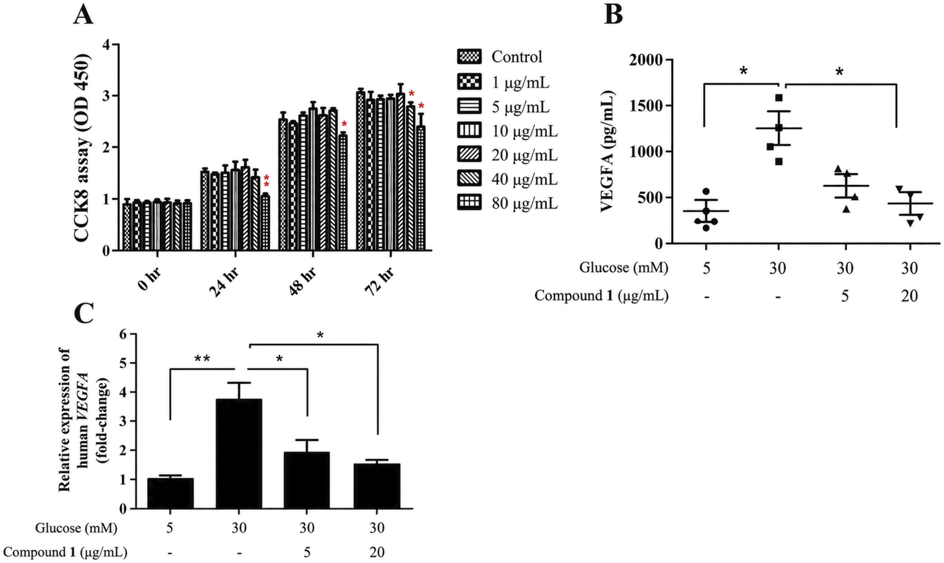
Molecular Docking
The general in silico molecular docking framework, including protein preparation and energy grid generation using Discovery Studio, was guided by validated computational methodologies.
11
Molecular docking was performed to elucidate the structural basis underlying the inhibitory differences among compounds
Docking simulations were carried out using the LigandFit module, a shape-directed cavity-based docking algorithm.
12
Energy grid calculations were generated using Dreiding, CFF, and PLP1 scoring potentials. For each ligand, the top 100 docking poses were ranked using the Ludi_3 empirical scoring function, which evaluates hydrogen bonding, hydrophobic interactions, and entropic contributions to binding affinity.
13
Torsion angle analysis was performed to classify poses with dihedral angles < 90° as cis- and > 90° as trans- conformations. The best-scoring conformations were compared across compounds
Statistical Analysis
All experimental data are presented as the mean ± standard error of the mean (SEM) from three independent biological replicates (n = 3). For the CCK-8 assay, each biological replicate included three technical replicates; for ELISA, each biological replicate included two technical replicates; and for qRT-PCR, one measurement was obtained from each biological replicate without technical replicates. For assays with technical replicates, the mean of the technical replicates was treated as a single biological replicate for statistical analysis. Statistical comparisons between two independent groups were performed using the unpaired Student’s t-test. Exact p-values for the comparisons related to Figure 1 are provided in the Results section. A value of p < 0.05 was considered statistically significant.
Results
Chemical Constituents of the Bioactive n-Butanol Fraction
In this study, the constituents of the bioactive n-butanol fraction obtained from the leaves of T. mairei were investigated, as this fraction exhibited the most pronounced anti-α-glucosidase and anti-α-amylase activities among all solvent partitions. Successive chromatographic separation of the n-butanol extract yielded one new compound, 3-hydroxy-2-(3-hydroxy-4-methoxyphenyl)propyl 2-(4-hydroxyphenyl)acetate ( Structures of compounds 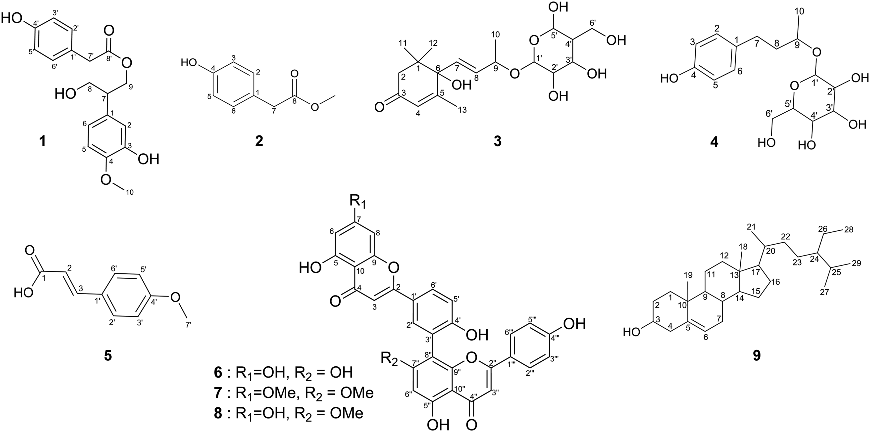
Structural Elucidation of Compound 1
Compound
1H (400 MHz, Pyridine-d
5
) and 13C (100 MHz, Pyridine-d
5
) NMR Data of
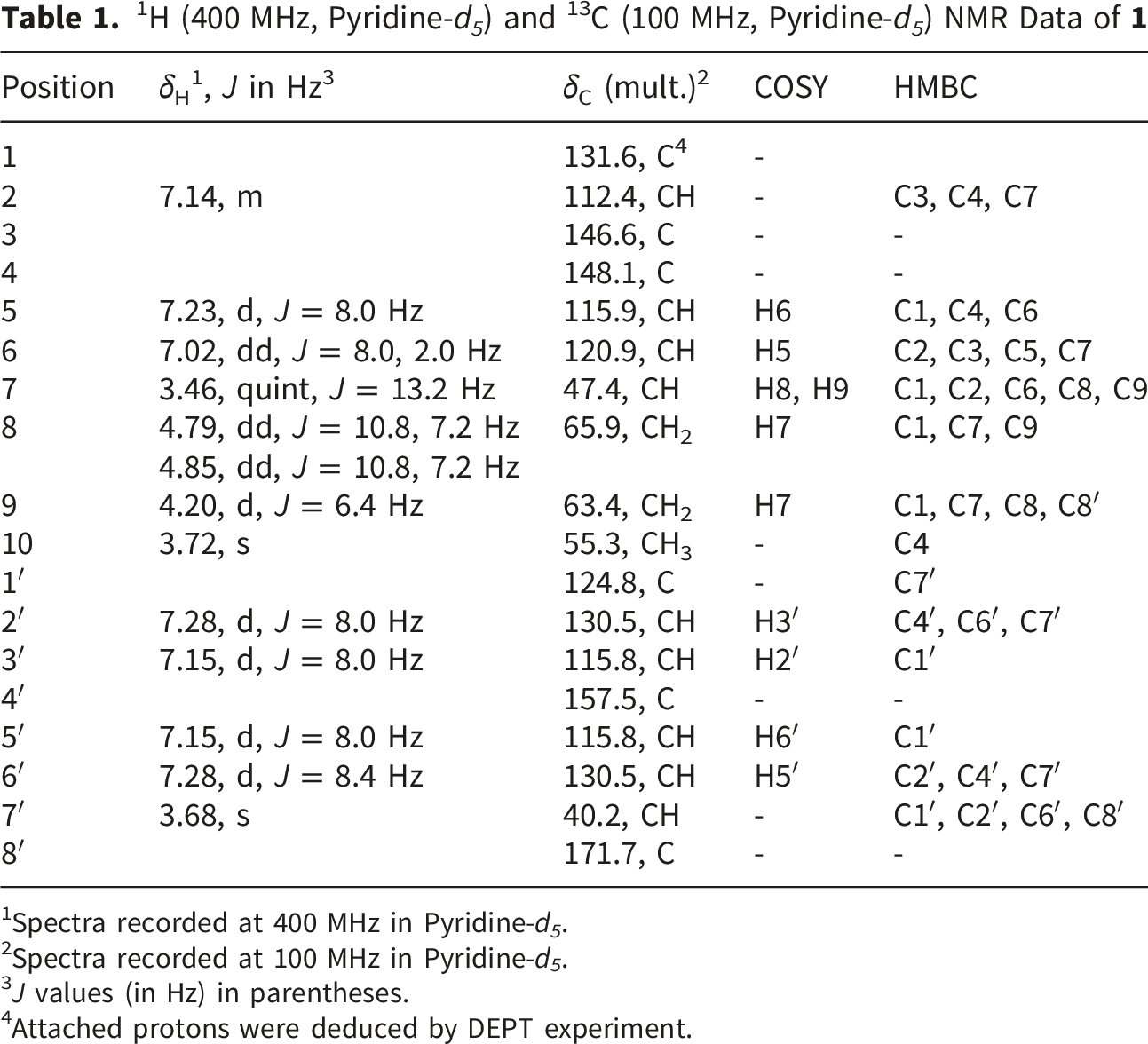
1Spectra recorded at 400 MHz in Pyridine-d 5 .
2Spectra recorded at 100 MHz in Pyridine-d 5 .
3J values (in Hz) in parentheses.
4Attached protons were deduced by DEPT experiment.
Figure 3 illustrates the key COSY and HMBC correlations used for establishing the planar structure of Key COSY and HMBC of 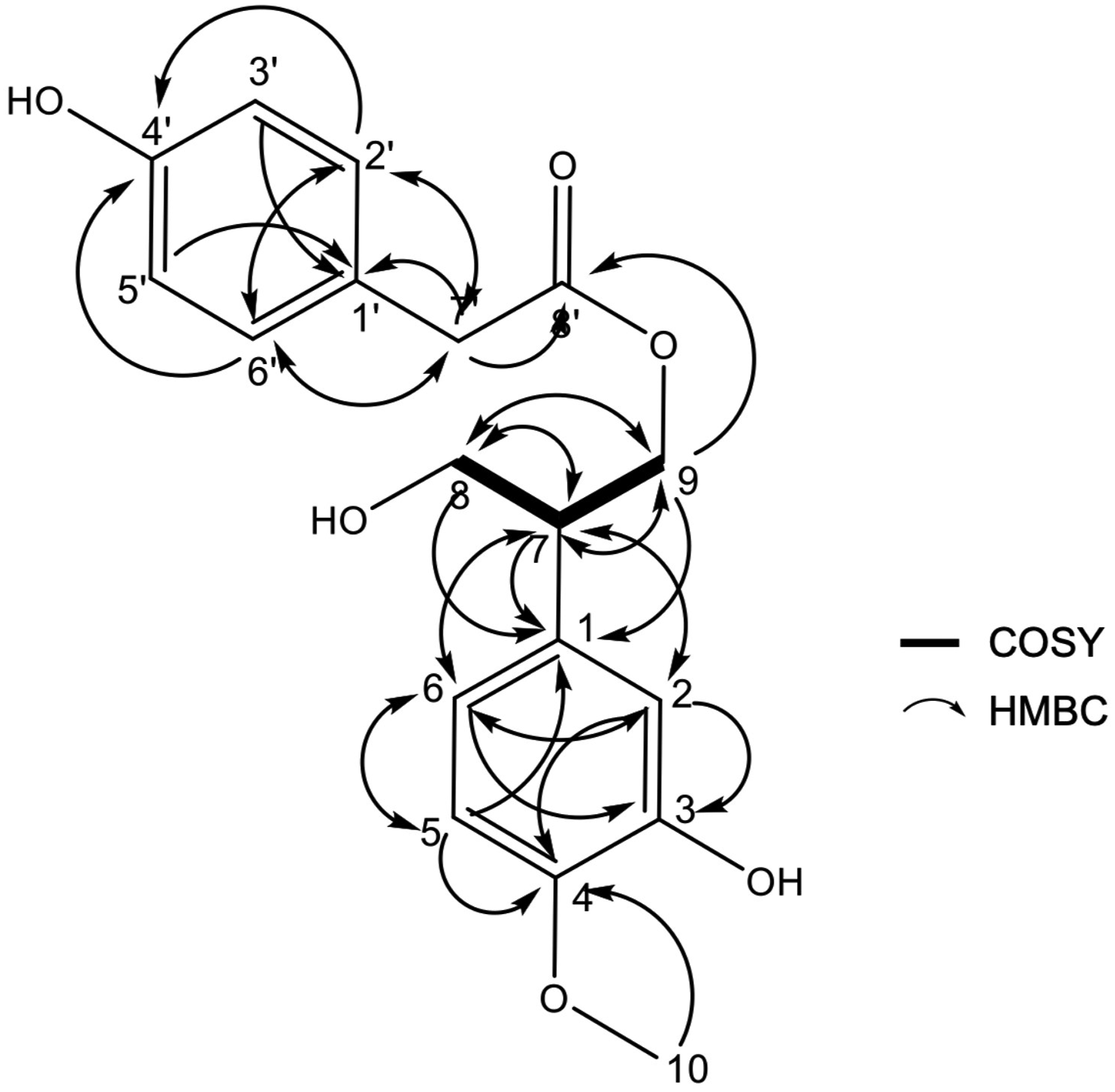
Taken together, these MS, IR, and comprehensive NMR analyses unequivocally established the structure of
Along with the new compound (
Inhibitory Effects of Compounds 1 -9 on α-Glucosidase and α-Amylase
Inhibition of α-Glucosidase and α-Amylase Activity by Compounds
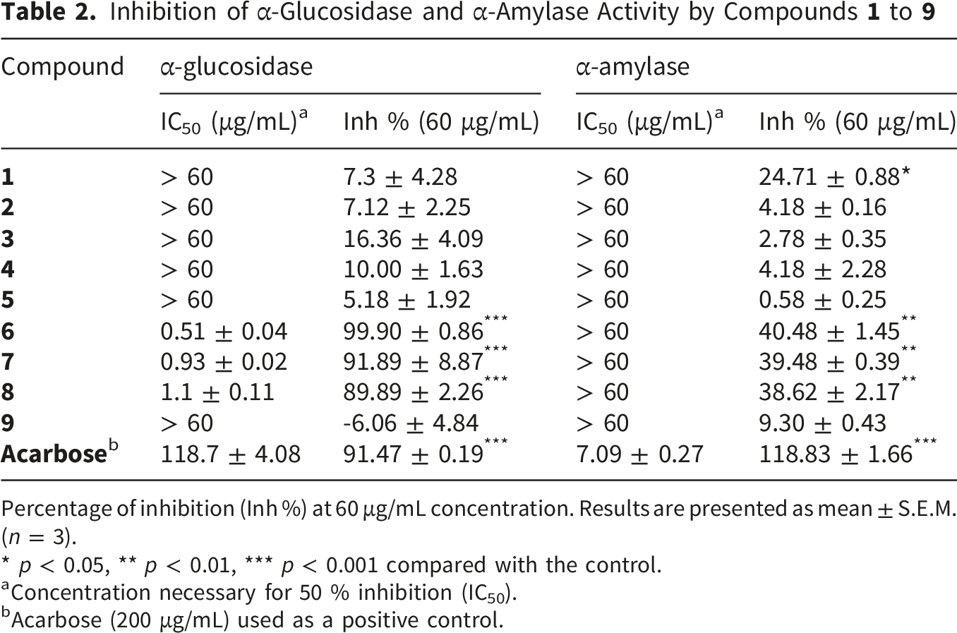
Percentage of inhibition (Inh %) at 60 μg/mL concentration. Results are presented as mean ± S.E.M. (n = 3).
* p < 0.05, ** p < 0.01, *** p < 0.001 compared with the control.
aConcentration necessary for 50 % inhibition (IC50).
bAcarbose (200 μg/mL) used as a positive control.
For α-glucosidase, compounds
For α-amylase, compounds
Taken together, the biflavonoids amentoflavone (
Molecular Docking Analysis
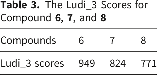
The markedly different inhibitory potencies of compounds
Docking results indicated that all three biflavonoids, amentoflavone (
Amentoflavone (
Sotetsuflavone (
The Ludi_3 Scores for Compound
Protective Effects of Compound 1 on ARPE-19 Cell Viability and VEGF-A Expression and Secretion
To evaluate the potential cytoprotective and anti-angiogenic effects of compound
We next examined whether compound
Discussion
Diabetes management involves both the regulation of postprandial glucose metabolism and the protection of vulnerable retinal tissues from hyperglycemia-induced injury. In this context, our study revealed two mechanistically distinct yet complementary bioactivities among the isolated compounds from T. mairei. While the biflavonoids amentoflavone (
High-glucose exposure is well established to trigger oxidative stress, mitochondrial dysfunction, and pathological VEGFA upregulation in retinal cells-key events in the early development of diabetic retinopathy.14-16 Consistent with these mechanisms, compound
In contrast, compounds
Molecular docking analyses further supported the structure–activity relationships of these biflavonoids. Compounds
Importantly, molecular docking in the present study was restricted to α-glucosidase, and no docking analysis was conducted for α-amylase. Human α-amylase was used in the in vitro enzyme inhibition assay; however, the computational component of this work was specifically designed to examine the structural basis of α-glucosidase inhibition among compounds
Collectively, our findings indicate that T. mairei contains two classes of bioactive compounds with divergent but synergistic antidiabetic potentials. The biflavonoids (
Amentoflavone, one of the major biflavonoids identified in the present study, has been previously reported to possess antidiabetic activity. In an earlier study, amentoflavone isolated from Cycas pectinata showed inhibitory activity against both α-glucosidase and α-amylase, with IC50 values of 8.09 ± 0.023 μM and 73.6 ± 0.48 μM, respectively. A review on amentoflavone also summarized both its in vivo antidiabetic activity in diabetic mice and its digestive enzyme inhibitory activity, while a more recent systematic review reported α-glucosidase IC50 values ranging from 3.28 to 24.43 μM across four studies. In the present study, compound
Flavonoids are recognized as an important class of natural α-glucosidase inhibitors. A review by Yin et al summarized 411 natural α-glucosidase inhibitors from medicinal plants, including 103 flavonoids, supporting the flavonoid scaffold as a major source of inhibitory activity. In this context, the strong α-glucosidase inhibitory activity of compounds
In a comparative context, several agents have been reported to suppress VEGF/VEGFA-related responses under glucose stress in retinal cells. In the present study, compound
However, the present study has several limitations. First, the isolated compounds were evaluated individually without combination experiments, precluding the assessment of potential synergistic or antagonistic interactions on α-glucosidase or α-amylase inhibition. Additionally, differences in solubility and bioavailability were not rigorously controlled beyond nominal assay concentrations, and retinal safety evaluation was performed only for compound
Conclusions
Taxus species are known to contain a chemically diverse array of flavonoids, including flavones, biflavones, flavonols, dihydroflavones, dihydroflavonols, and flavanols, many of which have been associated with important biological functions. Among these constituents, amentoflavone (
Supplemental Material
Supplemental Material - Isolation of a New Phenolic Ester and Biflavonoids From Taxus mairei Leaves: Dual Mechanisms Targeting α-Glucosidase and High-Glucose-Induced VEGF Expression
Supplemental Material for Isolation of a New Phenolic Ester and Biflavonoids From Taxus mairei Leaves: Dual Mechanisms Targeting α-Glucosidase and High-Glucose-Induced VEGF Expression by Mon-Der Cho, Shang-Yu Chou, Yu-Ming Hsu, Chi-Ying Li, Yang-Chang Wu, Yi-Hong Tsai, Fang-Rong Chang in Natural Product Communications
Footnotes
This research was financially supported by grants from the National Science and Technology Council (NSTC), the Drug Development and Value Creation Research Center at Kaohsiung Medical University, and the Kaohsiung Municipal Min-Sheng Hospital Project. The authors gratefully acknowledge Jing Koou Enterprise Co., Ltd. (No. 4, Aly. 23, Ln. 288, Juemin Rd., Sanmin District, Kaohsiung, Taiwan; TEL: +886-7-3854349) for their technical assistance and support in resource coordination that contributed to this work. The authors also appreciate the Center for Research Resources and Development in Kaohsiung Medical University for the assistance in protein identification, flow cytometry and confocal image analysis. We also sincerely thank Ho Ni Cheng and part-time project assistants in Prof. Fang-Rong Chang’s lab for their valuable assistance in the experimental work conducted in this study.

Ethical Considerations
Ethical approval is not applicable for this article.
Consent to Participate
There are no human subjects in this article and informed consent is not applicable.
Author Contributions
Conceptualization, M.D. Cho, F.R. Chang, and Y.H. Tsai; methodology, Y.M. Hsu; software, S.Y. Chou; validation, Y.H. Tsai, F.R. Chang, and Y.C. Wu; formal analysis, Y.H. Tsai; investigation, M.D. Cho; resources, S.Y. Chou; data curation, M.D. Cho and Y.M. Hsu; writing—original draft preparation, M.D. Cho and C.Y. Li; writing—review and editing, Y.H. Tsai and F.R. Chang; visualization, Y.H. Tsai; supervision, Y.C. Wu and F.R. Chang; project administration, M.D. Cho and S.Y. Chou; funding acquisition, F.R. Chang and Y.C. Wu. All authors have read and agreed to the published version of the manuscript.
Funding
The authors disclosed receipt of the following financial support for the research, authorship, and/or publication of this article: This study was partly supported by funds from the National Science and Technology Council (113-2320-B-127-001, 113-2321-B-037-002, 112-2321-B-037-007, 111-2321-B-037-004, 115-2320-B-127-001), Ministry of Science and Technology (MOST 111-2320-B-037-020-MY3), and the Drug Development and Value Creation Research Center, Kaohsiung Medical University (KMU-TC112A03-3). This work was also supported by the Kaohsiung Municipal Min-Sheng Hospital Project (KMSH-11303), awarded to Mon-Der Cho.
Declaration of Conflicting Interests
The authors declared no potential conflicts of interest with respect to the research, authorship, and/or publication of this article.
Data Availability Statement
The data presented in this study are available in the article and the Supplementary Materials.
Statement of Human and Animal Rights
This article does not contain any studies with human or animal subjects.
Institutional Review Board Statement
Ethical review and approval were not required for this study because no human participants, human-derived clinical specimens, identifiable personal data, or animal experiments were involved.
Supplemental Material
Supplemental material for this article is available online.
Appendix
References
Supplementary Material
Please find the following supplemental material available below.
For Open Access articles published under a Creative Commons License, all supplemental material carries the same license as the article it is associated with.
For non-Open Access articles published, all supplemental material carries a non-exclusive license, and permission requests for re-use of supplemental material or any part of supplemental material shall be sent directly to the copyright owner as specified in the copyright notice associated with the article.
