
Abstract
The female reproductive system is highly complex, making it essential for applied research and translational medicine to accurately model its intricate physiological functions or develop strategies for restoring them. However, significant structural and functional differences between human and animal models, along with the limitations of static 2D cell culture technologies, underscore the need for more dynamic and sophisticated in vitro platforms, as well as in vivo therapies. These advancements are critical for deepening our understanding of reproductive biology and supporting clinical applications. Recent advancements in additive manufacturing technology have opened new frontiers in the study of the female reproductive system. By introducing diverse preclinical models and expanding the range of potential applications, this field has reached new heights, with the rapidly evolving research paradigm reshaping the scientific landscape. This review aims to summarize the growing body of evidence surrounding bioengineering strategies, platforms, and therapies in female reproductive medicine, with the goal of advancing our understanding of female reproductive biology and providing new avenues for fertility restoration. Specifically, we will examine the historical development, technological innovations, and scientific research related to the creation of 3D-engineered tissues for reconstructing the female reproductive system.
Impact Statement
This review aims to summarize the growing body of evidence surrounding bioengineering strategies, platforms, and therapies in female reproductive medicine, with the goal of advancing our understanding of female reproductive biology and providing new avenues for fertility restoration. Specifically, the historical development, technological innovations, and scientific research related to the 3D-engineered tissues for reconstructing the female reproductive system were summarized. This review would help the audience, especially bioengineers who study the female reproductive system disease, as well as obstetricians and gynecologists, understand the possible application of additive manufacturing and acquire the strategies to engineer the female reproductive system in vitro.
Keywords
Introduction
The female reproductive system is a highly dynamic physiological system, which consists of ovary, fallopian tube, uterus, cervix, and vagina. 1 Amid rapid aging and declining fertility rates, protecting women’s reproductive, maternal, newborn, and child health is of critical importance. 2 Traditional treatments for diseases of the female reproductive system, such as endometriosis and premature ovarian failure, typically involve pharmacotherapy, surgical interventions, or hormone replacement therapy.3–5 However, these methods only provide temporary symptom relief and may be accompanied by significant side effects, such as hormonal imbalances or the need for repeated surgery. In addition, many treatments lack targeted mechanisms, which cannot effectively solve the root causes of the disease, resulting in a high recurrence rate. With advances in regenerative medicine and tissue engineering technology, the emerging therapies utilizing biomaterials and cell-based strategies provide innovative treatment options for these diseases.6,7 These engineered tissues can replace or repair damaged reproductive tissues, not only reducing recurrence but also restoring fertility. The field of tissue engineering has emerged as a revolutionary approach in regenerative medicine, offering the potential to restore damaged or lost tissues and organs. 8 In particular, the reconstruction of the female reproductive system represents a critical area of focus due to its complex structure and essential roles in reproduction, hormonal regulation, and overall health. 9 The proper function of this system is crucial for fertility, sexual health, and the overall well-being of women. Reconstructive surgery for the female reproductive system has traditionally been challenging, often relying on synthetic materials or donor tissues that may not fully integrate with the patient’s body or restore the organ’s native function. 10 These limitations have driven the search for more effective and biocompatible solutions, leading to tissue engineering as a promising alternative. By leveraging advances in cellular biology, materials science, and engineering, researchers are developing novel strategies to reconstruct organs such as the uterus, ovaries, and vagina. These developments hold great promise for improving outcomes for women affected by congenital anomalies, cancer, trauma, or other conditions that compromise reproductive health.
Tissue engineering aims to create biological substitutes that can replicate the natural structure and function of the female reproductive organs. 11 Three-dimensional (3D) tissue engineering is an interdisciplinary field that combines biology, engineering, and materials science to develop biological tissue models. Its goal is to generate tissues that anatomically and functionally resemble natural ones, improving in vitro research and potential in vivo applications. While animal models provide valuable insights, they are costly, time-consuming, and may not accurately reflect human biology due to species differences. The key advantage of 3D tissue engineering is its ability to create scaffolds that support cell growth in three dimensions, closely mimicking the natural extracellular matrix (ECM) compared with two-dimensional (2D) cultures. This 3D environment is crucial for proper cell proliferation, differentiation, and communication. In medical research, 3D tissues offer significant advancements in disease modeling, drug testing, and regenerative therapies, as they more accurately reflect human physiological and pathological conditions. To achieve this, it is essential to recreate cellular microenvironments, incorporating essential signals from neighboring cells and the ECM. 12 Additive manufacturing technology enables the creation of complex reproductive tissue models by precisely arranging cells and materials, enabling the personalized reconstruction of organs such as the uterus, ovaries, and fallopian tubes. This approach provides a new platform for functional reconstruction and drug effect studies in the female reproductive system.
Considering the complex structure and dynamic characteristics of the female reproductive system, researchers have made various summaries. Over the past decade, additive manufacturing technology has advanced rapidly, 13 emerging as a pivotal innovation in biomedical engineering, particularly in research focused on the female reproductive system. Although extensive work has been done on the 3D-engineered tissue for the female reproductive system, there, to the best of our knowledge, appears too little review paper in this field.14,15 In this article, we have explored the current state of additive manufacturing as it pertains to the reconstruction of the female reproductive system. Then we discussed the key components and techniques involved, the challenges faced by researchers, and the future directions of this cutting-edge field. By understanding the advancements and hurdles in this area, we can better appreciate the transformative potential of tissue engineering in enhancing women’s health and reproductive capabilities.
Evolution of Additive Manufacturing Applied to Female Reproductive System
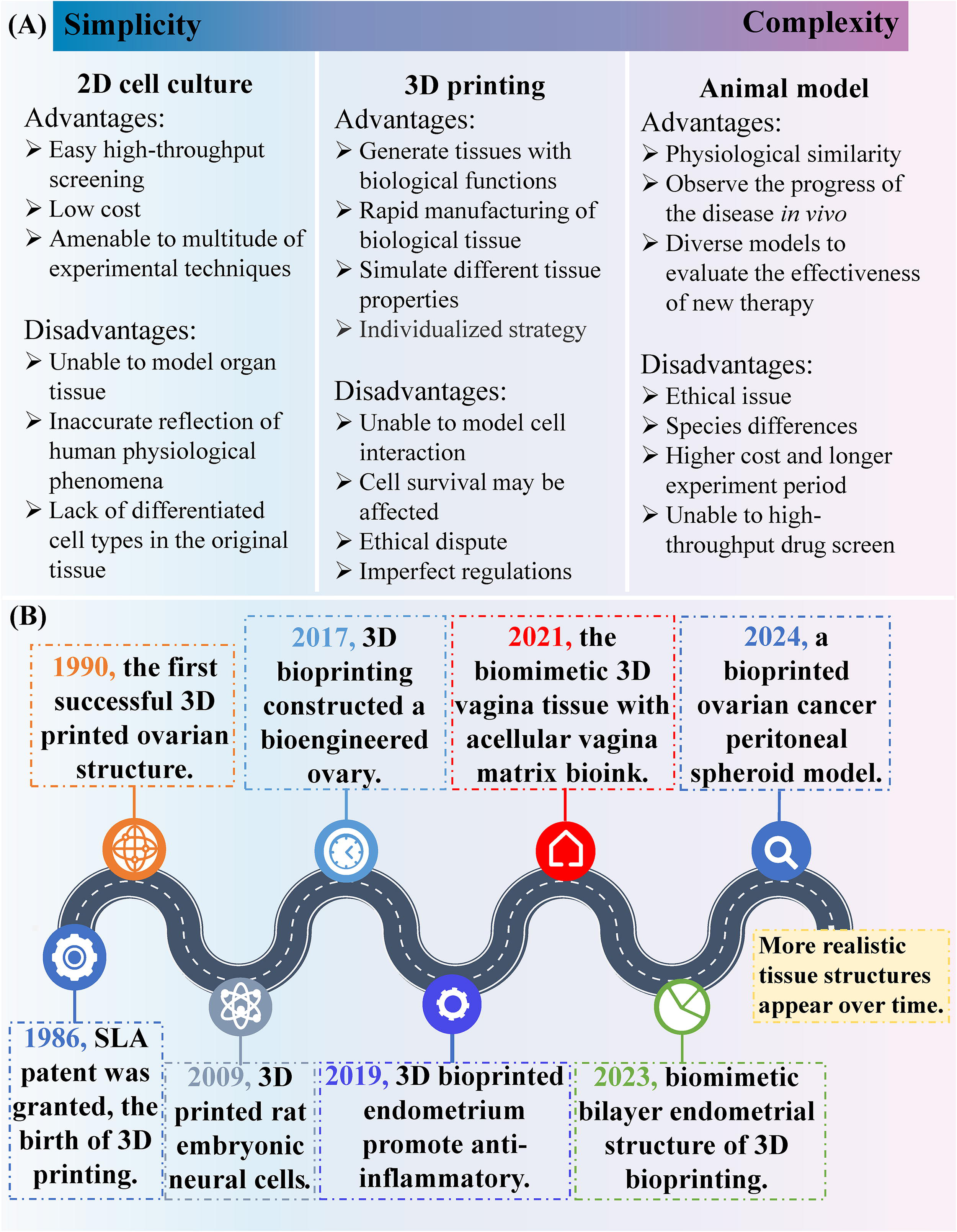
Animal models are valuable for studying female reproductive system, as they can mimic certain characteristics and provide insights into disease mechanisms, diagnostic approaches, and treatment strategies.16,17 However, they have limitations due to significant species differences and challenges related to technical complexities and ethical concerns, which can affect their accuracy and applicability. Initially, studies relied heavily on 2D cell cultures, which, although informative, lacked the complexity of in vivo environments. Specifically, cell culture based on petri dishes misses the basic cell functions existing in tissues, which limits its ability to accurately predict cellular responses in real organisms. 18 In contrast, 3D in vitro models offer a more accurate representation of in vivo tissues by incorporating biophysical cues. Furthermore, the advent of 3D tissue engineering has enabled researchers to more effectively replicate the structural and functional characteristics of female reproductive organs, including the ovaries, uterus, and fallopian tubes. Traditional tissue engineering methods encounter numerous challenges in constructing complex female reproductive system structures, including the complexity of the structure, the reproduction of the cellular microenvironment, and the regeneration of functional tissues (Fig. 1A).
Evolution of 3D-engineered female reproductive system.
The evolution of 3D-engineered tissues applied to female reproductive system research has been driven by several key milestones (Fig. 1B). Since Chuck Hull publicly proposed this technology in 1984 and patented it in 1986, the technology has greatly advanced various fields of medicine by replicating in vivo conditions in the laboratory, representing a significant breakthrough for healthcare applications. 19 The groundbreaking work by Telfer et al. in 1990 marked the first successful 3D-printed ovarian structure in murine models. 20 This achievement was later followed by the printing of human ovarian preantral follicles, which were successfully implanted in utero in immunodeficient mice. In the early 2000s, advancements in scaffold-based techniques enabled the construction of 3D structures that more accurately represented the architecture of tissues such as the endometrium and ovaries. 21 In 2009, Lee et al. proposed a direct cell printing technique to pattern nerve cells in a 3D multilayer collagen gel. 22 In 2015, a groundbreaking system was developed to monitor the interaction between the follicle and fallopian tissue in real time, capturing both the follicular and luteal phases of the menstrual cycle. 23 The following year, further research led to the development of coculture models combining fallopian reproductive tissues with ovarian follicles, offering a valuable tool for understanding fertility and exploring the mechanisms behind reduced fertility. 24 Three-dimensional bioprinting technology follows the standard 3D printing method and uses bioink instead of metal or plastic as the material. Another landmark event was the creation of a 3D-bioprinted model of the endometrium in 2017, which provided unprecedented insights into the implantation process and the early stages of pregnancy. 25 In 2017, Laronda et al. used 3D bioprinting technology for the first time to construct a bioengineered ovary made of gelatin-based bioink and successfully implanted and maintained in mice, restoring normal follicle development and hormone secretion function. 26 In 2019, Paul et al. introduced an innovative treatment strategy for vaginal wall repair, utilizing bioprinting of endometrial mesenchymal stem cells encapsulated in a hydrogel, combined with a 3D melt electrospun mesh to create a tissue engineering construct. 27 This study represents the first report of bioprinting mesenchymal stem cells derived from the female endometrium to aid in the treatment of pelvic organ prolapse. In 2021, Hou et al. demonstrated the application prospect of biomimetic 3D vaginal tissue in vaginal reconstruction, believing that this method has great potential. 28 In 2023, Nie et al. described a 3D-printed endometrial construct that features a dense upper layer comprising endometrial epithelial cells and a lower layer consisting of endometrial stromal cells, effectively replicating the natural bilayer structure and cellular makeup of the endometrium. 29 Although this research is still in the early stages, it lays the foundation for future whole-organ printing of the reproductive system. Recently, Xiang et al. constructed an ovarian cancer model macrophage spheroid by bioprinting based on digital-light-processing. 30 These milestones have greatly enhanced our understanding of female reproductive biology and opened new avenues for research and therapeutic development.
Multiscale Simulation the Female Reproductive System
Additive manufacturing represents a groundbreaking approach in tissue engineering, enabling the creation of tissues for disease modeling and drug testing, as well as the development of tissues and organs for therapeutic repair, regeneration, and transplantation. 31 However, additive manufacturing technology provides new possibilities for constructing complex reproductive organ models through its precise spatial positioning ability and coprinting of multicell types. 32 In this part, we will summarize the in vitro simulation of vagina, uterus, ovary, and fallopian tube from macroscale and microscale dimensions, respectively.
From vagina structure to 3D-engineered models of vagina
The 3D structure of the vagina exhibits complex anatomical and organizational levels that support its versatility. The innermost mucosal layer of the vaginal wall, composed of squamous epithelial cells, lacks keratinization and forms folds that enhance extensibility during sexual activity and childbirth. Beneath the mucosa, an extensive network of blood vessels and lymphatic structures provides nutritional support and immune defense. The muscular layer, composed of smooth muscle, facilitates contraction and regulates vaginal tone. It includes inner circular muscle layer and outer longitudinal muscle layer, providing elasticity and enabling expansion during delivery. The outer fibrous layer, made of connective tissue with collagen and elastic fibers, enhances strength and flexibility, helping the vagina maintain its structure under mechanical stress. The interaction between these layers makes the vagina a dynamic rather than a static organ. The mucosal folds increase surface area, aiding moisture retention and mechanical adaptation. The contractility of the muscular layer plays a crucial role in regulating functions during sexual activity, childbirth, and menstruation. In modern biomedicine, studying the 3D structure and function of the vagina is essential for understanding disease mechanisms and developing treatment strategies.
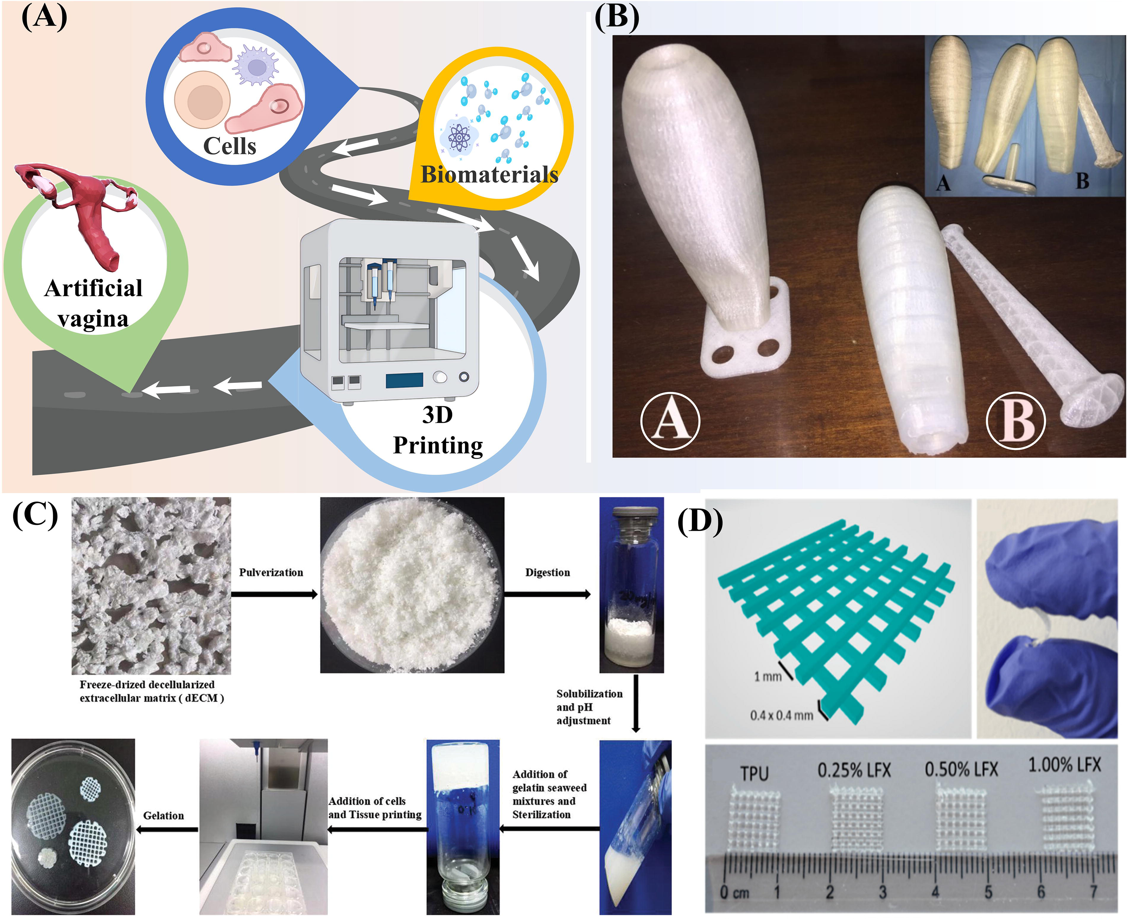
Vaginal reconstruction aims to create an integrated structure and a functional vagina. Advances in additive manufacturing and tissue engineering have led to the creation of biologically similar vaginal tissue models, providing new avenues for researching drug applications, infection processes, and tissue regeneration (Fig. 2A). Acién et al. used the method of polylactic acid (PLA) prosthesis using 3D-printing technology to create a macroscale vagina (vaginoplasty). 33 This PACIENA prosthesis can obtain good anatomical and functional results (Fig. 2B). In contrast, some researchers have developed a macroscale 3D-printed lumenal scaffold that encapsulates exosomes, potentially offering a cell-free therapeutic approach for vaginal reconstruction. 35 Recent advancements in bioprinting have also facilitated the development of decellularized ECM (dECM)-based bioinks for 3D bioprinting, which provide a biocompatible microenvironment for tissue regeneration. 36 In another study, Tian et al. used acellular vaginal matrix to reconstruct vagina in Bama miniature pigs. 37 However, it is still unclear whether the acellular vaginal matrix is the best biomaterial. The use of a cell-laden hydrogel derived from an acellular vaginal matrix could potentially be the optimal approach for microscale vaginal reconstruction. 38 The work shows that encapsulating bionic 3D vaginal tissue with acellular vagina matrix bioink holds great promise for the in vitro reconstruction of the vagina (Fig. 2C). 28 These models effectively simulate the vaginal microenvironment, mechanical properties, and tissue interactions.
Simulation of the vagina with 3D-engineered models.
In order to develop effective strategies for cervical cancer, 3D printing was used to prepare a patient-centered personalized disulfiram sustained-release vaginal membrane as a potential method for anticervical cancer. 39 Darwesh et al. have crafted a 3D-printed nanofiber/film composite for vaginal administration of an antiretroviral drug cocktail. 40 Beyond drug delivery, 3D printing has been utilized to reinforce vaginal soft tissue. Researchers used thermoplastic polyurethane filament (TPU) to fabricate a 3D-printed vaginal mesh, evaluating its mechanical resilience, drug release kinetics, and antimicrobial efficacy to validate its clinical feasibility (Fig. 2D). 34 In addition, advancements in additive manufacturing have enabled the production of medical-grade TPU with tailored hardness and hydrophilicity, allowing for the fabrication of complex vaginal suppository structures that enhance drug incorporation and controlled release. 41 In one study, Tiboni et al. integrated antifungal drugs with TPU through 3D printing to create a drug-eluting vaginal ring, offering a sustained-release formulation for the treatment and long-term prevention of recurrent vulvovaginal candidiasis. 42 In addition, to replace the traditional vaginoplasty, Wang et al. developed a 3D-printed hydrogel scaffold that provides sufficient mechanical stability and biological functionality, thereby promoting postoperative healing of defects in rat models. 43 These advances highlight the transformative potential of 3D bioprinting in gynecological applications, bridging the gap between regenerative medicine and personalized therapy. As printing technology continues to evolve and interdisciplinary collaborations expand, biomimetic 3D-printed vaginal tissue constructs may emerge as highly effective solutions for patients requiring vaginal tissue repair and reconstruction.
From uterus structure to 3D-engineered models of uterus
The uterus is a specialized organ in the female reproductive system, essential for menstruation, pregnancy, and childbirth. It is a hollow, muscular organ with a three-layered wall, providing mechanical support and dynamic functionality. Its pear-shaped structure facilitates embryo implantation and fetal development, adapting to physiological changes during the reproductive cycle. Hormonal fluctuations regulate its function, affecting endometrial remodeling and myometrial activity. Microscopically, the uterus has three layers: the endometrium, which sheds cyclically during menstruation and regenerates from the basal layer; the myometrium, composed of smooth muscle for contraction during labor and postpartum; and the perimetrium, a connective tissue layer providing structural support. Advances in biofabrication now enable the creation of uterine tissue models, offering new platforms for studying implantation, reproductive disorders, and regenerative therapies.
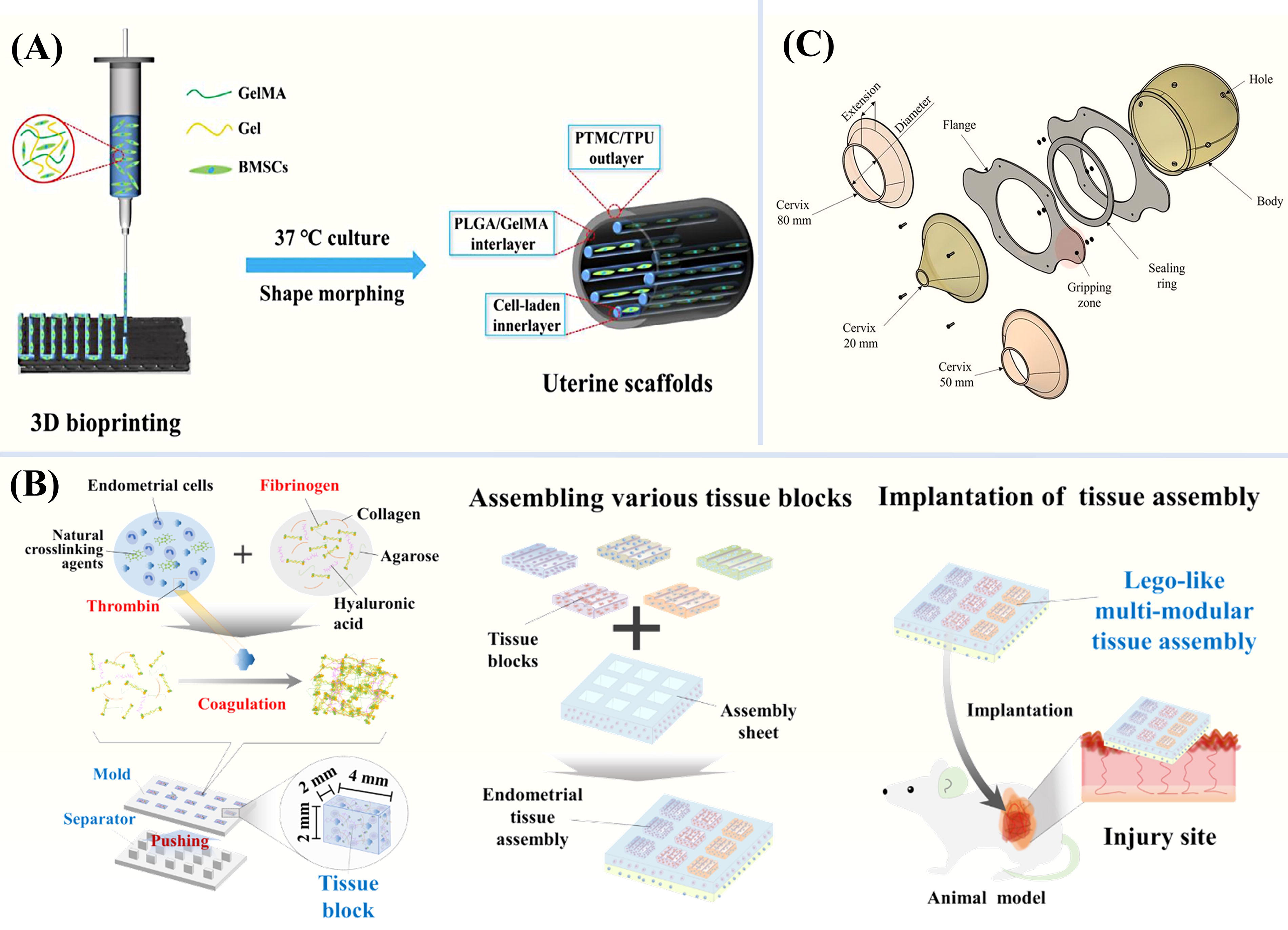
The development of a fully functional 3D-printed uterus is still in its early stages within the research community. 15 Scaffold-based tissue engineering provides an efficient approach for repairing uterine tissue defects and restoring fertility (Fig. 3A). 44 In one study, researchers utilized 3D printing to create a multimodular endometrial tissue assembly (Fig. 3B), which efficiently preserved key physiological traits of the endometrium, including responsiveness to sex hormones and reproductive hormone production. 45 When transplanted into mice, this bioengineered tissue significantly alleviated severe degenerative damage. Similarly, Park et al. developed a novel 3D-printed artificial endometrium seeded with endometrial stem cells, successfully mimicking the multicellular and multilayered structure of the native endometrium. 47 Further advancing the field, Nie and colleagues employed extrusion-based bioprinting to fabricate a bilayer endometrial construct designed to restore endometrial morphology and enhance its functionality. 29 Meanwhile, Zhao et al. developed a tissue-engineered uterus with biomimetic heterogeneous features and an ECM microenvironment. 48 Beyond endometrial regeneration, Souza et al. successfully established a 3D model of human uterine myometrial cells to investigate various pathological states impacting maternal well-being. 49 This system facilitates high-throughput assessment of various reagents and conditions and serves as a potent tool for examining human childbirth physiology. These 3D models offer a more realistic macroscale and microscale environment and providing new insights and tools for enhancing women’s reproductive health and treatment.
Simulation of the uterine with 3D-engineered models.
Severe endometrial damage often leads to infertility or pregnancy-related complications. Postpartum hemorrhage (PPH) is the predominant cause of maternal mortality in obstetrics globally. To address this, Candidori et al. presented a modular, flexible, and transparent 3D-printed uterus model (Fig. 3C), designed to evaluate medical devices aimed at preventing and treating PPH. 46 Using this 3D-printed uterine cavity model, the researchers tested the possibility of an innovative intrauterine balloon tamponade system to treat PPH. 50 Beyond PPH management, 3D printing has also improved preoperative planning for uterine fibroid surgeries. Patient-specific 3D-printed uterine models based on magnetic resonance imaging accurately depict the size, location, and distribution of fibroids, aiding in surgical decision-making and optimizing patient outcomes. In addition, 3D-bioprinted scaffolds featuring a hierarchical architecture and curved design, along with high elasticity and sustained estradiol delivery, show great potential for the regeneration of uterine tissue. 51 In the field of endometrial repair, Ji et al. confirmed that 3D-printed mesenchymal stem cells loaded hydrogel scaffold derived from human induced pluripotent stem cells may be a promising material for endometrial repair. 52 Similarly, Zhao and colleagues created a customizable cervical implant with drug-eluting capabilities, utilizing 3D-printing technology for implantation purposes. 53 Tests that these 3D-printed cervical implant models inhibit the isolated virus near the cervix. Cervical cancer accounts for a significant proportion of cancer-related deaths among women worldwide. 54 To advance in vitro cervical tumor modeling, Zhao et al. described a technique for fabricating an in vitro cervical tumor model through 3D printing of Hela cells embedded within a gelatin/alginate/fibrinogen hydrogel, providing a more physiologically relevant platform for uterine cancer research. 55 Collectively, 3D-printed models are revolutionizing the study of uterine diseases, enhancing disease comprehension, improving diagnostic accuracy, and facilitating the development of more effective, patient-specific treatment strategies.
From fallopian tube structure to 3D-engineered models of fallopian tube
The fallopian tube is essential for fertilization and embryo transport in the female reproductive system. It consists of four sections: the fimbriae, which capture the egg; the ampulla, the primary site of fertilization; the isthmus, guiding the fertilized egg toward the uterus; and the interstitial segment, ensuring smooth embryo transition into the uterine cavity. Its curved, folded 3D structure enhances efficiency in capturing and transporting gametes. At the microscopic level, the tube comprises three layers. The mucosal layer contains ciliated epithelial cells, which propel eggs and embryos, and secretory cells, which provide nutrients. The muscularis layer, with circular and longitudinal smooth muscles, generates peristaltic contractions for precise transport. The outer serosal layer, made of connective tissue, offers structural support and protection. Tubal function is regulated by hormonal signals, particularly estrogen and progesterone, which modulate ciliary motion and muscular contractions. Advances in tissue engineering are now enabling the development of bioengineered fallopian tube models.
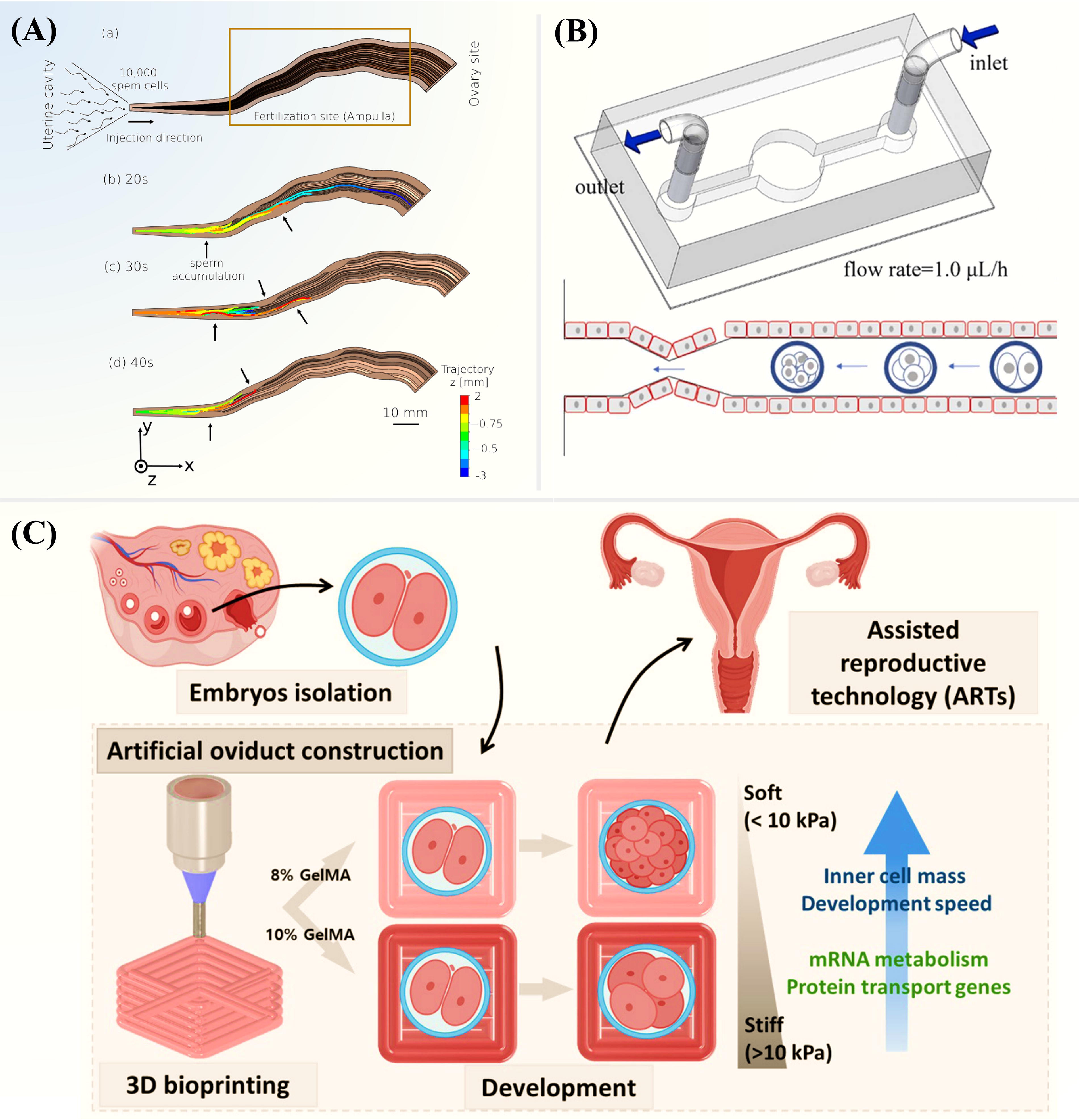
3D models of the fallopian tube are invaluable in reproductive research, helping to study interactions between eggs and sperm (Fig. 4A). 56 Researchers have proposed the development of a biomimetic 3D fallopian tube culture system to improve in vitro models for reproductive research. 59 A 3D-printed microchannel model, incorporating primary oviduct epithelial cells, was designed to create an embryo culture environment that closely replicates in vivo conditions (Fig. 4B). 57 The engineered systems offer a more physiologically relevant platform for studying fertilization, early embryo development, and reproductive disorders. To further enhance the accuracy of fallopian tube models, Ferraz et al. introduced the “fallopian tube on a chip” using 3D-printing technology, which successfully mimicked the bovine fallopian tube in vitro. 60 This microfluidic system enables the study of dynamic interactions between gametes and embryos, improving the understanding of sperm-egg transport, embryo implantation, and tubal pathologies such as ectopic pregnancy. However, the lack of a folded tubular geometry in conventional culture models has remained a major challenge. To address this, researchers have combined Transwell cell culture systems with 3D-printing techniques, effectively replicating the native architecture and microenvironment of fallopian tube epithelial cells. 61 The tubular arrangement of polarized and differentiated oviduct epithelial cells allows for controlled fluid perfusion, real-time imaging, and functional studies of ciliary activity, making them highly suitable for assisted reproductive technologies and fertility research. Recent advances in 3D-bioprinting materials have further improved the biofabrication of fallopian tube models. Koo et al. used GelMA as the material and constructed a hydrogel Petri dish simulating the tubular structure of fallopian tubes through 3D-bioprinting technology (Fig. 4C). 58 This study systematically evaluated the mechanical properties of GelMA hydrogels with varying stiffness, identifying 10 kPa hydrogels as the optimal condition for promoting embryo development, blastocyst formation, and hatching efficiency. As 3D-bioprinting continues to evolve, integrating patient-specific stem cells, ECM components, and bioactive factors into these models could open new avenues for treating tubal infertility, understanding fallopian tube disorders, and advancing reproductive medicine.
Simulation of the fallopian tube with 3D-engineered models
The intricate structure of the fallopian tube plays a crucial role in oocyte transport, fertilization, and early embryo development, and its recovery after injury is challenging. Advances in 3D-printing technology enable the fabrication of biomimetic scaffolds and engineered tissues that closely resemble the native fallopian tube structure, thereby facilitating functional restoration. 62 However, the application of additive manufacturing in tubal disease treatment remains in its early stages. One promising approach involves the use of high-precision 3D printing to create microchannel structures that function as biological catheters, potentially reestablishing tubal patency in vitro. 63 Due to individual variations, patients may exhibit different pathological characteristics. To address such conditions, personalized scaffolds can be developed using 3D printing, integrating patient-specific medical imaging data with the patient’s own cells. These bioengineered constructs are designed to meet the patient’s physiological needs while minimizing the risk of immune rejection following transplantation. Additionally, 3D-printed models of tubal diseases can simulate the microscale environment of fallopian tubes, which serve as valuable tools for drug screening and optimizing treatment strategies.
From ovary structure to 3D-engineered models of ovary
The ovary is a dynamic organ with a distinct structural composition, featuring a collagen-rich, firmer cortex and a more flexible medullary layer. 64 The outer cortex houses ovarian follicles at various developmental stages—from primordial to mature follicles—each playing a crucial role in hormone production, including estrogen and progesterone. The inner medulla, composed of connective tissue, blood vessels, nerves, and lymphatics, supplies nourishment and support to the ovary. Ovarian follicles, including primordial, primary, secondary, and mature follicles, release eggs during ovulation. The outermost layer, the surface epithelium, consists of a single layer of cuboidal or columnar epithelial cells, offering protection and contributing to ovarian physiology. The ovary primarily secretes estrogen, progesterone, and small amounts of androgens, all of which are vital for female reproductive health and endocrine regulation (Fig. 5A).
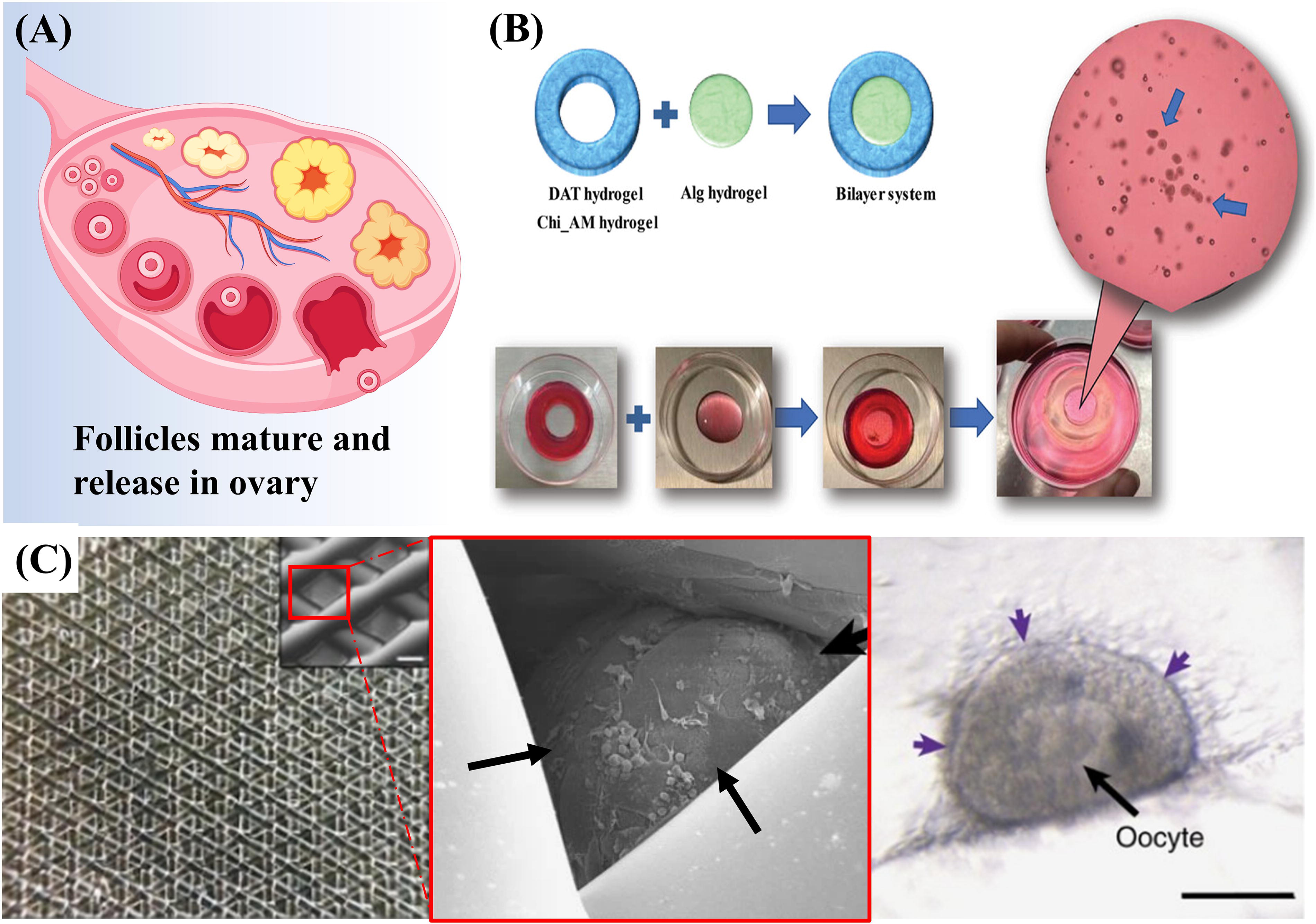
Simulation of the ovary with 3D-engineered models
Simulating the ovary in vitro is crucial for understanding its role in reproduction, health, and fertility. 66 Researchers have developed a biomimetic 3D model using a bilayer hydrogel system, incorporating hydrogels with tunable mechanical properties to better mimic the native oocyte microenvironment and enhance in vitro fertilization efficiency (Fig. 5B). 65 To identify the fibrin composition that most accurately replicates the ovarian microenvironment, a fibrin-based artificial ovary prototype has been developed. 67 In addition, a GelMA-based 3D-printing culture system has emerged as a promising platform for follicle growth, maturation, and transplantation. 68 While this scaffold demonstrates excellent shape fidelity and hygroscopicity, it remains unsuitable for creating 3D-printed structures with primary ovarian cells using extrusion-based bioprinting technology. To investigate the viability and growth of human preantral follicles in vitro, researchers have designed an alginate-encapsulation culture system. 69 Furthermore, modifying the density and elasticity of collagen hydrogels is critical for maintaining follicular phenotype and function in 3D microscale environments. 70 By perfusion of blood into microchannels, hormones can be released into the systemic circulation from vascularized hydrogel with ovarian hormones, and the endocrine function can be promoted, which indicates that 3D printing has played an unprecedented role in replacing ovarian function. 71 The emergence of 3D-printed ovaries may open new avenues for the transplantation of in vitro-grown ovarian follicles, offering hope for patients with infertility or ovarian insufficiency.
Three dimensional-printed artificial ovary may restore ovarian function and protect the fertility of young women who have undergone oophorectomy or have ovarian dysfunction. In pursuit of this objective, Laronda et al. employed 3D-printed human ovarian tissue to foster follicular development and reestablish ovarian function after ovariectomy. 72 They demonstrated that 3D-printed follicular seed scaffolds, upon implantation in sterilized mice, evolved into highly vascularized structures and effectively restored the mice’s ovarian function. Most notably, they successfully mated naturally and gave birth to healthy offspring (Fig. 5C), indicating hormone recovery of follicular hormone function. 26 The 3D-bioprinted engineered ovary showed significant potential in reviving compromised ovarian function in rats with premature ovarian failure, potentially mitigating some of the drawbacks of drug-free in vitro activation. 73 Zheng et al. fabricated a 3D porous cylindrical ovarian structure utilizing a biological ink derived from the pig ovaries' dECM to encapsulate primary ovarian cells, thereby addressing ovarian dysfunction in mice. 74 This study indicated that the group treated with the 3D model incorporating primary ovarian cells exhibits superior outcomes in terms of angiogenesis, cell proliferation, and cell survival, as compared to the group that solely utilizes the 3D model. Baka et al. reported that 3D-printed hydrogels, composed of sodium alginate and GelMA and containing ovarian follicles, can serve dual purposes: as an alternative therapy for infertility and as models for drug screening in ovarian cancer. 75 For the advancement of more potent therapeutic strategies, establishing dependable models that accurately reflect the intricacies of the ovarian disease microenvironment is essential.
Outlook and Challenge
Research on the female reproductive system and its repair must establish a comprehensive, multidimensional technological framework that integrates immune regulation, dynamic biomimetic systems, gene-material codesign, and real-time monitoring alongside existing research priorities. Functional biomaterials and targeted delivery systems can modulate immune responses to support tissue repair, while physiologically responsive smart materials and organoid platforms can mimic native tissue dynamics.76,77 Incorporating gene editing technologies can enhance key cell functionality and material compatibility, while advanced imaging and sensing technologies enable dynamic tracking and optimization of repair processes.78,79 These approaches focus not only on technical advancements (e.g., gene-editing targets and material response mechanisms) but also emphasize a closed-loop clinical translation process—from material implantation to long-term functional monitoring. Through interdisciplinary collaboration in immunology, synthetic biology, and biosensing, along with technological innovations such as dynamic materials and organ-on-a-chip systems, this holistic approach aims to provide systematic solutions for the repair of the female reproductive system, accelerating the transition from foundational research to clinical applications.
In the realm of tissue engineering, properties such as tissue stiffness, elasticity, viscoelasticity, and porosity are paramount. 80 3D models offer greater reproducibility and standardization compared to traditional 2D cultures, leading to more reliable and translatable research outcomes. The integration of 3D tissue engineering into female reproductive system research represents a monumental advancement in both basic science and clinical applications. These innovations have not only revolutionized how we model reproductive tissues but have also provided invaluable tools for understanding complex reproductive processes, studying diseases, and developing more personalized and effective treatments for women. Throughout Table 1, we have seen how 3D-engineered tissues are applied to different areas of female reproductive research. These models allow clinicians to test how different therapies will work on an individual patient cells, leading to more tailored and effective treatment plans. The development of new materials and technologies helps to restore the fertility of female reproductive system. Xin et al. have created a collagen scaffold populated with mesenchymal stem cells derived from human umbilical cords, which can be utilized for the regeneration of the endometrium. 81 In addition, the exosomes derived from mesenchymal stem cells were used to study the mechanism of endometrial regeneration through macrophage immunomodulation, which proved the novel feasibility of a cell-free treatment strategy. 82 In vitro follicular growth also has great potential, and Khunmanee et al. provided an alternative method to preserve fertility. 83 In addition, soft hydrogels derived from ECM effectively enhanced in vitro follicular culture outcomes, maintaining follicular morphology and growth, and fostering oocyte maturation. 84 In vivo bioprinting enables the direct fabrication of 3D structures within living organisms, facilitating the repair or reconstruction of tissues. Zhao et al. introduced a uniquely engineered gel rivet for underwater hydrogel patch printing, addressing the issue of premature membrane rupture, which refers to the amniotic sac breaking before childbirth. 85 Furthermore, the study indicates that the engineered PEG-based hydrogel is capable of facilitating the functionality and repair of artificially created mouse ovarian tissue. 86 These patches replicate the mechanical properties of natural tissues, exhibit robust tissue adhesion, and can efficiently seal ruptures to extend the duration of pregnancy.
Commonly Used Biomaterials for Reconstructing 3D Female Reproductive System
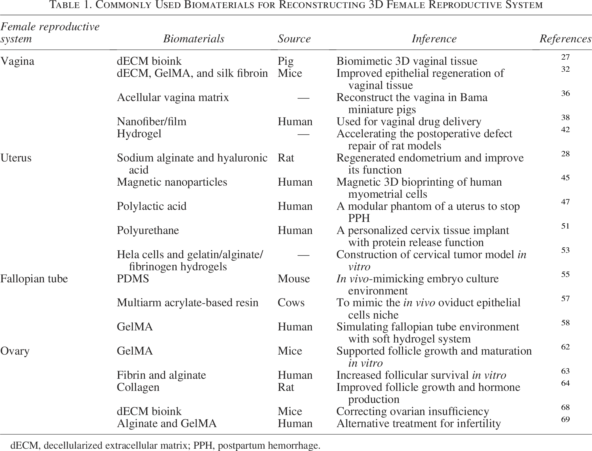
dECM, decellularized extracellular matrix; PPH, postpartum hemorrhage.
One of the primary challenges in 3D tissue engineering is scalability, creating large-scale, functional tissues that maintain consistency across experiments and clinical applications. While researchers have developed small-scale models of reproductive tissues, scaling these to construct larger structures, such as full-sized ovaries or uteri, presents significant obstacles. 87 A major challenge in constructing full-size tissue is replicating the vascular network, which is crucial for delivering nutrients and oxygen. In smaller constructs, diffusion can provide sufficient support, but as tissue size increases, this method becomes inadequate. Without a functional vascular system, larger tissues struggle to survive long-term and maintain functionality. To address this challenge, researchers are exploring several strategies.88,89 These include incorporating preformed vascular networks into tissue scaffolds and utilizing bioprinting to create intricate vascular structures. Advances in biofabrication, where vascular cells are printed alongside other tissue components, show promise but still represent a bottleneck in scaling 3D models for clinical use. Another significant challenge is creating functional tissues that accurately mimic the complexity of human reproductive organs. While progress has been made with simpler tissues such as ovarian follicles and endometrial layers,58,69 replicating the intricate structures and interactions of entire organs, such as the uterus or fallopian tubes, is far more complex. These organs comprise multiple cell types that interact and respond to hormonal signals. For example, the uterus undergoes dynamic changes during the menstrual cycle driven by this cellular interplay, 90 making it essential to recreate this diversity in vitro. 91 Finally, translating 3D-bioprinted tissues from the laboratory to clinical applications remains a challenge. The regulatory pathways for approving 3D tissue models are still developing, with ongoing challenges in establishing safety and efficacy standards necessary for personalized medicine and regenerative therapies. In addition, the production of patient-specific 3D tissues remains costly and time-intensive, requiring substantial advancements in cost efficiency and scalability to facilitate broader clinical adoption. Surfacing the hurdles in enhancing organ-level functional 3D tissue constructs, both in vitro and in vivo, will eventually lead to a lifesaving advancement in the biomedical domain.
Conclusion
Recent progress in tissue engineering has resulted in the creation of reliable tissue models that aim to emulate the intricate architecture of human tissues. Future research should aim to use complementary methods, ensure stability, and develop consistent protocols to improve therapy evaluation and create better predictive in vitro models for women’s health. The vision for the future of 3D tissue engineering in reproductive research is a world where complex reproductive diseases can be modeled with precision, where personalized treatments are the norm, and where even the most challenging fertility issues can be overcome with bioengineered tissues. In the coming years, we can expect to see more personalized and effective treatments for female reproductive health issues, greater success in fertility preservation, and potentially the creation of bioengineered reproductive organs for clinical use. These advancements promise to improve the quality of life for millions of women around the world, offering new hope for those who face infertility, cancer, or chronic reproductive health conditions. In conclusion, 3D tissue engineering represents a paradigm shift in reproductive health research, with applications that span from basic science to clinical therapies. Its integration with personalized medicine, advanced bioprinting, and gene editing technologies will continue to revolutionize the field, making the future of female reproductive health more promising than ever before.
Authors’ Contributions
Z.D.: Writing—original draft (lead); investigation (lead); writing—review and editing (equal). P.F.: Investigation (equal); writing—original draft (equal). L.Y.: Conceptualization (lead); writing—review and editing (lead). J.H.: Conceptualization (equal); writing—review and editing (equal). X.D.: Writing—original draft (equal). Yaohui W.: Writing—original draft (equal). Yujie W.: Writing—original draft (equal). Yulong W.: Writing—original draft (equal). L.L.: Writing—original draft (equal).
Footnotes
Author Disclosure Statement
The authors declare they have no competing interests.
Funding Information
This work was supported by the National Natural Science Foundation of China (No. 81973596) and the Science and Technology Research Program of Henan Province (No. 212102310341).