
Abstract
Coaxial extrusion-based bioprinting (EBB) is an emerging technology that enables the fabrication of biomimetic tissues with precise structural and biological complexities. This three-dimensional bioprinting technique utilizes specialized concentric nozzles to facilitate the simultaneous extrusion of distinct biomaterials, enabling the fabrication of layered constructs that closely resemble native tissues. Unlike traditional extrusion-based methods, coaxial printing allows for independent control over core and shell materials. This enables multimaterial integration, and tailored microenvironments that conventional extrusion methods cannot achieve. Recent technical innovations in coaxial EBB also include improved nozzle designs and bioink formulations, which have contributed to enhanced functional mimicry of native tissues and mechanical integrity of printed constructs. Coaxial EBB has demonstrated potential in spinal cord injury repair, perfusable small-diameter vessel engineering, accurate tumor microenvironment replication for oncology research, and complex organoid systems for personalized medicine. Despite these advancements, persistent challenges in coaxial EBB include maintaining cell viability under shear stress, optimizing bioink rheology, preventing nozzle clogging, and managing regulatory considerations. Future research directions involve the development of predictive computational models and the incorporation of innovative biomaterials for dynamic functionality. Addressing these challenges would allow the full therapeutic and clinical potential of coaxial bioprinting in regenerative medicine to be achieved. This review discusses and summarizes these advancements and limitations in coaxial EBB over the last decade, with an emphasis on applications in regenerative medicine.
Impact Statement
Coaxial extrusion-based bioprinting (EBB) is an innovative technology that is transforming regenerative medicine. Its design allows for simultaneous coextrusion of two or more bioinks to create three-dimensional structures that promote tissue-like function. It has been applied in nerve conduit fabrication, tumor modeling, and organoid development. This review highlights recent technological and biological advancements of coaxial EBB and challenges like maintaining cellular viability or optimizing bioink extrusion properties. Future innovations discussed also include exploring dynamic biomaterials to achieve coaxial EBB’s transformative potential to inform future translational preclinical and clinical work.
Introduction
Bioprinting has emerged as a transformative technique in regenerative medicine by enabling the fabrication of biomimetic tissue constructs with precise spatial organization.1,2 Bioprinting is an additive manufacturing technique for fabricating constructs that can be used as in vitro models to study disease or drug action or to treat and repair tissue or organ damage in vivo. 3 The origins of this printing modality date back to 1988 when Robert J. Klebe introduced the “cytoscribing” technique to fabricate two-dimensional (2D) tissue constructs using inkjet printing and later three-dimensional (3D) tissues from stacked cell-seeded collagen sheets. 4 Broadly, bioprinting approaches can be categorized into three types, each with distinct advantages and limitations. 5 Laser-assisted bioprinting relies on photopolymerization of structures via a laser source. It is advantageous in that it is highly precise and biocompatible, yet remains limited by the cost and availability of photosensitive polymers. 6 Alternatively, jetting-based bioprinting methods selectively eject droplets of photocurable materials from nozzles to achieve high resolution with rapid deposition. 7 However, this approach requires low-viscosity inks and often produces constructs with limited mechanical strength. 8 Finally, extrusion-based bioprinting (EBB) is achieved by extruding a solid or semisolid filament through a heated nozzle and depositing it layer-by-layer onto a substrate.3,9,10 This approach is well suited for creating large, mechanically durable structures from biodegradable polymers.11,12 Furthermore, among these approaches, EBB has significantly gained popularity due to its affordability and relatively low energy demands.11,13 However, special attention should be given to material and printing parameters, as balancing structural fidelity and printability remains a challenge.
As demands for engineered tissues with greater biological and structural complexity have increased, novel strategies within EBB have gained attention.3,10 One such approach is coaxial bioprinting. First described by Gao et al. 14 in 2015, this extrusion-based technique relies on specialized concentric nozzles that are capable of simultaneously extruding two or more distinct biomaterials. These systems allow for the concomitant deposition of either hollow or bulk fibers, while facilitating sophisticated control over the mechanical and biological microenvironments of tissue constructs (Fig. 1). 15 Like traditional extrusion-based methods, printing performance depends on parameters such as nozzle gauge size, ink viscosity, and extrusion pressure. However, additional considerations include core-to-sheath diameter ratio (annular gap), pressure balance, and flow rate ratio between channels. Nozzle gauge size refers to the standardized inner diameter of each tube in the coaxial assembly, whereas the annular gap is the radial space between the outer wall of the core nozzle and the inner wall of the sheath nozzle through which the shell material flows. This gap controls the thickness of the shell layer during printing and influences shear stress, pressure drop, and required extrusion force.
Schematic of coaxial extruder.
These factors influence not only print fidelity but also cell viability, as shear stress at the nozzle walls and fluid–fluid interfaces can damage cells.16,17 In practice, the sheath often uses a lower viscosity, cell-laden bioink, while the core may use a more highly viscous sacrificial material for structural support. The rheological behavior of bioinks used in coaxial extrusion can often be described by non-Newtonian power-law models. The shear-thinning behavior of bioinks can be described through the apparent viscosity (
Coaxial EBB offers significant advantages over traditional extrusion-based methods by enabling the creation of multilayered, structurally sophisticated constructs in a single printing step. By separating the mechanically robust sheath from the biologically active core, coaxial EBB helps reduce shear-induced cell damage during extrusion. 20 The dual-layer design allows for independent optimization of the core and shell layers. Key parameters including wettability, mechanical strength, and biocompatibility can each be tailored to better replicate native tissue properties. Furthermore, the multilayered design allows for the encapsulation and delivery of cells, therapeutic agents, or signaling molecules in a spatiotemporal manner. These capabilities create tailored microenvironments that better mimic the native tissue and ultimately enhance tissue functionality and integration. 3 These capabilities are simply not achievable with traditional single-nozzle extrusion. Clinically, coaxial bioprinting shows promise across various medical specialties (Fig. 2). Recent advances include the fabrication of vascular grafts and nerve conduits that mimic native tissue morphology and function.22,23 Applications extend to organ engineering, organoid development, and tumor modeling.21,24 Emerging triaxial and quadruple coaxial techniques further enhance the ability to replicate complex tissue architectures. 25 This review aims to provide a summary of recent technological and biological advancements in coaxial EBB, focusing on emerging applications in tissue engineering and highlighting critical research gaps.
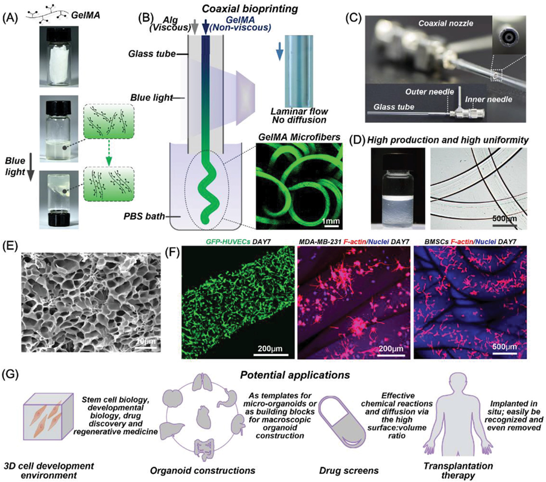
Fabrication of gelatin methacrylate (GelMA) microfibers and potential applications.
Methods
The scope of this comprehensive review encompassed a range of fields, including (1) nerve conduits, (2) vascular surgery, (3) tumor models, (4) organoids, and (5) multilayer coaxial bioprinting. A literature search was performed using the PubMed database, utilizing a combination of keywords, Medical Subject Headings, and Boolean operators to optimize the retrieval of relevant studies. Independent search queries, restricted to the last 10 years (2015–2025), were developed for each area of interest to ensure targeted coverage of the literature. All search queries incorporated the core terms “coaxial,” “core-shell bioprinting,” and “coaxial extrusion.” The search strategies for all the fields were collectively reviewed by members of the review team (I.D.G., T.J., S.E.M., M.S.T., and V.V.N.). Articles were included if they were peer-reviewed full-text studies in English that focused on coaxial extrusion for clinical or regenerative medicine. Select references from identified articles were cross-referenced, and if they met the inclusion criteria, they were manually added to the final dataset. Articles were excluded for the following reasons: (1) did not involve coaxial bioprinting as the primary fabrication approach; (2) lacked biological or clinical relevance in fields of interest; (3) abstract or letter to the editor; or (4) non-English study. Four independent reviewers (I.D.G., T.J., S.E.M., and M.S.T.) conducted the screening process. Any disagreements were resolved through discussion, and if needed, a fifth reviewer (V.V.N.) was consulted to reach consensus.
Results
Screening and selection
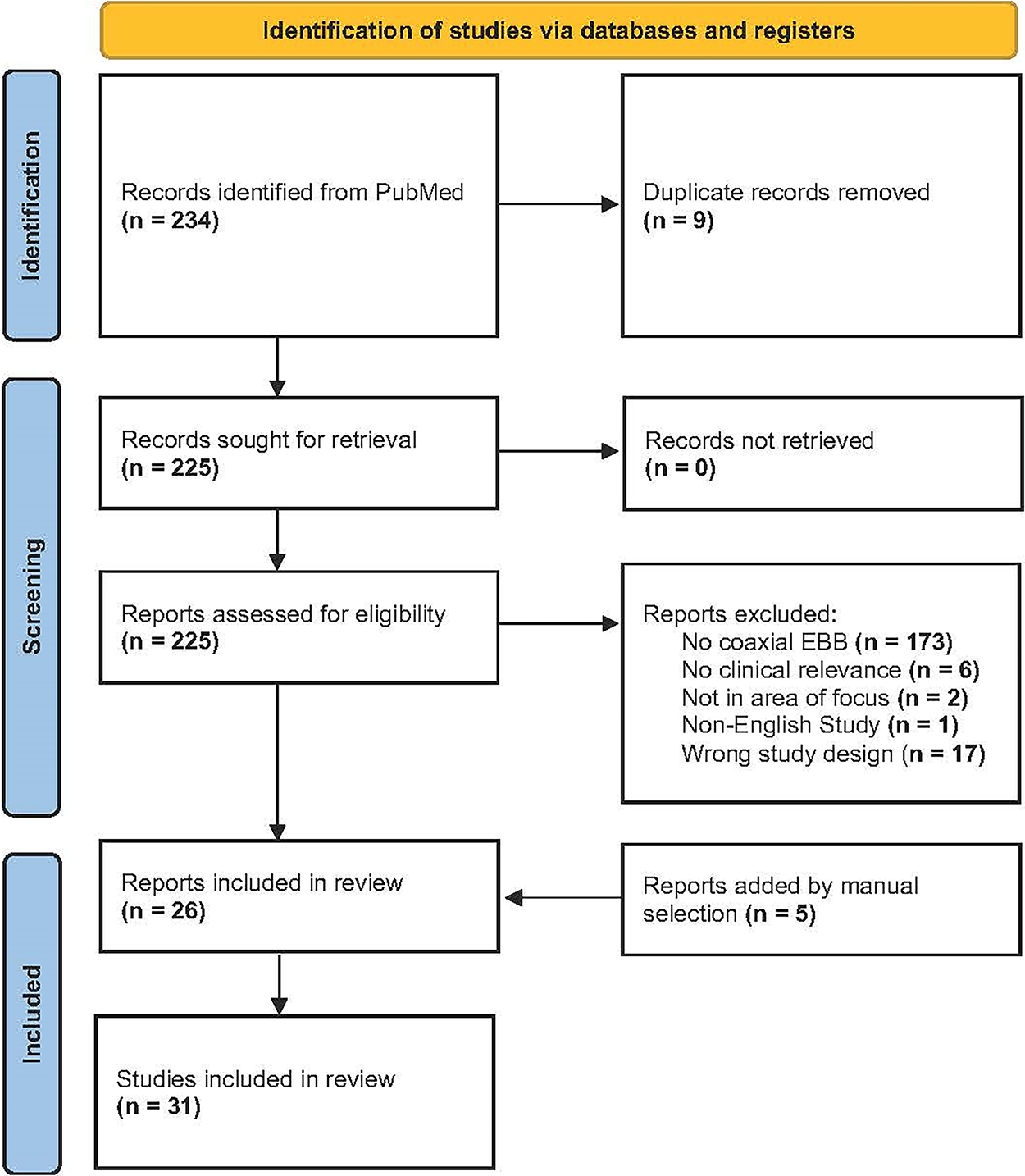
Our initial search identified 234 articles (Fig. 3). After the removal of 9 duplicates, 225 articles underwent full-text review and 199 articles were excluded. Cross-referencing of select articles led to the manual inclusion of 5 articles. Ultimately, 31 studies were deemed eligible and included (Table 1).
Flow diagram for screening and selection of studies.
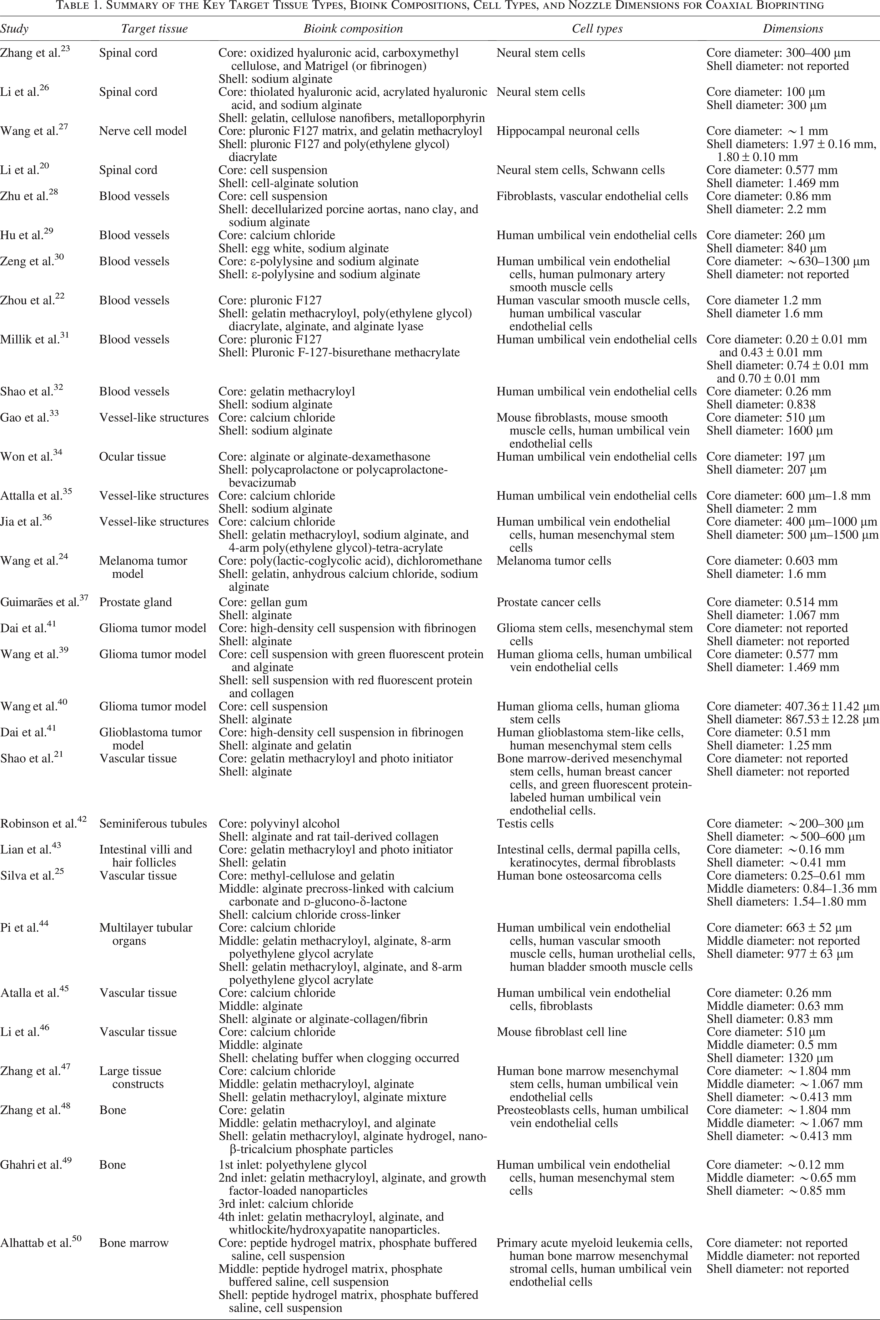
Summary of the Key Target Tissue Types, Bioink Compositions, Cell Types, and Nozzle Dimensions for Coaxial Bioprinting
Nerve conduits
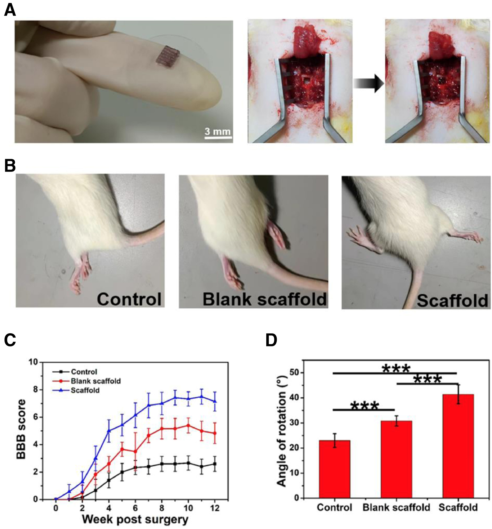
Coaxial EBB has emerged as a promising technique for the treatment of spinal cord injuries (SCIs) by enabling the fabrication of nerve conduits that integrate multiple cell types and bioactive materials. This approach allows for structured, multifunctional scaffolds with precise spatial organizations. For example, Li et al. seeded Schwann cells on an alginate shell and fabricated a hydrogel core containing neural stem cells (NSCs). This enhanced neurotrophic signaling, promoted NSC proliferation and differentiation, and supported axonal regeneration. 20 Additionally, coaxial constructs can also incorporate sacrificial cores that are removed postprinting to form perfusable, vascular-like channels, improving nutrient diffusion and cellular viability within dense neural models. 27 Beyond structural support, coaxial scaffolds have been engineered to deliver biochemical cues that mitigate the hostile postinjury environment.23,26 For example, one study demonstrated that outer layers containing superoxide scavengers reduced oxidative stress, while inner cores functionalized with cell surface proteins enhanced NSC differentiation. 26 Together, these contributed to improved motor function recovery in vivo (Fig. 4). Similarly, constructs utilizing sodium alginate, oxidized hyaluronic acid, and carboxymethyl chitosan have demonstrated the ability to reduce inflammatory markers, increase NSC differentiation, and promote neuroprotection in SCI models. 23 These studies collectively highlight the versatility of coaxial EBB in combining mechanical integrity, spatial cell patterning, and biochemical modulation to create regenerative microenvironments. While current designs show significant promise in neural repair and functional recovery, challenges remain in ensuring long-term scaffold integration, axonal extension, and sustained cell–cell interaction. Continued optimization of these systems may also expand their utility in pharmacological testing and disease modeling for various neurological conditions. 27
Vascular surgery
Coaxial EBB has become an increasingly valuable tool in vascular surgery, offering advanced capabilities for fabricating blood vessels and perfusable scaffolds that mimic the structure and function of native vasculature. These systems allow for the spatial separation of materials and cells, enabling the design of constructs that support endothelialization, mechanical stability, and long-term functionality. 52
Mechanical stability and biocompatibility
Achieving a balance between mechanical strength and biocompatibility is particularly important for successful application of coaxially printed vascular grafts. Bioink formulations play a central role in this aim, since natural components such as collagen and decellularized extracellular matrix (ECM) have been shown to significantly enhance cell adhesion and viability.28,31,36,53 For example, Zhu et al. combined ECM with nano clay and alginate in hybrid bioinks and demonstrated improved cell proliferation, adhesion, and biocompatibility. 28 However, these soft hydrogels often lack sufficient mechanical integrity, prompting the development of dual-crosslinking strategies. 36 Bioinks that undergo both rapid ionic and covalent crosslinking demonstrate improved shape fidelity, strength, and cell viability, while supporting early vascular maturation. 36 Additionally, to further support graft survival, perfusable microchannels have been integrated into scaffolds to enhance oxygen and nutrient exchange. 35 These internal networks have the potential to reduce the risk of necrosis in thick constructs and improve long-term functionality. 35
Small-diameter vessel challenges
Engineering small-diameter vascular grafts (<6 mm) remains challenging due to the high risk of thrombosis and lumen collapse, which are complications largely attributed to inadequate endothelialization post-transplantation. 30 Traditional microfiber approaches often lack the complexity and biocompatibility needed to support endothelial cell migration and lumen formation. 30 Thus, the improved structural complexity provided by coaxial structures can promote endothelialization. For example, Shao et al. used the liquid rope-coil effect to create gelatin methacrylate (GelMA) fibers encased in alginate shells with various morphologies that guided endothelial cells toward the fiber periphery. 32 Other approaches have focused on the delivery of bioactive molecules to promote immediate antithrombogenicity with subsequent epithelialization. 30 However, prior printing techniques faced challenges due to a lack of spatiotemporal control over the release of factors. 30 Zeng et al. aimed to combat this by utilizing coaxial printing to create a sequential release system, where charged bioinks were utilized to enable the layer-by-layer assembly of a luminal coating with heparin (for immediate antithrombogenicity) and polypeptides (for delayed endothelialization). 30 This spatiotemporally controlled release strategy enhanced both short- and long-term graft function.
Multilayered and multimaterial approaches
Native blood vessels have a multilayered organization of endothelial and smooth muscle cells that traditional fabrication methods fail to replicate. As such, multiple cell types have been incorporated to better replicate the cellular architecture of native vessels. 22 Furthermore, beyond cellular organization, reproducing the multilayered composition (intima, media, and adventitia) of vessels has driven the use of multimaterial and multilayer coaxial bioprinting. For example, Gao et al. used coaxial printing around a rotated rod to create multichannel systems featuring central macrochannels for support and peripheral microchannels for diffusion. 33 Another advanced construct incorporated electrospun polycaprolactone-collagen nanofibers with alginate and egg white hydrogel to better replicate the multilayer mechanical and cellular microenvironment of native vessels, resulting in scaffolds with enhanced durability and biocompatibility. 29 Triple-layered coaxial printing, using three separate bioinks, has also been employed to enable immediate crosslinking and organization of distinct tissue layers.25,36
Drug delivery constructs
Core-shell designs have been used to spatially control dual-drug release. For example, Won et al. developed a system that provided fast delivery of dexamethasone from a hydrogel core and sustained release of bevacizumab from a polycaprolactone shell. 34 Their design reduced inflammation and pathological angiogenesis in vivo and ultimately improved integration and remodeling of vascular grafts. 34 Such multifunctional platforms hold promise for therapeutic vascular interventions and engineered tissue integration.
Tumor models
Coaxial bioprinting has advanced tumor models, which aim to simulate the microenvironment of tumors for the investigation of tumor growth, cell-to-cell interactions, and drug therapies. Coaxial EBB provides distinct advantages over traditional 2D models, as they are more physiologically accurate and allow for better assessment of tumor behavior and treatment response. 54 Specifically, coaxially printed tumor models have shown promise for studying various malignancies, including glioblastomas, prostate cancer, and melanoma. Glioblastomas are biologically complex, malignant tumors that arise from the central nervous system and have a poor prognosis. 55 A major contributor that drives tumor progression and treatment resistance is the multicellular microenvironment. Surrounding cells and stromal elements continuously interact and influence glioblastoma cell behavior by promoting proliferation, angiogenesis, and metastasis.56,57 Hence, understanding and accurately modeling this complex microenvironment is critical for developing more effective therapeutic strategies. By using coaxial EBB, one study demonstrated that fusion between glioma stem cells and mesenchymal stem cells (MSCs) produced tumor-like traits. 38 Another study found that tumor cells can recruit blood vessels from a xenograft host, contributing to angiogenesis. 39 In glioblastoma coaxial EBB research, several studies have incorporated an alginate-based bioink to create scaffolds that mimic functions, like angiogenesis, of the tumor microenvironment (Fig. 5).38,39,57
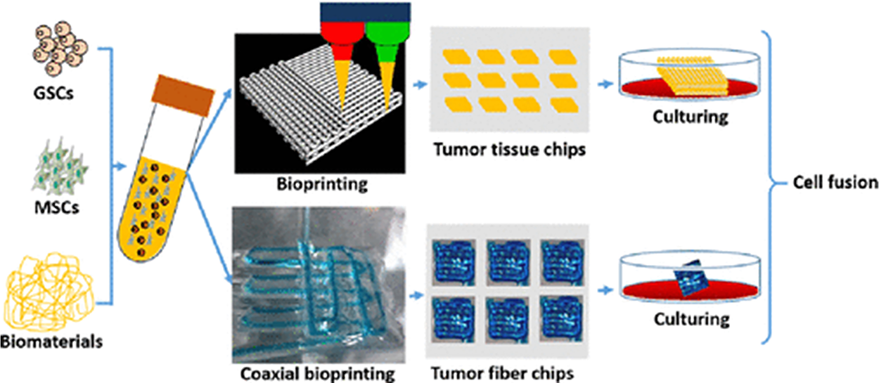
Patterns of coaxial printing and low-temperature formation of multicellular tumor models. Reprinted with permission from ACS Applied Materials & Interfaces, Vol 14, Dai et al., “Fusion between Glioma Stem Cells and Mesenchymal Stem Cells Promotes Malignant Progression in 3D-Bioprinted Models.” © 2022 American Chemical Society. 38
Beyond studying tumor behavior, coaxial bioprinting offers a platform for drug testing and studying the therapeutic effects of anticancer drugs. In another study, X. Wang et al. utilized this technology to construct microfibers containing different glioma cell lines to evaluate drug resistance and sensitivity. 40 Additionally, Guimarães et al. modeled treatment-induced neuroendocrine prostate cancer using three cell lines in a physiologically relevant environment and tested responses to talazoparib and enzalutamide. 37 They found that coaxial bioprinting of layered biomaterials effectively recreated the tumor’s concentric architecture, enabling lumen-like models for drug testing. Similarly, H. Wang et al. fabricated a scaffold loaded with an AKT inhibitor and an immune agonist, which acted as an artificial lymph node, to achieve controlled, sequential release of immunotherapy medications. 24
Organoids
Organoids are miniature, self-organized 3D structures derived from cells of various origins that are utilized in drug discovery and precision medicine. Fabricating organoids from patient-specific tissues allows for retention of the original genetic expression, mutations, and biological functions of parent cells. 58 Dai et al. fabricated a 3D in vitro tumor to evaluate tumor–stroma interactions and ECM production. 41 The authors used coaxial EBB to create heterogenous brain tumor fibers containing human glioblastoma stem-like cells or human mesenchymal stem cells (hMSCs). This resulted in high cell viability, stromal–tumor interactions, and collagen production that mimicked native tumor tissue, highlighting coaxial EBB’s potential to create multilayered constructs for complex organoid formation.21,41
Coculture models and heterogeneous constructs
Coculture models grow two different cell types together to study cell–cell communication while modeling physiological conditions; however, they often lack vascularization and scalability. Organoid models better mimic development and disease. Coaxial bioprinting addresses coculture limitations by enhancing cell compartmentalization and supporting vascular network formation, offering a more relevant platform for tumor modeling. 59 In 2019, Shao et al. utilized coaxial EBB to create bioactive, cell-laden hydrogel microfibers designed for tunable compositions, multilayered constructs, and complex coculture models. 21 They created six bioink formulations with various cell types, using GelMA as the core and sodium alginate as the shell (Fig. 6). Specific cell lines included human umbilical vein endothelial cells (HUVECs) for vascular models, breast cancer cells for tumor models, and bone mesenchymal stem cells (bMSCs) for cryopreservation studies. Coculture of HUVECs and breast cancer cells led to angiogenic sprouting toward tumor signals, demonstrating successful fabrication of functional 3D microenvironments. The authors also achieved heterogenous filament fabrication through programmable switching of hydrogels, enabling gradient and spatial patterning within fibers (Fig. 6). This work highlights coaxial bioprinting’s versatility in building functional microenvironments, advancing coculture systems, and organoid engineering. 21
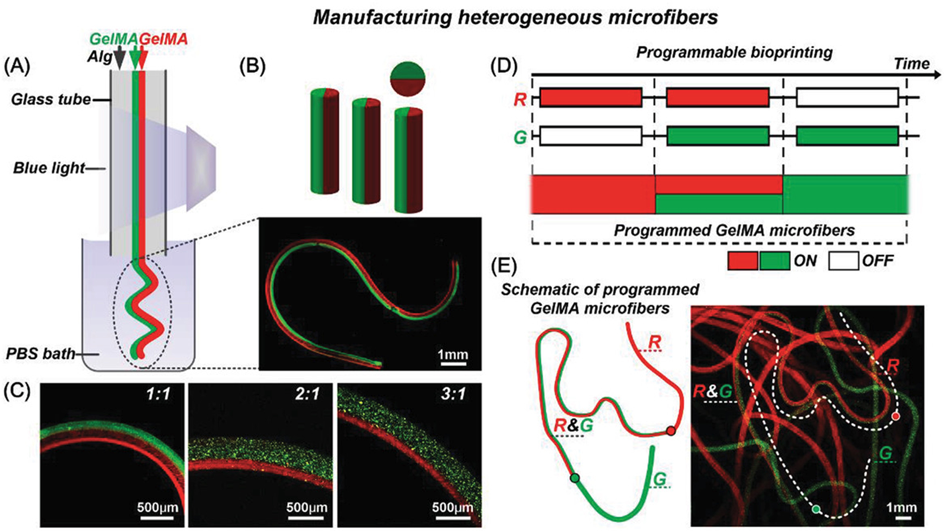
Fabrication of 3D constructs and heterogeneous GelMA microfibers.
Tubular and vertically embedded organoids
In 2022, Robinson et al. used coaxial EBB to fabricate patient-specific testicular tubules from patient-derived testis cells in a donor with nonobstructive azoospermia—a severe form of infertility caused by failed spermatogenesis.42,60 Cells were collected and then 3D bioprinted into tubular structures mimicking seminiferous tubules with a shell of alginate and collagen and a core of sacrificial polyvinyl alcohol. 42 Compared with nonbioprinted organoids, the printed constructs showed higher viability and greater upregulation of spermatogonial stem cell and spermatogenic gene expression after 12 days in culture. These findings underscore the potential of coaxial EBB for creating personalized, functional testicular structures and advancing in vitro models of spermatogenesis in both healthy and diseased states. 42 Coaxial EBB can also be utilized to form vertical structures with enhanced integrity. Constructs from conventional EBB methods struggle to maintain vertical fidelity due to gravitational collapse. To address this, Lian et al. developed a vertical embedded (VE) bioprinting strategy that created high-aspect ratio structures resembling intestinal villi and human hair follicles. 43 Using this strategy, the authors sequentially deposited human dermal papilla cells-laden GelMA and human keratinocyte-laden gelatin into a dermal fibroblast-laden GelMA base, forming hair-like constructs. Over time, these constructs exhibited morphological differentiation and increased cytokeratin expression, suggesting successful mimicry of native hair follicles. This approach shows coaxial VE’s promise in engineering linear, multicellular structures for applications in tissue regeneration and personalized medicine. 43
Multilayer bioprinting
Nozzle and flow rate innovations
Advancements in triaxial and quadruple axial nozzles have improved bioprinting capabilities. Silva et al. designed a triple-layer coaxial extruder optimized via computational fluid dynamics to minimize shear stress and preserve cell viability during extrusion (Fig. 7). 25 Using this nozzle, they printed hollow, tubular constructs mimicking vascular networks with over 80% cell viability postprinting. 25 The nozzle design facilitated precise layer control and rapid gelation of alginate bioinks upon extrusion, maintaining shape fidelity. 25 Similarly, Attalla et al. developed a scalable, polydimethylsiloxane-based triaxial microfluidic nozzle with needles of various sizes for one-step printing of three-layered hydrogel conduits. 45 Fluorescence showed distinct cell-laden layers with rising live-cell counts over 5 days and strong live-cell signals. 45 These studies show that custom nozzles can pattern perfusable, multilayered tissue constructs under cytocompatibility shear conditions, highlighting their potential in advancing multilayered tissue engineering.
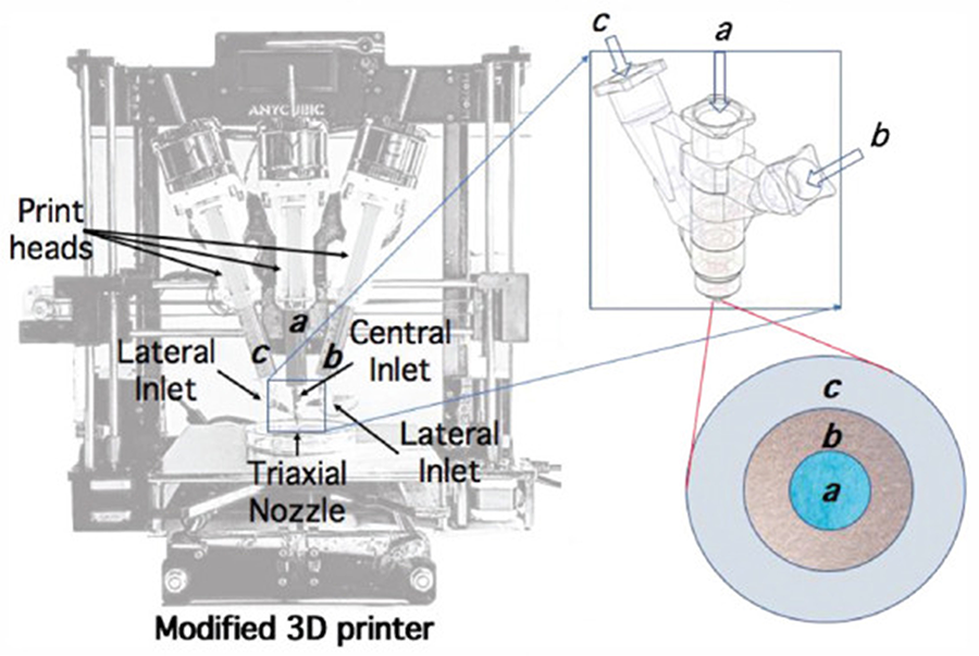
Bioprinting setup used by Silva et al. where 3 piston-driven extrusion printheads were mounted on a fused deposition modeling 3D printer and routed into a triple-channel coaxial nozzle, with each channel’s inlet and outlet clearly labeled for clarity. Reprinted under the terms of the Creative Commons Attribution—Noncommercial 4.0 License from Silva et al. (2020). Figure 2 in the article “Rational Design of a Triple-Layered Coaxial Extruder System: In Silico and In Vitro Evaluations Directed Toward Optimizing Cell Viability” in the International Journal of Bioprinting (doi: 10.18063/ijb.v6i4.282). 25
Perfusable tissue constructs
Native tissues rely on branched microvascular networks to sustain metabolism, as diffusion-driven transport alone only supports cells within ∼100–200 µm of a perfused vessel.61–63 Beyond this range, hypoxia leads to necrosis and graft failure. Traditional coaxial EBB yields straight, unbranched filaments, limiting its use in replicating complex vascular geometries. Conversely, multiaxial nozzle designs allow for the creation of three or more concentric flows to sculpt advanced lumen structures that incorporate perfusable, hierarchically branched channels.63–65 For example, Li et al. employed a triaxial nozzle to fabricate hollow fibers that were trimmed into Y-shaped branches. 46 These scaffolds supported media perfusion and fibroblast viability (∼97% at day 6), demonstrating their cytocompatibility and potential for vascular engineering. 46 Similarly, Pi et al. developed a multichannel coaxial bioprinter capable of printing single-, double-, or triple-layer tubular filaments using a tunable bioink system. 44 The printer alternated between vascular and urothelial models, controlling cell-specific layer deposition. The resulting cannular tissues were able to maintain structural integrity under perfusion. 44 Triaxial bioprinting can improve soft-tissue engineering. Zhang et al. showcased this by creating a large GelMA-based construct with built-in nutrient channels which improved biological outcomes, yielding high cell survival, proliferation, migration, and vascular network formation compared with solid controls. 47 Jia et al. further demonstrated direct printing of perfusable hollow tubes using triaxial extrusion and a ultraviolet/ionic crosslinking system without separate molding or sacrificial steps. 36
In bone tissue engineering, triaxial printing and quadruple axial printing enable simultaneous integration of vascular and mineralized compartments. Zhang et al. fabricated a vascularized bone model with an outer GelMA-alginate layer (with β-Tricalcium Phosphate (β-TCP) and preosteoblasts), a middle layer of HUVECs, and an inner gelatin layer. This resulted in high cell viability with concurrent microvessel and bone matrix formation. 48 Expanding on this, Ghahri et al. used a quadruple axial nozzle to develop an “osteon-like scaffold-cell” with four concentric layers which included a removable core (Haversian canal), a vasculogenic layer (HUVECs + vascular endothelial growth factor-loaded nanoparticles), a calcium chloride (CaCl2) crosslinking layer, and an outer osteogenic layer (hMSCs + mineralized particles) (Fig. 8). The construct achieved excellent cell viability (∼94% at 10 days) and supported prevascular tube formation and accelerated osteogenic activity. 49
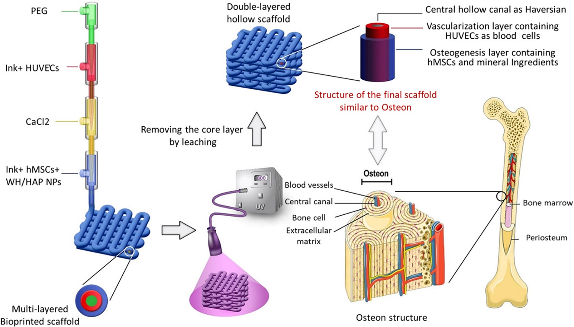
A schematic illustration showing the fabrication of a hollow scaffold-cell construct using a quadruple-layer core-shell nozzle and the comparison of the bio-printed scaffold with osteon in native bone. Reprinted with permission from Ghahri et al. (2023). Figure 1 in the article “Development of Osteon-Like Scaffold-Cell Construct by Quadruple Coaxial Extrusion-Based 3D Bioprinting of Nanocomposite Hydrogel” in Biomaterials Advances. 49
Disease-model systems
Multiaxial bioprinting further improves the accuracy of coculture models. One example is seen in the acute myeloid leukemia bone marrow niche created by Alhattab et al. using a quadruple-coaxial nozzle. 50 This system coextruded leukemic blasts, stromal, and endothelial cells into a 3D construct that mimicked the leukemia microenvironment, while increasing cell viability and chemoresistance. 50 Triaxial bioprinting and quadruple axial bioprinting have expanded regenerative medicine capabilities by replicating complex tissue architectures. However, challenges remain, including maintaining mechanical stability in thick prints and achieving functional vascularization.25,44,47 Future advances in nozzle engineering, bioink chemistry, and integrated tissue maturation are aimed at bridging the gap between printed constructs and native tissue architecture.51,66,67
Implementation into the Laboratory
Implementation of coaxial EBB into a laboratory environment requires consideration of several factors, such as equipment configuration and design principles. The printing setup must allow for precise coordination between core and shell extrusion systems, while printing parameters must be carefully tuned to achieve structural fidelity and biocompatibility. A coaxial EBB platform typically consists of a motion-controlled print bed, independent syringe pump extruders, and a specialized concentric nozzle assembly. 3 Each extrusion channel should have its own regulation system that can independently control extrusion pressures and flow rates. This is often achieved through pneumatic or mechanical systems. 3 Additional accessories such as automated bed-level sensors and heated or cooled build plates can further enhance performance.
Several commercial systems exist, yet these systems can be costly or may restrict nozzle geometry and bioink choices. 68 However, open-source and Do-It-Yourself approaches may offer a low-cost and highly adaptable alternative.69–71 The concentric nozzle is the defining component, as its geometry determines the achievable resolution as well as shear stress levels and flow profiles. Thus, existing extrusion-based bioprinters can be modified to incorporate specialized nozzle assemblies. For example, studies have demonstrated the feasibility in replacing the heating component from a traditional printer with syringe pump extruders and a coaxial nozzle.68,69 3D printing of these pieces allows for tailored design and improved machine adaptability, broadening the bioinks that can be employed. The designs for 3D printed hardware elements can be acquired from open-source licenses.68,72 Other nonprinted elements are standard hardware components that are inexpensive and easy to acquire.68,71 Finally, a plethora of open-source software is available that research labs can leverage to meet their individual needs.68,69,71,72
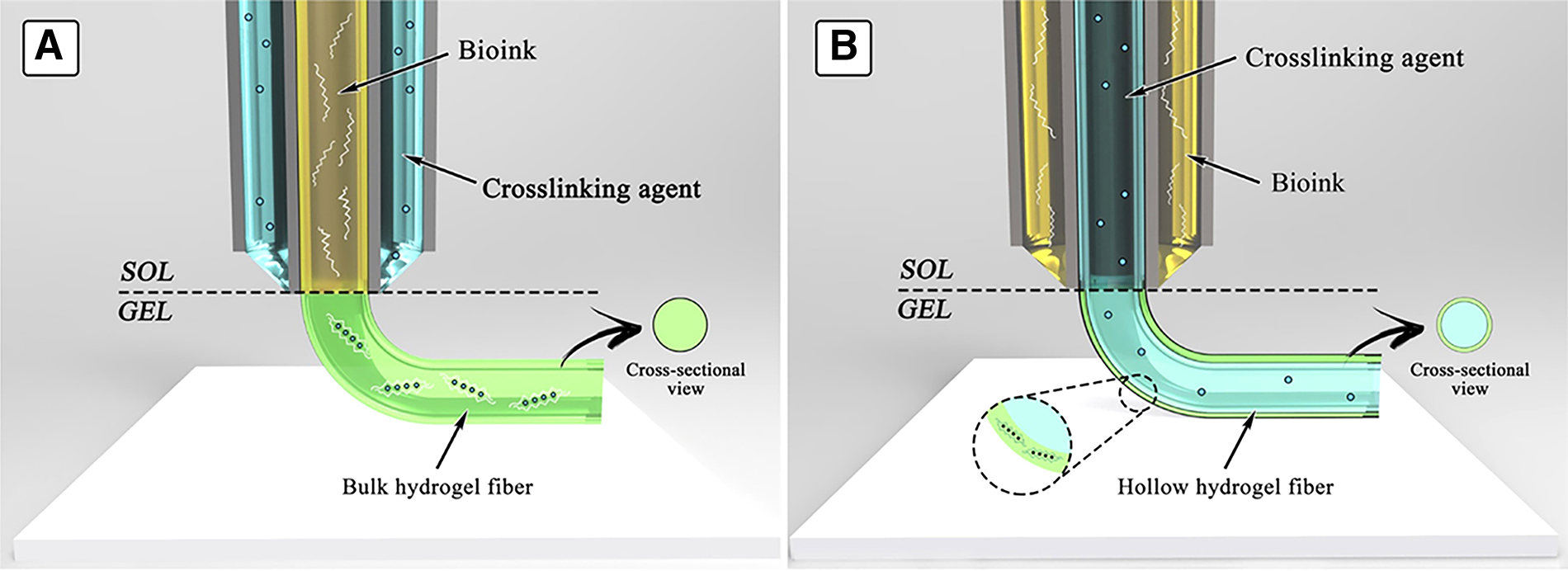
Beyond equipment configuration, successful coaxial printing requires attention to several design principles. A fundamental design consideration is the precise control of core-shell geometry. The bioinks selected must be chemically compatible yet differ in their gelation dynamics. Often, extrusion of materials directly into a crosslinking or colloidal bath allows for the decoupling of printability and structural fidelity parameters.15,73 These systems can be organized into various configurations depending on the desired construct structure. 3 For instance, the bioink may surround a core containing a crosslinking agent to produce hollow fibers. In the reverse configuration, the crosslinker encapsulates the bioink to produce bulk fibers. In addition, minimizing shear stress within the nozzle is essential for maintaining high cell viability. As such, nozzle dimensions, extrusion pressures, and flow conditions govern the resulting structural and biochemical properties, requiring careful selection for the intended application.
Unresolved Challenges and Future Directions
Coaxial bioprinting holds transformative potential for regenerative medicine, yet several challenges and opportunities remain. One of the central goals in tissue engineering is the fabrication of constructs that closely replicate the native tissue both structurally and functionally. For coaxial EBB, this means producing hollow, tube-like structures that can maintain shape fidelity, sustain cell viability, and ideally interact dynamically with the surrounding environment. Achieving this balance has proven difficult, since many of the natural materials used to promote biocompatibility often exhibit poor shape fidelity and result in shape deformation and post-printing bioink dispersion. 74 Increasing bioink viscosity can help address these challenges. However, highly viscous bioinks expose cells to greater shear stress during extrusion, which can reduce cell viability.75,76 Furthermore, viscous bioinks are prone to nozzle clogging and non-uniform flow. 77 Printing at higher extrusion pressures or with smaller nozzle diameters can help to improve resolution, yet these approaches often lead to a reduction in cell survival.17,78 Thus, an ongoing challenge in coaxial bioprinting is optimizing bioink rheological properties to balance printability, shape fidelity, and cell viability.
Advances in bioink formulation have focused on shear-thinning materials, cross-linkable hydrogels, and the incorporation of nanomaterials to overcome these hurdles.47,79–83 These approaches allow for a reduced viscosity when the flow rate is increased, allowing for improved structural fidelity while maintaining cell viability. While the benefits of these strategies are well recognized, optimal concentrations and bioink combinations are not as well established. 48 Recent efforts have focused on developing quantitative models that predict printing performance under variable conditions.84,85 One study modeled filament collapse during EBB using Euler–Bernoulli beam theory and established a strong linear correlation between Young’s modulus and filament radius. 86 They demonstrated that as filament radius decreased, material stiffness increased. Furthermore, this model enabled the identification of the optimal bioink composition that minimized filament collapse during and after deposition while maintaining printability. Thus, these predictive models have the potential to pre-screen materials and print parameters prior to fabrication to reduce trial-and-error optimization.
Despite these advancements, the applicability of some modeling approaches for coaxial EBB remains limited by several factors. The assumption of homogeneous, isotropic material properties does not accurately reflect the core-shell structures of coaxial bioprints. 87 Furthermore, dynamic crosslinking kinetics may not be incorporated despite their dramatic influence on mechanical properties. 86 Environmental factors such as humidity and temperature can alter material properties and deformation behavior yet may not be incorporated into models. 85 Addressing these limitations could significantly improve predictive accuracy and printing outcomes for clinical applications. In the future, integration of models with machine learning-based feedback systems could offer improved analysis of bioprinting parameters, allowing for more reproducibility of constructs. 88
Another area for improving shape fidelity lies in using smart materials which can respond to environmental stimuli over time. A subset of these are shape memory materials, which are characterized by their ability to “remember” and recover their original shape after deformation in response to certain stimuli. 89 For example, luminal stents with dual-phase structures have been designed to remain compact during implantation but later expand to anchor in surrounding tissues.90–92 Although the application of shape memory materials in coaxial bioprinting remains largely unexplored, the potential benefits of combining these technologies are substantial. Other smart materials may enable functional responses rather than purely structural ones. For example, various nanoparticles can serve as smart delivery materials, as they can release bioactive substances in response to various stimuli. 93 Coaxially printed vascular constructs have demonstrated this controlled, spatiotemporal release of bioactive molecules. 93 This shift toward dynamic bio constructs represents a step forward in replicating the functional complexity of native tissues.
Advancements in machine design are also essential to the evolution of coaxial EBB. 94 As translational needs grow, scalable production of larger, clinically relevant constructs is crucial. Since scaling up construct size prolongs fabrication time, integrating coaxial bioprinting with complementary manufacturing strategies may help streamline production while achieving structured tissue scaffolds with macro- and microscale features. Use of multinozzle extrusion systems enables more complex scaffold fabrication, and further innovations in nozzle design can improve spatial precision. 95 For example, stereolithography can be used to 3D print unique coaxial nozzles.31,35 However, increased nozzle complexity can compromise flow consistency and resolution across channels. 95
Finally, regulatory considerations for 3D bioprinting are becoming increasingly important. However, there is still uncertainty about how 3D bioprinted tissues and organs will be regulated. 96 In hopes of balancing safety and innovation, a process-based approach has been proposed. 97 Rather than creating an entirely new regulatory framework, this concept emphasizes regulation of the manufacturing process itself rather than only the final product. Overall, establishing widely accepted testing protocols will be crucial for moving coaxial bioprinting from the lab to clinical use. 97
Conclusion
This review examined emerging clinical applications of coaxial EBB in regenerative medicine. By utilizing specialized concentric nozzle systems, coaxial bioprinting facilitates the simultaneous extrusion of distinct biomaterials. By allowing for the incorporation and independent control of multiple layers, coaxial extrusion provides a level of structural and functional complexity that is not possible with traditional single-nozzle extrusion. This technique enables precise spatial organization of printed structures, mimicking the morphologies of native tissues. Recent advances in bioprinting include nerve conduits, vascular grafts, tumor models, organoids, and disease model systems. Future directions of coaxial EBB include addressing shape fidelity, enhancing printing resolution, and overcoming issues related to shear stress and nozzle clogging. Addressing these areas is crucial to bridging the current gaps, enhancing construct functionality, and realizing the full translational potential of coaxial bioprinting in clinical practice.
Authors’ Contributions
Conceptualization: L.W., V.V.N., and P.G.C.; Methodology: I.D.G., T.J., S.E.M., M.S.T., and H.S.; Software: I.D.G., T.J., S.E.M., M.S.T., and H.S.; Investigation: I.D.G., T.J., S.E.M., M.S.T., H.S., and V.V.N.; Resources: K.T., L.W., V.V.N., and P.G.C.; Data curation: I.D.G., T.J., S.E.M., M.S.T., and H.S.; Writing—original draft preparation: I.D.G., T.J., S.E.M., M.S.T., and H.S.; Writing—review and editing: K.T., L.W., V.V.N., and P.G.C.; Supervision: K.T., L.W., V.V.N., and P.G.C.; Project administration: K.T., L.W., V.V.N., and P.G.C. All authors have read and agreed to the published version of the article.
Footnotes
Author Disclosure Statement
The authors declare no conflict of interest.
Funding Information
This research received no external funding.