
Abstract
In bone regenerative medicine, scaffolds play a pivotal role in promoting cell adhesion, proliferation, and differentiation. Among the various materials employed for creating scaffolds, mixed materials composed of poly-
Impact Statement
These findings point to poly-L-lactic acid/hydroxyapatite (PLLA/HA) materials as alternatives to traditional titanium alloy for bone reconstruction, aiming to promote cell ingrowth. The biocompatible PLLA/HA composite matches the target tissue’s mechanical stability, reducing scaffold degradation mismatch and minimizing inflammation at the implantation site. Using PLLA/HA scaffolds with optimized pore sizes (e.g., 400 µm) may create a balanced flow field—supporting fluid shear force, nutrient delivery, and metabolite excretion. These aids seed cell survival, enhance cell interactions, and guide tissue-specific differentiation. These synergies could improve functional outcomes and patient recovery, offering insights for advanced orthopedic and regenerative biomaterials.
Introduction
Poly-
Hydroxyapatite (HA) is a naturally occurring calcium phosphate mineral. Its remarkable similarity to biological apatite endows it with exceptional biocompatibility, making it one of the most widely studied and utilized materials for bone tissue regeneration. The unique structural and chemical properties of HA allow it to integrate well with biological tissues, promoting bone growth and repair while minimizing adverse immune responses. The crystal structure of HA belongs to the hexagonal system, featuring a complex arrangement of calcium, phosphate, and hydroxyl ions. This structure is highly adaptable, permitting various ionic substitutions that can enhance its mechanical and biological properties. For instance, carbonate ions can replace phosphate or hydroxyl groups, increasing solubility and mimicking the composition of natural bone mineral. 11 Similarly, the incorporation of strontium, magnesium, or zinc ions can improve osteogenic activity and antibacterial performance, while fluoride substitution enhances acid resistance. 12 Biologically, HA excels owing to its osteoconductive and bioactive properties. When implanted in bone defects, it acts as a scaffold, facilitating the attachment and proliferation of osteoblasts and mesenchymal stem cells. 13 Over time, a carbonated apatite layer forms on its surface, strengthening the bond between the implant and surrounding bone tissue. 14 This biointegration is crucial for bone graft applications. Additionally, HA’s ability to dissolve and release calcium and phosphate ions supports natural bone remodeling processes, further enhancing its regenerative potential. 15 Furthermore, HA has emerged as a promising carrier for drug and gene delivery. Its high surface area and ion-exchange capacity allow it to adsorb and release therapeutic agents in a controlled manner. Antibiotics, anticancer drugs, and growth factors can be loaded onto HA nanoparticles, enabling the targeted treatment of bone infections, tumors, and degenerative diseases. 16
The combination of PLLA and HA forms a highly advantageous composite material that synergizes the beneficial properties of both components for biomedical applications. PLLA, a biodegradable polyester derived from renewable resources, contributes excellent processability and controlled degradation kinetics, while HA, as the primary mineral constituent of bone, provides outstanding osteoconductivity and bioactivity. When blended together, these materials could form a composite that overcomes the individual limitations of each component. The PLLA matrix enhances the mechanical flexibility and fracture toughness of the otherwise brittle HA, allowing the creation of scaffolds with improved load-bearing capabilities. 17 Simultaneously, the incorporation of HA particles into the PLLA polymer significantly improves the composite’s bioactivity by promoting cell adhesion, proliferation, and differentiation. 18 This combination also modulates the degradation profile of the material, as the presence of HA can buffer the acidic byproducts generated during PLLA degradation, thereby reducing potential inflammatory responses. 19 The composite’s surface properties can be precisely tuned by adjusting the HA content, enabling optimal protein adsorption and subsequent cellular interactions. 20 These characteristics make PLLA/HA composites particularly valuable for various orthopedic and dental applications, such as bone tissue engineering scaffolds.
Bone tissue engineering aims to repair or replace damaged bone tissues through the combination of cells, scaffolds, and signaling molecules. PLLA/HA scaffolds could serve as temporary templates that provide structural support and guidance for tissue regeneration. They are engineered to mimic the extracellular microstructure of native tissues, facilitating cell attachment, proliferation, and differentiation.21–23 Among the myriad scaffold properties, pore size is a critical determinant of cell behavior. 24 Cell attachment to scaffolds is the initial step. Cells adhere to scaffolds through specific interactions between cell surface receptors and ECM proteins present on the scaffold surface. 25 The pore size of scaffolds influences the accessibility of these adhesion sites, thereby affecting cell attachment efficiency. Small pores may restrict cell penetration and adhesion, whereas larger pores allow for better cell infiltration and interaction with the scaffold surface. 26 However, excessively large pores may result in loosely attached cells.27,28 Cell proliferation within scaffolds is crucial. 28 The pore size of scaffolds can impact cell proliferation by modulating nutrient and oxygen diffusion, waste removal, and cell–cell interactions. 24 Scaffolds with optimal pore sizes facilitate the efficient transport of nutrients and oxygen to cells, supporting their growth and proliferation. Conversely, scaffolds with too small or too large pores may impair cell proliferation due to insufficient nutrient supply or limited cell–cell contact. 29 Cell migration on scaffolds is essential, as it allows cells to populate the entire scaffold. The pore size of scaffolds plays a pivotal role in regulating cell migration. Small pores may hinder cell movement, while larger pores provide channels for cell migration. 30 Cell differentiation is the process by which cells acquire specialized functions and characteristics. 31 Scaffold pore size can influence cell differentiation by providing cues that guide cellular fate decisions. 32 Scaffolds with specific pore sizes may mimic the microenvironments of native tissues, promoting the differentiation of stem cells into desired cell types. 28 Additionally, pore size can affect the microenvironment hydromechanics of scaffolds, which in turn regulate cell differentiation through mechanotransduction pathways. 33
The design and fabrication of scaffolds for bone tissue engineering play a crucial role in supporting cell attachment, proliferation, and differentiation. Among various factors influencing scaffold performance, pore size has emerged as a significant determinant of cell behavior. This study delves into the intricate relationship between pore size and cell attachment, proliferation, and differentiation. By understanding these interactions, it can tailor PLLA/HA scaffolds designs to optimize bone tissue regeneration outcomes.
Materials and Experiments
Cells and culture
Human bone marrow mesenchymal stem cells were acquired from National Engineering Research Center, including authentication, sterility, and mycoplasma for Human Stem Cells. Cells were cultured with Dulbecco's Modified Eagle Medium (DMEM)/F12 containing 10% fetal bovine serum (FBS) at 37°C, 5% CO2.
PLLA/HA scaffolds fabrication
The scaffold contained 30 wt.% HA. PLLA and HA powders were mechanically mixed through ball mill. The scaffold was fabricated with selective laser sintering (SLS) system. The carbon dioxide laser of SLS system was for sintering successive layers of powder. First of all, a layer of powder was spread onto the workplace by the counter-rotating roller. Preheat the powder to a temperature slightly below its melting point. The laser beam tracked the surface of the powder, heating it to the sintering temperature, thereby causing the scanned powder to bond together. The unscanned powder remained in its original position, serving as a support for the next layer of powder and reducing deformation. When the cross-section of one layer of scaffolding was completed, the roller flattened another layer of powder onto the sintered one layer of powder for the next stage, until the scaffolding was finished.
Biocompatibility
PLLA/HA scaffolds with different pore sizes (300/400/500/600 µm) were placed in a 24-well plate and seeded with cells. After 24/72/120 h culture, scaffolds were removed. The treated cells were stained with LiveDye and NucleiDye reagent. Finally, cells were observed with the fluorescence microscope, and images were taken.
Cell adhesion
PLLA/HA scaffolds with different pore sizes were placed in 24-well plate and seeded with cells. After 24 h of culture, the culture medium was removed. It was washed with PBS and then fixed with 4% paraformaldehyde for 1 h. Then cells were stained with 4′,6-Diamidino-2′-phenylindole (DAPI) solution. Finally, cells were observed with a fluorescence microscope, and images were taken.
PLLA/HA scaffolds with different pore sizes were placed in 24-well plate and seeded with cells. After 48 h of culture, the culture medium was removed. Scaffolds with cells were washed with PBS and then fixed with 2.5% glutaraldehyde aqueous solution for 1 h. Cells were dehydrated by gradient ethanol. Finally, cells on scaffolds were observed with scanning electron microscopy (SEM), and images were taken.
Cell proliferation
PLLA/HA scaffolds with different pore sizes were placed in a 24-well plate and seeded with cells. After 24/72/120 h culture, cells were removed to a 96-well plate for 12 h culture. Subsequently, the culture medium was removed, and detection buffer (10 µL cell counting kit-8 reagent with 190 µL fresh medium) was added for 60 min incubation. Finally, the absorbance was measured with Microplate Reader at 450 nm wavelength.
Cell differentiation
PLLA/HA scaffolds with different pore sizes were placed in a 6-well plate and seeded with cells. When the cell over 80%, the culture medium was removed, and the osteogenic differentiation medium (10−7 M dexamethasone, 50 mg/L ascorbic acid, 10−2 M β-glycerophosphate disodium in DMEM/F12 containing 10% FBS) was added. At day 14, the treated cells were fixed for alkaline phosphatase (ALP) staining. At day 21, the treated cells were fixed for Alizarin red (ARS) staining.
Real-time fluorescence quantitative PCR
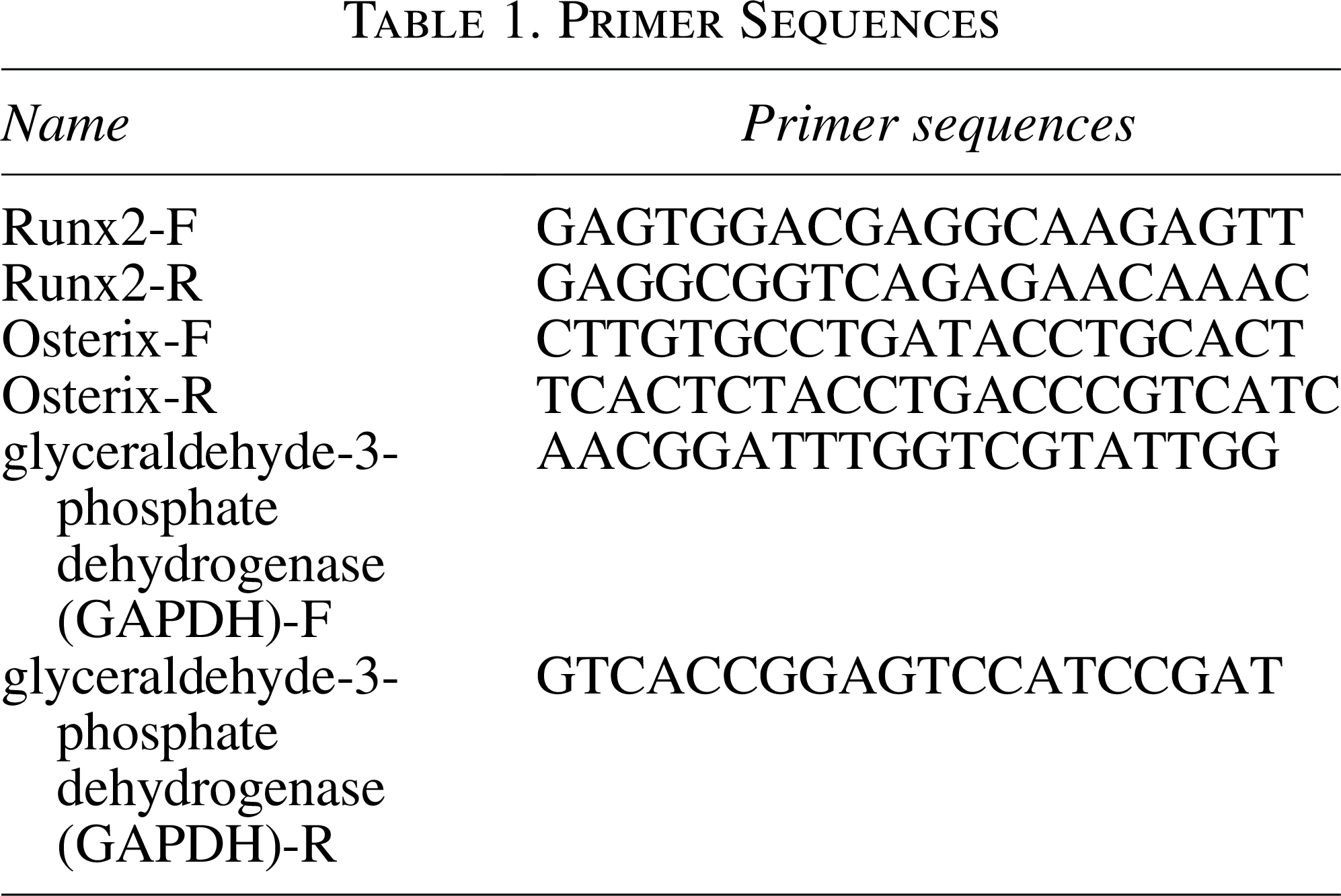
The purified RNA was obtained according to the protocol of RNAeasy™ Animal RNA Isolation Kit. The reverse transcription reaction of 1.5 µg purified RNA was operated under the instructions of cDNA Synthesis Kit. Then the real-time quantitative polymerase chain reaction (RT-qPCR) was conducted according to the protocol of 2×SYBR Green qPCR Master Mix kit. The Primer sequences are shown in Table 1.
Primer Sequences
Flow field microenvironment simulation
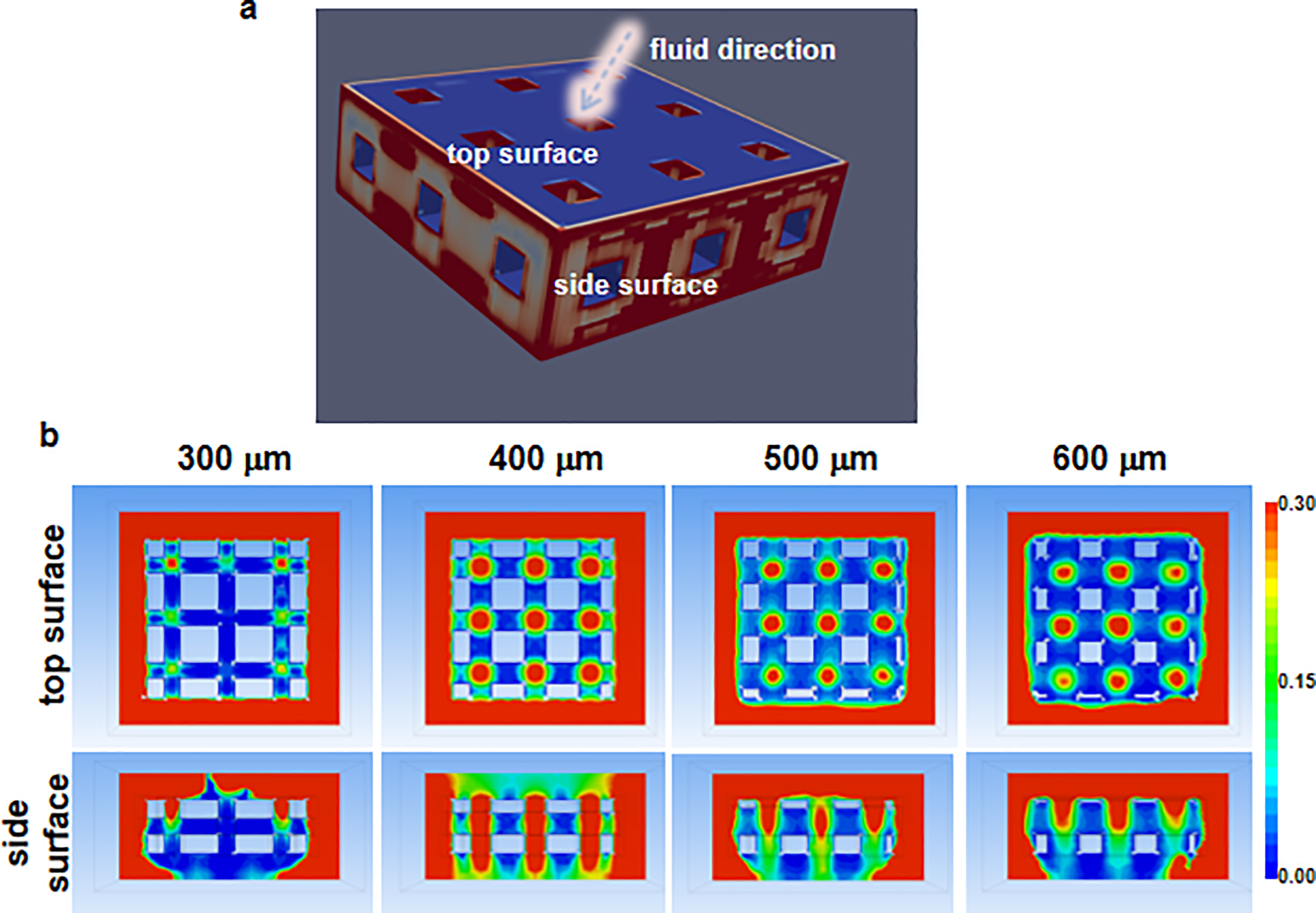
The hydrodynamic response of the support was evaluated through Computational Fluid Dynamics (CFD) analysis, with the scaffold model (9 mm in length and 3.5 mm in height) defined as a rigid, nondeformable solid region. The fluid domain was constructed and meshed in Hypermesh, incorporating a porous model at its center to minimize boundary effects. Key interfaces, including the inlet, outlet, and side surfaces, were explicitly defined to govern fluid entry and exit. The meshed model was subsequently imported into ABAQUS for CFD simulation, with the fluid properties configured to mimic DMEM at 37°C, featuring a viscosity of 1.45 × 10⁻³ Pa·s and a density of 1000 kg/m3. The outer boundaries of the fluid domain and the fluid–solid interfaces were treated as impermeable rigid walls to enforce no-slip conditions. A laminar flow regime was assumed due to the low Reynolds number (Re < 10), with a uniform inlet velocity of 1 mm/s. The outlet boundary was assigned a zero-pressure condition to represent free outflow. This setup ensured a physiologically relevant simulation of fluid–structure interaction under controlled hydrodynamic loading.
Statistical analysis
The data were all presented as mean values ± standard deviations (n = 3). The one-way analysis of variance was used for statistical analysis. The p value <0.05 was considered to be statistically significant.
Results and Discussion
Fabrication of PLLA/HA scaffolds
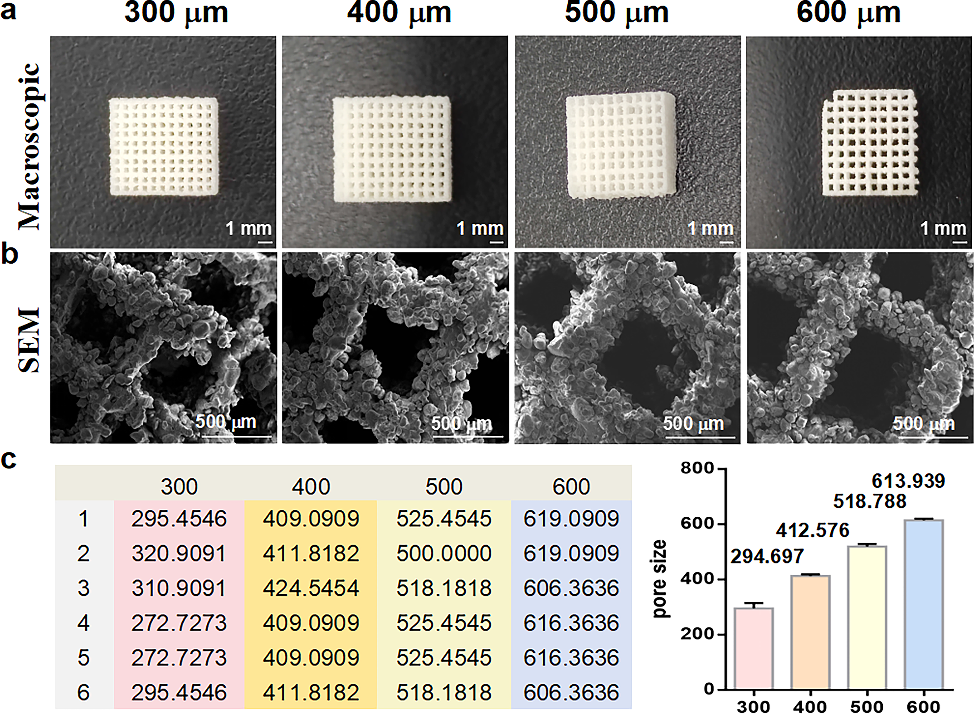
The SLS was used for the fabrication of PLLA/HA scaffolds (Fig. 1a) with tailored pore sizes (300, 400, 500, and 600 µm), as evidenced by the results obtained through SEM analysis (Fig. 1b). The SEM images illustrated the architecture of the scaffolds, revealing pores of varying diameters that were engineered according to the desired specifications. The pores were uniformly distributed across the entire scaffold structure. This uniformity ensures efficient cell infiltration and nutrient transport, which are crucial for bone regeneration, reflecting the high precision and control achieved during the SLS process. As shown in Figure 1c, the pore sizes of scaffolds were presented. The error between the actual size and the design size of each scaffold was not more than 10%, and the error between the average size and the design size of each group of scaffolds was less than 4%. This suggested that the pore size was controllable when SLS was used to fabricate the scaffold.
Characterization of scaffolds.
Biocompatibility
The Live/Dead staining assay was performed after the cells were treated with scaffolds to evaluate the biocompatibility of PLLA/HA scaffolds (Fig. 2). The large number of green fluorescent spots on all groups were exhibited, indicating that the scaffolds with different pore sizes were all with good biocompatibility (Fig. 2). Additionally, a few red dots were observed in the 300 µm scaffold group at day 1, indicating a few dead cells in this group. Good biocompatibility facilitated cell recognition and adhesion to the surface of the scaffold, and then migration into the interior through the pore structure. This was an important initial step for soft tissue to growth into the scaffolds.
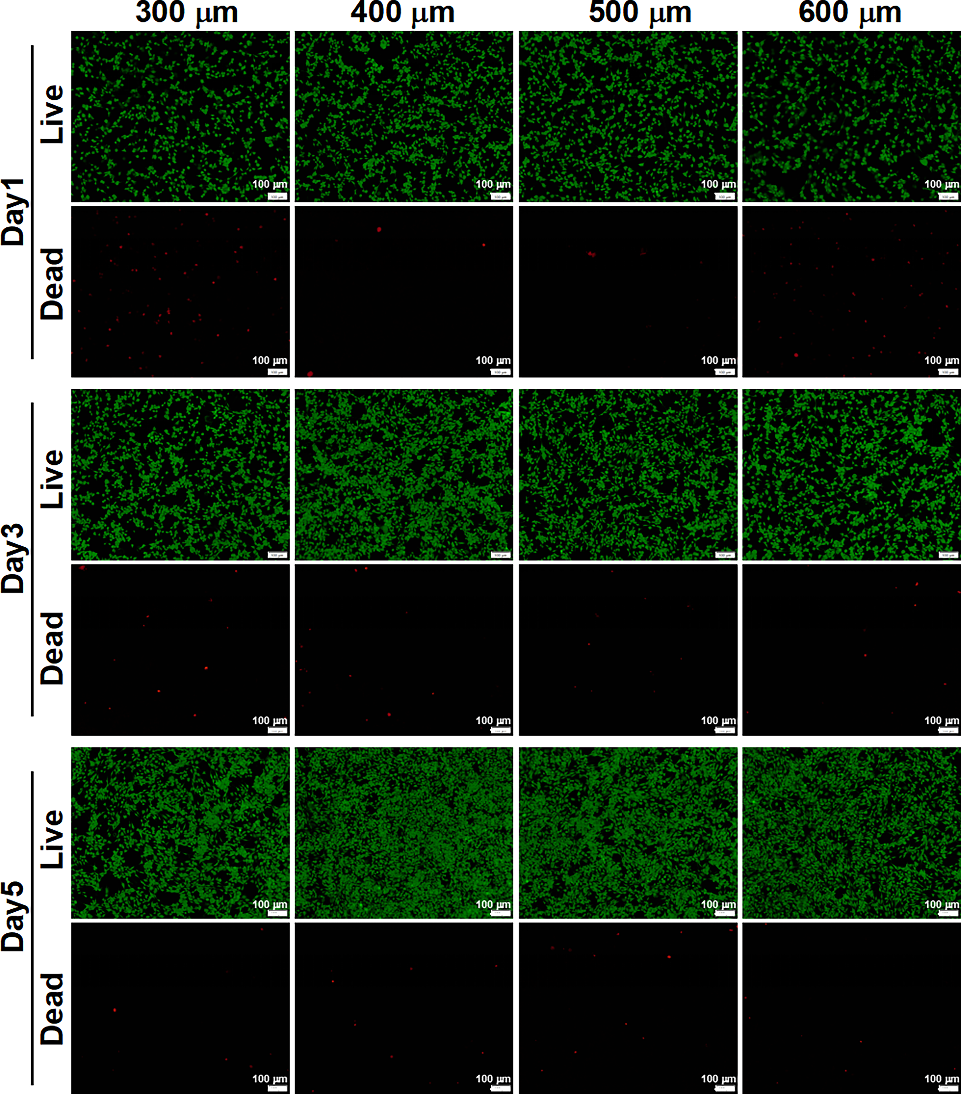
Biocompatibility of scaffold. Live and dead staining assay for cells treated with scaffolds with different pore sizes at days 1, 3, and 5. Green, live cells; red, dead cells. Scale bar, 100 µm.
Cell adhesion
As illustrated in Figure 3, DAPI staining images showed cells on the surface and within the pores of different scaffold groups. SEM images not only captured the morphology of cells adhering to PLLA scaffolds with varying pore sizes but also emphasized the presence of cellular extensions, such as filopodia and lamellipodia. These extensions represented critical aspects of cell behavior and function. 34 Filopodia were slender, actin-rich projections that extended from the cell membrane, acting as sensory organelles that explored the surrounding microenvironment. They were involved in cell–cell and cell–matrix interactions, playing a crucial role in cell migration, adhesion, and signaling. 35 Lamellipodia, on the contrary, were broader, sheet-like extensions that formed at the leading edge of migrating cells. They were rich in actin filaments and were responsible for cell motility and the generation of traction forces.36,37 The presence of filopodia in the SEM images indicated that the cells were actively sensing and responding to cues within the pore walls of the scaffolds. The SEM images also showed lamellipodia spanning, suggesting that the cells were actively migrating. These cellular extensions not only facilitated cell migration and adhesion but also contributed to the overall stability of the tissue construct. Additionally, scaffolds with the pore size of 400 µm showed the highest level of cell adhesion. Scaffolds with the pore size of 500 µm had a moderate level of cell adhesion. Meanwhile, those with the pore size of 300 or 600 µm exhibited the lowest number of adhered cells. In conclusion, .5the SEM observation clearly demonstrated the presence of filopodia and lamellipodia on PLLA scaffolds with varying pore sizes, highlighting active cell signaling and migration. Adequate cell adhesion could ensure the stable attachment of cells to the scaffold, which facilitated continuous migration of cells deeper into the pores, thereby providing a “cell reserve” for tissue infiltration and regeneration.

Cell adhesion of scaffolds.
Cell proliferation
Microscopic observations and CCK-8 assays of cell proliferation on scaffolds with different pore sizes provide valuable insight into how cellular behavior is influenced by the microstructure of scaffolds (Fig. 4). On scaffolds with smaller pore sizes (300 µm), cell proliferation was notably slower. Microscopic images revealed a relatively sparse distribution of cells with limited clustering. Scaffolds with larger pores (400 µm) exhibited a marked increase in cell proliferation with dense clusters of cells visible under microscopy. This difference might be attributed to the confined space and limited nutrient diffusion within smaller pores, which could restrict cell growth and proliferation. The larger pores, on the contrary, facilitated nutrient transport and waste removal, thereby creating a more favorable microenvironment for cell growth. Additionally, the increased surface area for cell attachment and the more open pore structure could allow for better cell–cell interactions, which further stimulated proliferation. Excessively large pore sizes in scaffolds can be detrimental to cell growth.24,38 Microscopic observations have shown that when pores were too large (500 or 600 µm), they could disrupt the cell-to-cell interactions and the formation of a cohesive cellular network. This can lead to reduced cell proliferation, as cells struggle to find adequate attachment points and lose the benefits of cell–cell communication. Moreover, large pores can create areas of low nutrient concentration and inefficient waste removal, compromising the metabolic health of the cells. The resulting nutrient deprivation and accumulation of waste products can further inhibit cell growth and viability.39,40 Sufficient cell proliferation could increase cell density, thereby enhancing the tight bond with scaffold materials through surface adhesion molecules (e.g., integrins). The ECM secreted simultaneously formed a “transition layer” at the interface between the scaffold and the tissue, reducing the gap between the two, enhancing the mechanical bonding strength, and preventing the tissue from detaching from the scaffold.
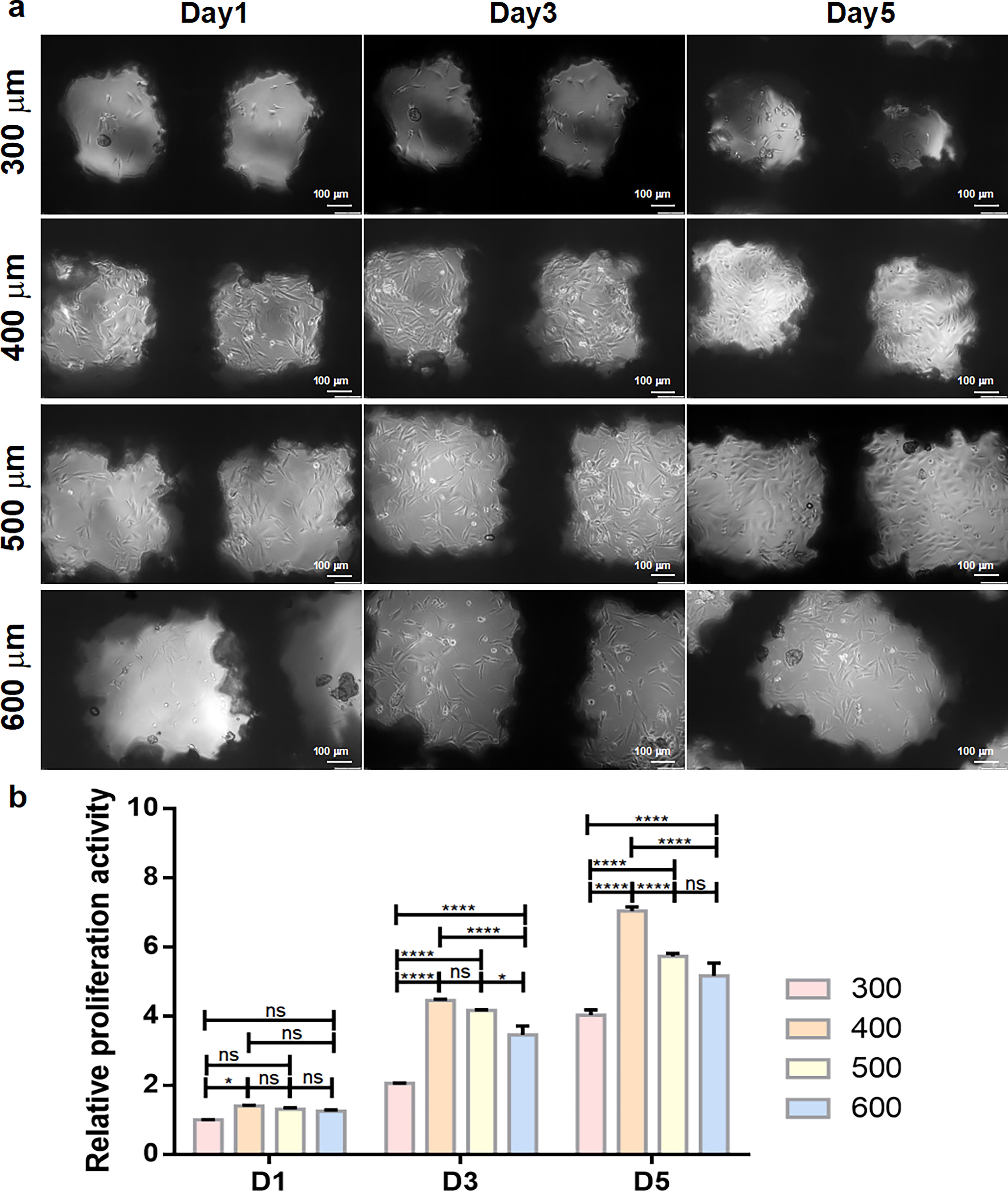
Cells proliferation of scaffolds.
Interestingly, the microscopic images also reveal that the shape and morphology of the cells vary depending on the pore size of the scaffolds. Cells on scaffolds with smaller pores (e.g., 300 µm) tended not to be outstretched and conform to the pore walls, while cells on scaffolds with larger pores (e.g., 400 or 500 µm) were more spread out and exhibited a fusiform morphology. In conclusion, although pores are necessary for nutrient and waste transport, excessively small or large pore sizes can have adverse effects on cell growth. Therefore, it is crucial to carefully select an appropriate pore size that balances nutrient transport and cell–cell interactions to optimize cell behavior on scaffolds.
Osteogenic differentiation
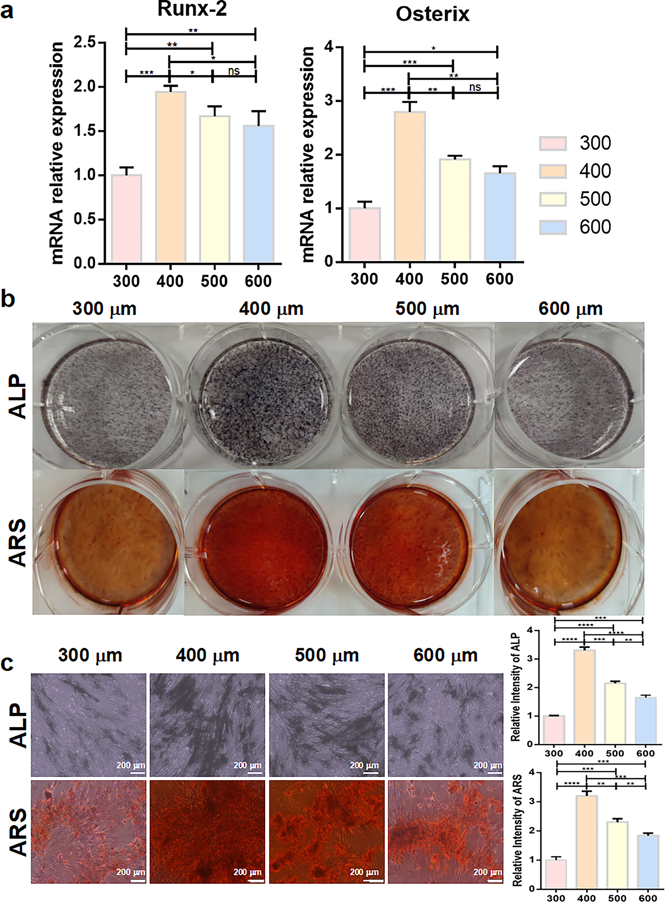
To analyze the influence of scaffolds with different pore sizes on osteogenic differentiation, the mRNA expression levels of key osteogenic factors, Runx-2 and Osterix, were analyzed by RT-qPCR in cells treated with scaffolds with different pore sizes. The results showed that scaffolds with 400-µm pore size exhibited higher mRNA levels of Runx-2 and Osterix, which indicated the stronger ability to promote osteogenic differentiation. While the mRNA level of Runx-2 and Osterix in scaffolds with 300-µm pore size was lower, which suggested a reduced osteoinductive capacity (Fig. 5a). Furthermore, the ALP and ARS staining were performed to evaluate the influence of scaffolds on osteogenic differentiation. As shown in Figure 5b, macroscopic ALP and ARS staining images presented the staining differences of scaffolds with different pore sizes. Scaffolds with a 400-µm pore size exhibited significantly darker and more extensive purple-black staining, indicating higher ALP activity, which was a key early marker of osteogenic differentiation. In contrast, other groups showed relatively lighter staining, suggesting lower ALP activity. Similarly, the ARS staining results demonstrated that scaffolds with a 400-µm pore size presented a prominent intense red color, signifying abundant calcium deposition, a crucial late indicator of osteogenic differentiation. However, other groups displayed a paler red hue, implying less calcium accumulation. Microscopic observation provided further evidence (Fig. 5c). At the cellular level, scaffolds with a 400-µm pore size showed more intense and widespread ALP-positive granules, reflecting vigorous osteogennesis-associated enzymatic activity. Other groups showed sparser and weaker ALP-positive staining. Regarding ARS staining, the scaffolds with a 400-µm pore size had denser and more clustered red stained calcium nodules, demonstrating enhanced matrix mineralization ability. Conversely, the other groups exhibited relatively scattered and smaller calcium nodules. Quantitative analysis of these staining verified a consistent trend. In addition, various biological factors secreted during osteogenic differentiation could directly or indirectly promote the interaction between soft tissue cells and the scaffold, such as transforming growth factor-β and insulin-like growth factor. These factors could activate receptor-mediated signaling pathways on the surface of soft tissue cells (e.g., fibroblasts) and enhance their migration ability.
Cell osteogenic differentiation of Scaffolds.
Flow field microenvironment simulation
In order to analyze the differences in hydrodynamic behavior within the pores, the flow field environments of scaffolds with different pore sizes were simulated (Fig. 6). When the pore size was relatively small (300 µm), fluid penetration into the pores was limited, resulting in high shear stress and obvious laminar flow characteristics. The diffusion of nutrients was restricted when the pore size was relatively small, which might lead to the accumulation of local metabolic waste. 41 With a medium pore size (400 µm), the flow field distribution was more balanced. The fluid generated moderate vortices and mixing effects, which not only maintained sufficient shear force to stimulate the mechanical response of cells but also ensured the effective transport of nutrients and the clearance of metabolic products. This dynamic balance precisely simulated the fluid microenvironment in natural bone tissue. 42 When the pore size increased (500 or 600 µm), the flow field gradually transitioned to a turbulent state with low shear stress. Although the overall flux increased, the interaction between the fluid and the scaffold surface weakened, which might lead to insufficient mechanical signals perceived by cells. Additionally, the streamline distribution was sparse and irregular, which was unfavorable for cell adhesion, proliferation, and directional differentiation. The differences in the characteristics of this multiscale flow field profoundly revealed the indirect influence of pore size on cell fate by regulating the hydrodynamic microenvironment. Similarly, the fluid shear force, nutrient delivery, metabolite excretion, and other factors in the flow field environment can also affect the growth of soft tissues into scaffolds.
Flow field microenvironment simulation of scaffolds.
Conclusion
In summary, our research on PLLA/HA scaffolds with tailored pore sizes (300, 400, 500, 600 µm) fabricated using the SLS technology, yielded insights into the relationship between scaffold pore sizes and cellular responses, as well as the underlying regulatory mechanisms.
Scaffolds with tailored pore sizes exhibited favorable biocompatibility, as evidenced by Live/Dead staining assays. The majority of cells in each group showed green fluorescence, indicating high viability. These results confirmed that the scaffold material does not induce significant cytotoxicity, which is a fundamental prerequisite for its application in bone tissue engineering.
More importantly, pore size significantly modulated cell behavior, with the 400-µm scaffold emerging as the optimal candidate across multiple key indicators. In terms of cell adhesion, the 400-µm scaffold supported the highest number of adhered cells, accompanied by abundant cellular extensions such as filopodia and lamellipodia. These structural features not only reflected active cell sensing and migration but also provided a stable foundation for subsequent cellular activities. In contrast, scaffolds with other pore sizes exhibited fewer adhered cells, possibly due to the overly confined space in smaller pores restricting cell spreading and the insufficient attachment points in larger pores weakening cell anchorage.
Cells on the 400-µm scaffold exhibited fusiform morphology and the most vigorous proliferation, forming dense cell clusters, as confirmed by proliferation assays. This possibly was attributed to the balanced pore structure, which provided sufficient space for nutrient diffusion and waste removal, avoided the nutrient transport limitations of small pores (300 µm), and prevented the disruption of cell–cell interactions caused by excessively large pores (500 and 600 µm).
In terms of osteogenic differentiation, the 400-µm scaffold showed the strongest promotional effect. RT-qPCR analysis revealed significantly higher mRNA expression levels of key osteogenic markers Runx-2 and Osterix compared to other groups. Consistently, ALP staining exhibited stronger activity, and ARS staining showed more abundant mineralized nodules in the 400-µm group, confirming its ability to effectively induce osteogenic lineage commitment.
Furthermore, flow field simulation provided a mechanistic explanation for the superior performance of the 400-µm scaffold. The flow field within 400 µm pores displayed balanced distribution, generating moderate vortices and shear stress. This not only ensured efficient nutrient transport and waste clearance (addressing the diffusion limitations in 300 µm pores and the irregular distribution in 500/600 µm pores) but also provided appropriate mechanical stimulation to activate cellular signaling pathways related to adhesion, proliferation, and differentiation. In contrast, the high shear stress and laminar flow in 300 µm pores inhibited cellular activities, while the turbulent flow and weak fluid–scaffold interaction in 500/600 µm pores failed to provide sufficient mechanical cues.
Taken together, these findings demonstrated that while all PLLA/HA scaffolds fabricated by SLS possessed good biocompatibility, the 400-µm pore size stood out as the most favorable, as it comprehensively enhanced cell adhesion, proliferation, and osteogenic differentiation by balancing structural support, nutrient transport, and mechanical stimulation. This optimization of pore size has significant implications for the design and application of scaffold materials in bone tissue engineering.
Future Directions and Challenges
The future of PLLA/HA in regenerative medicine looks promising. With ongoing research into potential applications, PLLA is poised to revolutionize bone tissue engineering and regenerative therapies.
The field of bone tissue engineering has placed significant emphasis on optimizing scale pore sizes to control cell adhesion, proliferation, and differentiation. Specifically, multiscale pore size distributions have emerged as a potential avenue for improving the efficacy of scaffolds used in bone tissue engineering. Incorporating pores of varying sizes within the same scaffold more accurately mimics the complex and hierarchical architecture of native tissues, which not only facilitates cell migration and infiltration but also promotes the differentiation of various cell types into their appropriate lineages. 43 Moreover, the presence of multiple pore sizes within a scaffold can enhance nutrient and waste transport, ensuring that cells receive adequate nourishment and are able to efficiently eliminate metabolic byproducts. 44 Ultimately, by embracing multiscale pore size distributions, the field of bone tissue engineering stands to make significant strides in creating more functional and physiologically relevant constructs that better serve the needs of patients.
Bone scaffold integration with biological factors for antibacterial, anti-inflammatory, and antitumor applications represents a frontier in regenerative medicine. By incorporating growth factors, biomolecules, and advanced materials, these scaffolds aim to enhance bone regeneration while addressing complications. The integration of antibacterial agents within the scaffolds effectively reduces the risk of postimplantation infections,45,46 promoting a sterile healing environment. Anti-inflammatory components help modulate the immune response, minimizing tissue damage and accelerating recovery. 47 Furthermore, the incorporation of antitumor components aims to prevent or inhibit the growth of malignant cells,48,49 ensuring the safety and efficacy of the regenerative process. This multifaceted approach leverages the synergistic effects of biology and materials science, paving the way for advanced therapeutic strategies that not only restore bone function but also safeguard patient health comprehensively.
Authors’ Contributions
C.S.: Writing—review and editing, project administration, conceptualization, funding acquisition. T.H.: Writing—original draft, methodology, conceptualization, data curation, investigation, formal analysis. Z.T.: Data curation, formal analysis. S.P.: Methodology, investigation, formal analysis. X.B.: Investigation, data curation, writing—review and editing.
Footnotes
Funding Information
This study was supported by the following funds: (1) National Key Research and Development Program of China (Grant No. 2023YFB4605800); (2) the Natural Science Foundation of China (U24A20120, 52475362, 52275393, 52275395); (3) JiangXi Provincial Natural Science Foundation of China (20224ACB204013); (4) the Project of State Key Laboratory of Precision Manufacturing for Extreme Service Performance, Central South University; and (5) Jiangxi Province early career youth science and technology talent training project (20244BCE52188).
Data Availability Statement
All data generated or analyzed during this study are included in this article, which are available upon request by contact with the corresponding author.
Disclosure Statement
The authors have nothing to declare.