
Abstract
Skin aging involves changes in extracellular matrix components, such as wrinkles and pigmentation. Caviar extract (CE) is a promising compound for skin rejuvenation, but effective topical delivery requires optimized carriers. This study evaluated polyvinyl alcohol/carboxymethyl chitosan (PVA/CMC) hydrogels loaded with CE at concentrations of 2%, 3.5%, and 5% as scaffolds to influence the epithelial differentiation of adipose-derived mesenchymal stem cells (ADMSCs). Hydrogels were synthesized using a freeze-thaw method and characterized by Fourier-transform infrared spectroscopy (FTIR), scanning electron microscopy, swelling and degradation tests, and mechanical analysis. Biocompatibility and cell migration were assessed using MTT and scratch assays; at the same time, expression of cytokeratin-18 (CK-18) and pan-cytokeratin (pan-CK) was measured via reverse transcription-quantitative polymerase chain reaction and immunocytochemistry (ICC), respectively. FTIR confirmed successful CE incorporation, and SEM revealed a porous structure. Hydrogels with 3.5% and 5% CE demonstrated a good balance between swelling and degradation over 336 h. The biocompatibility tests showed that 5% CE supported enhanced long-term cell growth. The scratch assay indicated improved cell migration, and transcriptional analysis revealed significantly higher CK-18 levels in ADMSCs treated with PVA/CMC/CE 5% (p < 0.001). ICC results showed significantly higher pan-CK expression at 3.5% CE (41.82%) and 5% CE (48.16%), suggesting that CE promotes repair processes. These findings suggest that 5% CE-loaded PVA/CMC hydrogel could be an effective option for skin regeneration and antiaging.
Impact Statement
Caviar extract (CE) was considered a bioactive ingredient, along with polyvinyl alcohol (PVA) and carboxymethyl chitosan (CMC) polymers, to prepare a functional and practical hydrogel without hazardous components for anti-aging and cosmetic applications. In the present study, the PVA/CMC hydrogel contains various concentrations of CE (3.5% and 5%), is biocompatible, and enhances cellular viability and migration of adipose-derived mesenchymal stem cell. Our results demonstrated that the synergistic effect of CE and CMC could promote the expression of cytokeratin-18 gene and pan-cytokeratin protein and play a critical role in stimulating skin regeneration.
This is a visual representation of the abstract.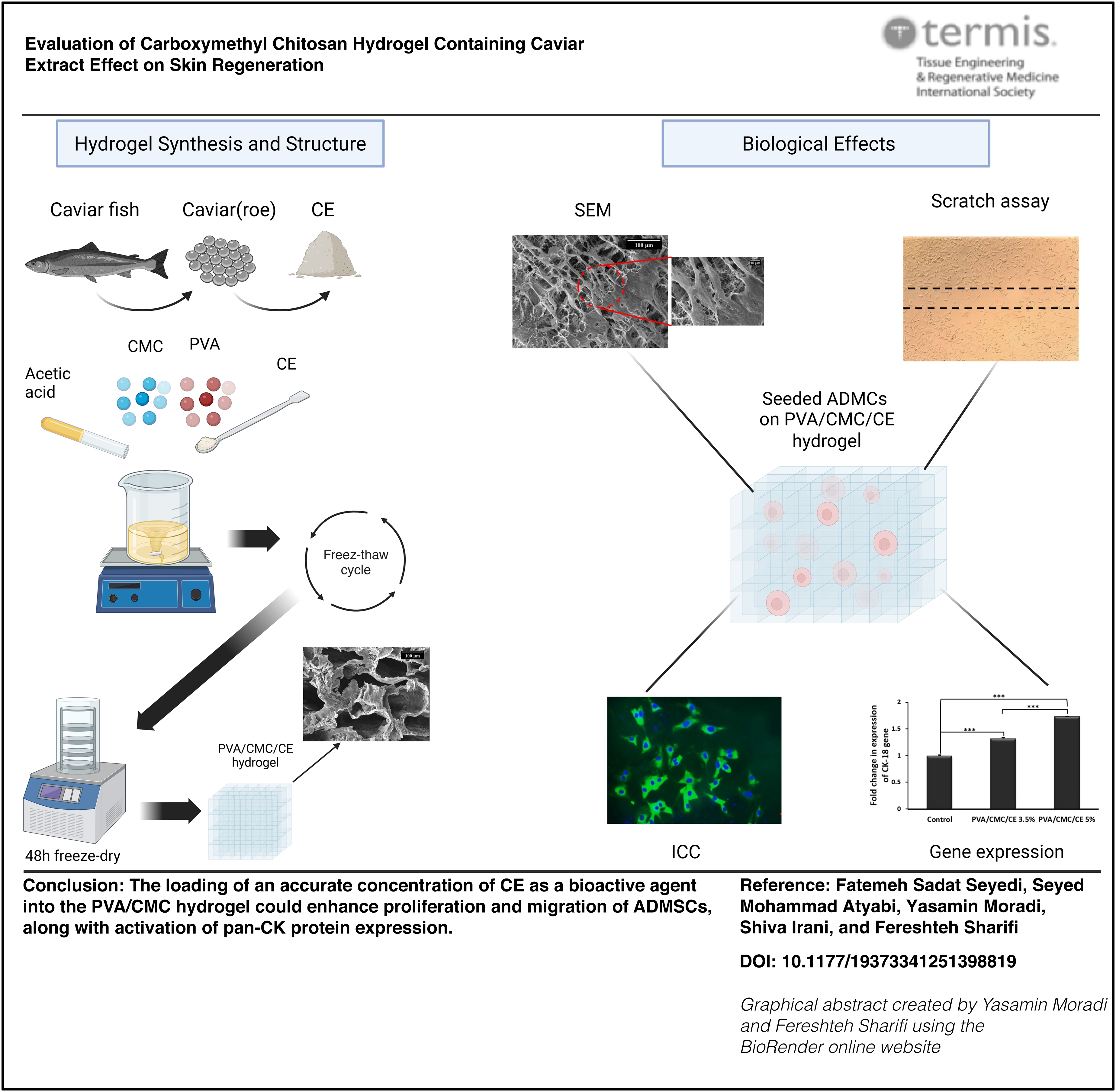
Introduction
Skin aging is a complex biological phenomenon characterized by structural and functional changes in extracellular matrix (ECM) components such as proteins (collagens and keratin) and glycosaminoglycans (hyaluronic acid and dermatan sulfate).1,2 The alteration in these factors collectively contributes to the signs of aging, including wrinkles, reduced elasticity, and hyperpigmentation. 3 The demand for effective rejuvenating cosmetic ingredients and formulations has led to the investigation of diverse natural and synthetic compounds that can address the visible signs of aging.4,5 Caviar extract (CE) has attracted considerable interest due to its rich nutrients, including amino acids and lipids, which are believed to benefit the skin.5–7 Additionally, the potential of CE to combat oxidative damage from environmental aggressors like UV radiation offers hope for skin care applications. The bioactive components in CE may interact with filaments of proteins, such as keratin and cytokeratin, potentially enhancing their durability and resilience to stress, leading to more effective skin healing and shorter recovery times.8,9 However, the efficacy of CE in topical applications relies on its delivery system, which ensures its impressive efficacy in skin repair. 10 Hydrogel is a potentially effective carrier for bioactive cosmeceutical ingredients due to its flexibility, high water content, and ability to control the release of encapsulated ingredients.11–14 Polyvinyl alcohol (PVA) and carboxymethyl chitosan (CMC) are promising biomaterials that have been studied for their potential use in cosmetics. 15 PVA is known for its outstanding biocompatibility, biodegradability, mechanical stability, and ability to form hydrogels.16,17 In contrast, CMC, derived from chitosan, is a natural polysaccharide with mucoadhesive properties that enhance skin permeation and possess antioxidant and anti-inflammatory characteristics.18,19 Developing a hydrogel that integrates the favorable characteristics of both PVA and CMC yields desirable mechanical and therapeutic properties for cosmeceutical applications. 20 This study builds upon this foundation by evaluating the efficacy of CE encapsulated in a PVA/CMC hydrogel delivery system, focusing on its role in enhancing skin repair and regeneration in an in vitro model.
Experiment
Materials
Human adipose-derived mesenchymal stem cells (ADMSCs) were acquired from the Iranian Biological Resource Center (Iran). Dulbecco’s Modified Eagle’s Medium (DMEM), fetal bovine serum (FBS), phosphate-buffered saline (PBS), and trypsin-EDTA (0.25%) were obtained from Bioidea (Iran). Caviar was prepared from fresh Caspian Sea caviar obtained from sturgeons of the genus Acipenser, commonly known as caviar fish. Chitosan with a 90% degree of deacetylation and medium molecular weight was bought from Bio Basic (Canada). PVA with an average molecular weight of 85,000–124,000 kDa, 3-(4,5-dimethylthiazol-2-yl)-2,5-diphenyltetrazolium bromide (MTT), dimethyl sulfoxide (MW 78.13), 4,6-diamidino 2-phenylindole (DAPI), and chloroform (99.0–99.4%) were supplied from Sigma-Aldrich (Germany). Formic acid (MW 96.03, 98–100%), monochloroacetic acid (MW 94.50, >99.0%), acetic acid (100%), isopropanol, ethanol (96%), and sodium hydroxide (NaOH, molar mass: 40 g/mol) were bought from Merck (Germany). Primary (orb1294309) and secondary (orb688925) antibodies specific to pan-cytokeratin (pan-CK) were obtained from Biorbyt (UK).
Methods
Extraction of caviar using the maceration method
Finely ground caviar (10 g) was soaked in 100 mL of distilled water (DW) and stored at room temperature for 1 week in an aluminum foil-covered container placed on an orbital shaker platform. After 1 week, the content was filtered through qualitative filter paper. 21 The solvent was then removed from the extracted solution via distillation at 60°C, yielding the final crude CE. The extracted CE was subsequently freeze-dried (DENA ACUUM Industry, Iran) to obtain a stable, dry sample for further use in hydrogel preparation.
Gas chromatography–mass spectrometry analysis of CE composition
About 100 mg of freeze-dried CE was dissolved in 1 mL of ethanol and thoroughly mixed to ensure complete dissolution. The solution was transferred to a gas chromatography–mass spectrometry (GC/MS) vial for analysis using a PolarisQ (Quadrupole Ion Trap GC/MS system, Thermo Scientific, USA) equipped with a DB-5MS capillary column (30 m × 0.25 mm i.d., 0.25 μm film thickness, Agilent Technologies). Standard GC/MS parameters were employed to analyze small molecules and volatile compounds, including electron ionization mode and a full scan range of 30–300 m/z. Compound identification was accomplished by comparing the obtained mass spectra with the NIST17 mass spectral library. The relative abundance of each compound was determined based on the peak area percentage (Area%). The components of CE obtained by GC–MS are listed in Table 1.
The Components of Caviar Extract Gained by GC–MS
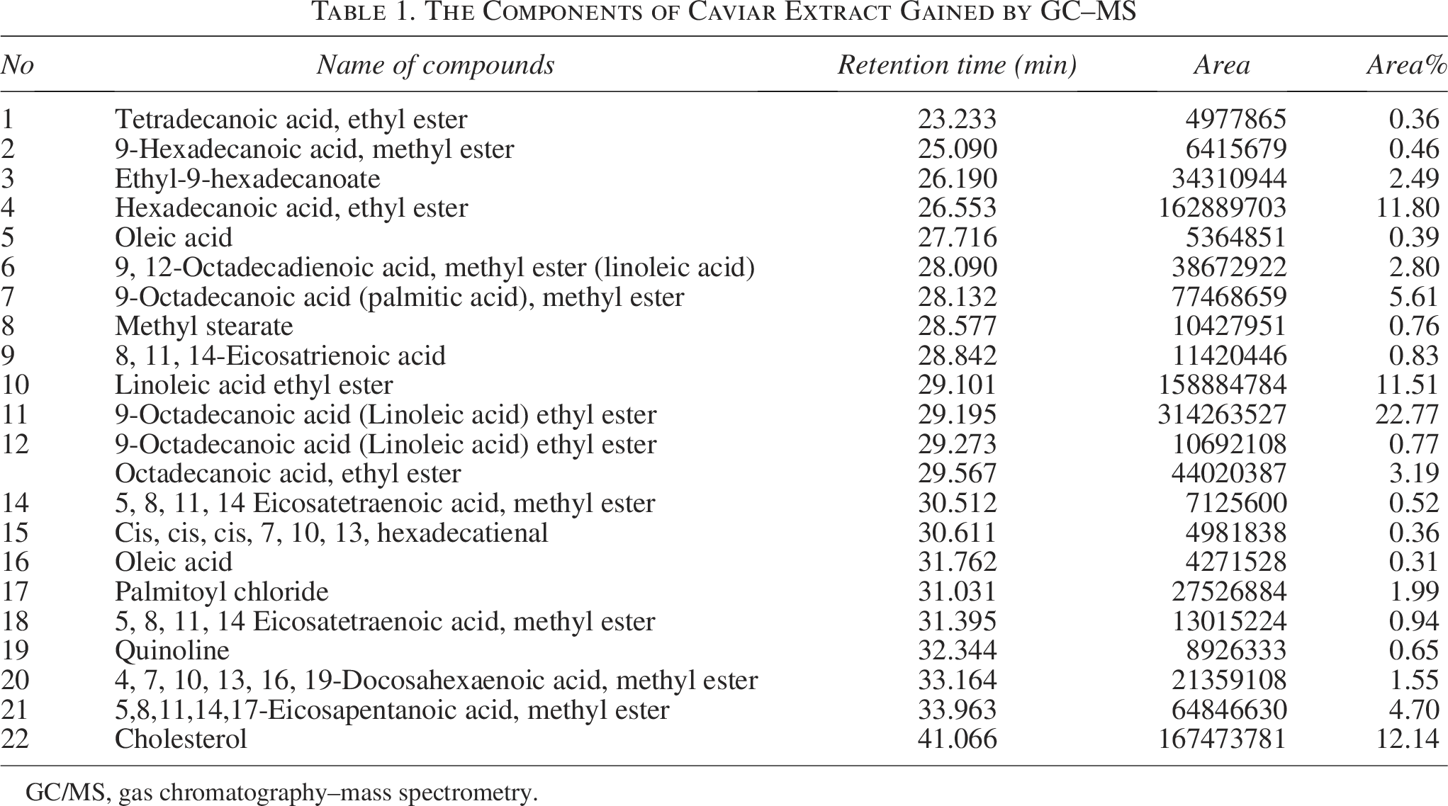
GC/MS, gas chromatography–mass spectrometry.
Hydrogels preparation
The CMC was prepared based on our previous study. 22 CMC was dissolved in 1% acetic acid, and the pH was adjusted to 5 with NaOH (10 N). The CMC solution was added to the PVA solution and mixed for 2–3 h. The resulting solution underwent five cycles of freezing (−20°C for 24 h) and thawing (25°C for 2 h). After the final cycle, the synthesized hydrogels were freeze-dried for 48 h. 22
Loading of CE onto prepared hydrogel
To load CE (PVA/CMC/CE), a stock solution was first formulated by dissolving 200 mg of CE in 5 mL of double-DW. The 2%, 3.5%, and 5% concentrations of this stock solution relative to the dry weight of PVA were integrated into the PVA solution. The mixtures of PVA and CE were stirred for about 2 h. The CMC solution was added to the PVA/CE mixture and stirred until completely homogeneous. The freeze-thawing cycles were performed, and the samples, including PVA/CMC/CE 2%, PVA/CMC/CE 3.5%, and PVA/CMC/CE 5% hydrogels, were freeze-dried and then autoclaved (Fedegari, Switzerland) at 121°C for 20 min. The compositions of the prepared hydrogels are summarized in Table 2.
The Composition of Different Hydrogels
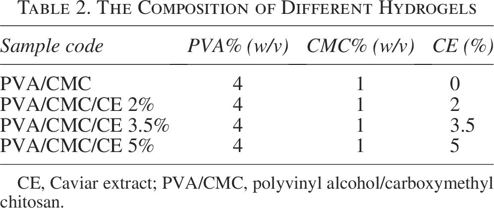
CE, Caviar extract; PVA/CMC, polyvinyl alcohol/carboxymethyl chitosan.
Scanning electron microscopy
To study the morphological properties of the hydrogel structure and cell–hydrogel interaction, SEM (SERON TECHNOLOGY, South Korea) was utilized. A cross-section of the hydrogel samples was conducted to analyze the hydrogel structure. For cell–hydrogel interaction assessment, the encapsulated ADMSCs 104 in PVA/CMC/CE 3.5% and 5% are immersed in glutaraldehyde (2.5% v/v) solution for 2 h at 4°C. The prepared samples were stored at −80°C and lyophilized for 24 h at −60°C. The samples were coated with a gold layer and placed on specimen holders using double-sided tape. 22
Pore size measurement
The pore size of PVA/CMC/CE 5%, PVA/CMC/CE 3.5%, PVA/CMC/CE2%, and PVA/CMC hydrogels was measured based on SEM images using ImageJ software (NIH, USA). The results expressed as mean ± SD represent measurements of 50 pores.
Fourier-transform infrared spectroscopy analysis
Fourier-transform infrared spectroscopy (FTIR) testing of PVA/CMC/CE 5%, PVA/CMC/CE 3.5%, PVA/CMC hydrogels, and CE was conducted according to protocol to evaluate the hydrogel molecular structure. 23 The analysis was performed using an Avatar FTIR instrument (Thermo Scientific, USA) with a wavelength of 4000–400 cm−1.
Swelling assay
The swelling behavior of the PVA/CMC/CE (2%, 3.5%, and 5%) and PVA/CMC hydrogels was subjected to specific intervals, 0.5, 1, 2, 4, 24, 48, 96, 168, and 336 h (n = 3). The hydrogels were weighed in their dry state (Wd0) and then immersed in 1 mL PBS at 37°C. The hydrogels were removed from the PBS, gently blotted to remove excess surface water, and weighed to determine the swollen weight (Wdt). The swelling ratio was calculated using Equation 1(E1).
24

Degradability assay
The degradability of PVA/CMC/CE (2%, 3.5%, and 5%) and PVA/CMC samples was initially weighed in their dry state (Wd0). The samples were then immersed in PBS at 37°C with gentle agitation. At specific time intervals (0.5, 1, 2, 4, 24, 48, 96, 168, and 336 h) (n = 3), the hydrogels were removed from the PBS, gently blotted to remove excess surface water, and freeze-dried before weighing (Wdt). The degradation percentage was calculated using E2.
25

Mechanical strength assessment
The mechanical properties of the uniaxial tensile strength of the PVA/CMC/CE (2%, 3.5%, and 5%) and PVA/CMC hydrogels were evaluated. 26 Rectangular strips measuring 1 × 3 cm2 were cut and mounted onto an STM-20 tensile testing machine (SANTAM, Iran). The maximum replacement of the crosshead was 1 mm with a speed of 0.1 mm/min. Tensile strength and elongation at break were recorded by stretching the samples across the head at 5 mm/min with a 10 N load cell (n = 3).
Release of CE from hydrogel
A calibration curve of CE was first drawn to quantify the release percentage of CE from PVA/CMC/CE (3.5% and 5%) hydrogels. Serial concentrations of 1, 5, 10, 25, 50, and 100 ppm of CE dissolved in PBS were prepared. Absorbance at 274 nm was measured using a spectrophotometer (Shimadzu UV-1800 UV-Vis, Japan). PVA/CMC/CE (3.5% and 5%) samples were cut into 1 × 1 cm2 samples and immersed in 5 mL of PBS in an incubator set to 37°C. At 0.5, 1, 2, 4, 24, 48, and 96 h, PBS was collected, and its absorbance was read at 274 nm using the UV spectrophotometer. 27
Biocompatibility assays
The ADMCs were cultured in DMEM, containing 10% FBS and 1% penicillin–streptomycin in an incubator at 37°C under a humidified CO2 (5%) atmosphere. ADMCs (104) on the third passage were seeded into the wells of a 96-well plate onto the PVA/CMC/CE 3.5% and 5% hydrogels for 0.5, 1, 2, and 4 h, and 1, 3, 7, and 10 days (n = 3). An inverted microscope was used to check the cytotoxicity, and then the cell viability was assessed via the MTT test using an ELISA plate reader (BioTek EL × 800) operating at a wavelength of 570 nm. 28 A tissue culture plate was used as a control.
Scratch assay
For the migration assay, an artificial wound or “scratch” is made in a confluent cell monolayer, and the migration into the cell-free zone is observed. In this study, 104 cells were seeded in six-well plates and cultured until confluent (∼24 h). A scratch was made through the cell layer using a sterile 200-µL pipette tip. Then, the scratch wells were treated with PVA/CMC/CE 3.5% and 5%, and a control. The cellular migration was assessed after 0, 24, and 72 h under an inverted microscope by imaging the same fixed reference areas.
Reverse transcription-quantitative polymerase chain reaction
ADMCs (105) were seeded on the 24-well plates for the control and treatment groups with PVA/CMC/CE 3.5% and 5% hydrogels for 10 days. Based on the Yektatajhiz kit (Iran) instructions, cells were detached via trypsinization and centrifuged at 350 g for 5 min. The supernatant was removed, and RiboEX lysis buffer was added. Chloroform was used to extract RNA into the aqueous phase. RNA quality/purity was assessed using a Nanodrop spectrophotometer (Thermo Scientific, NANODROF 2000, USA), measuring absorbance at 240, 260, and 280 nm. cDNA was prepared using a kit (Parstous, Iran) from 1 µL of RNA to determine the cytokeratin-18 (CK-18) expression, and GAPDH reference gene sequences were retrieved from the NCBI gene database. These primers were purchased from Gene Fanavaran (Iran) for reverse transcription-quantitative polymerase chain reaction (RT-qPCR) analysis (Table 3).
Primer Sequences (Forward and Reverse) Used for Amplification of the CK-18 and GAPDH Genes
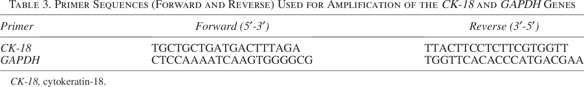
CK-18, cytokeratin-18.
Immunocytochemistry
ADMCs (104) were cultured for 10 days (n = 3) on PVA/CMC/CE (3.5% and 5%) and a control group. Samples were fixed with 4% paraformaldehyde for 20 min, washed with PBS, and permeabilized using 0.3% Triton-X for 30 min. Nonspecific binding was blocked via 45 min of incubation in 10% goat serum. Primary antibodies (1:1000) against pan-CK were applied overnight at 4°C. After washing, secondary antibodies (1:100) conjugated to fluorescent probes were added for 1.5 h. Nuclei were stained with DAPI for 20 min. Samples were imaged on a fluorescent microscope (Labomed, PCM400, Denmark, 440×). The immunocytochemistry (ICC) result was quantified to ensure consistency and accuracy in the findings.
Statistical analysis
Graph Pad Prism Statistics software (version 6.04 for Windows) used T-tests and one-way ANOVA to compare means and determine the significance of detected variations. The statistical significance was assigned as * for p ≤ 0.05, ** for p ≤ 0.01, and *** for p ≤ 0.001.
Results
GC/MS analysis
GC/MS chromatograms of CE revealed the presence of a wide range of lipid-based compounds, summarized in Table 1. The compounds were identified based on their respective retention times and Area% (Table 1). The chromatogram (Fig. 1) shows 22 distinct peaks, each of which represents bioactive substances that were identified by comparing mass spectra with the comprehensive NIST library.
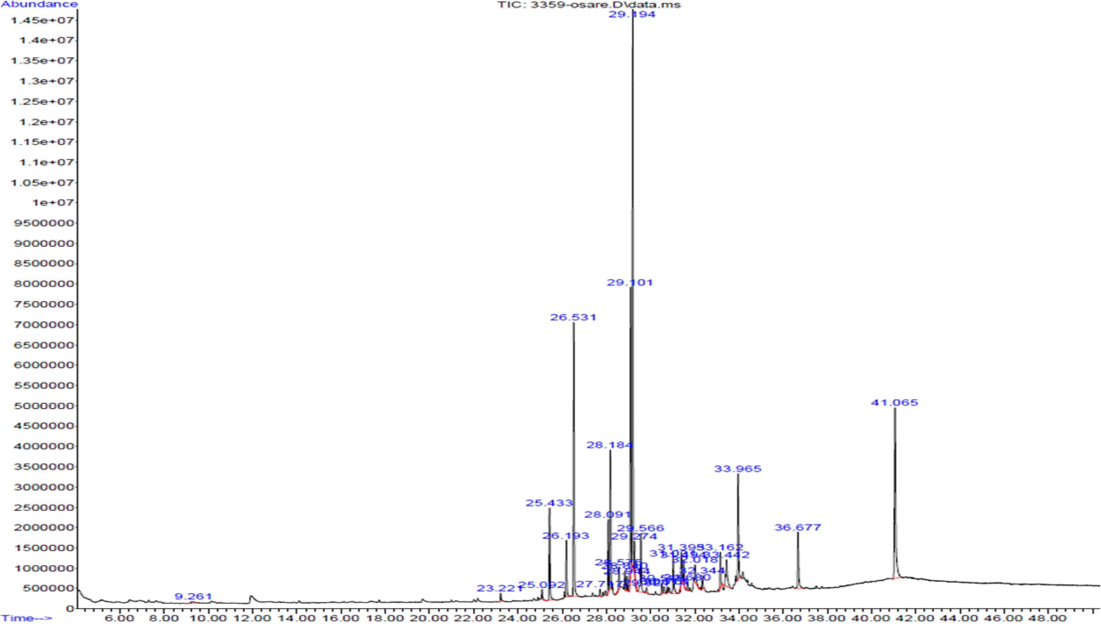
GC–MS spectral chromatogram of CE. The chromatogram displays the chemical composition of the CE, with distinct peaks corresponding to individual lipid-based compounds. CE, Caviar extract; GC/MS, gas chromatography–mass spectrometry.
PVA/CMC/CE hydrogels analysis
SEM
The morphological characteristics of the basic PVA/CMC hydrogels with CE (2%, 3.5%, and 5%) were analyzed using SEM (Fig. 2A). The SEM images illustrate a well-defined porous network. As the CE concentration increased, the pore size also grew accordingly (Fig. 2B).
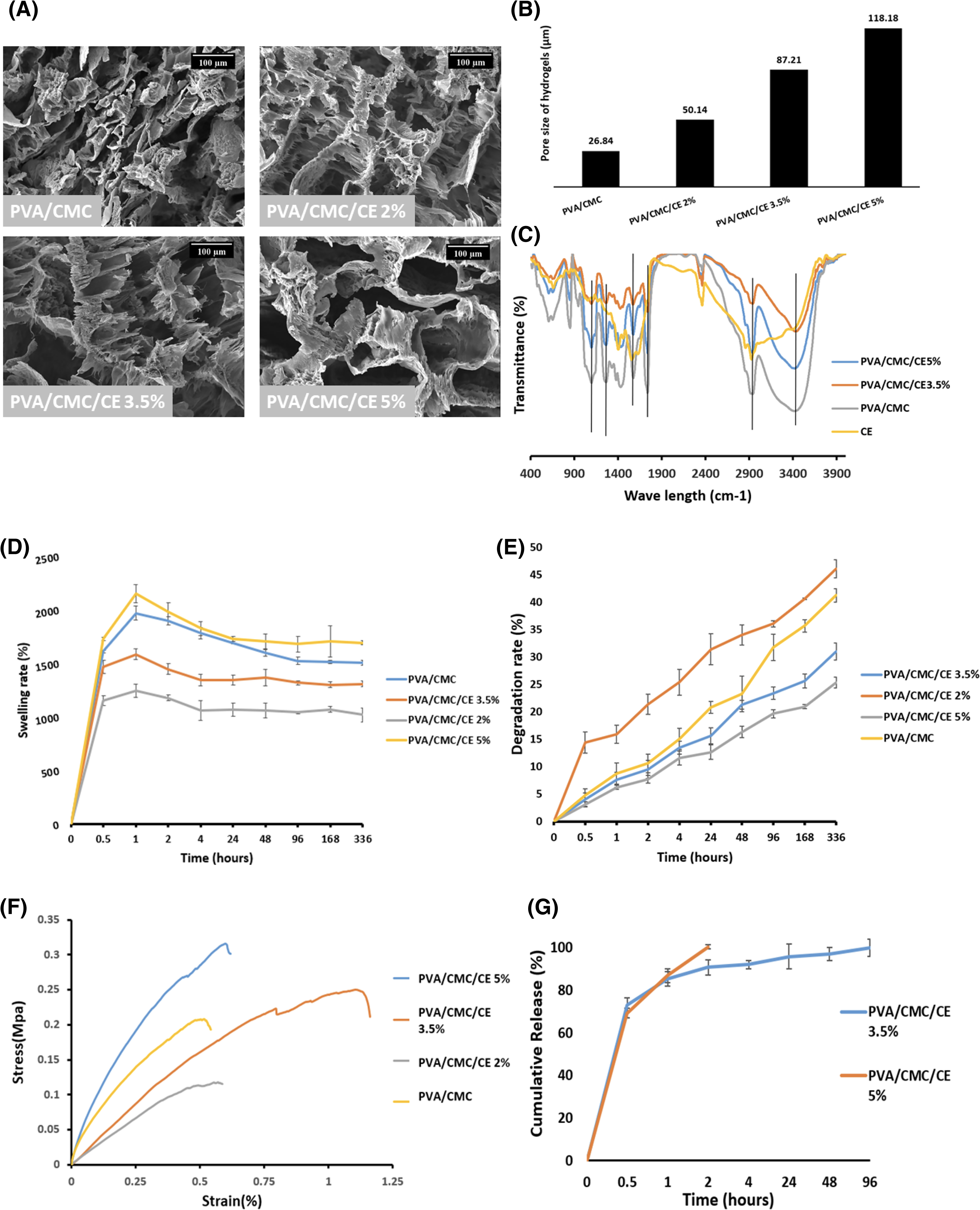
Characterization of PVA/CMC hydrogels loaded with CE.
FTIR
FTIR spectroscopy was performed to characterize hydrogel functional groups and the chemical interactions in the hydrogel matrix (Fig. 2C). Vibrations from N-H and O-H stretches were seen at approximately 3300 cm−1. A peak at 1700 cm−1 corresponding to carboxylate functionalities introduced by CMC confirmed its presence in the gels. 25 Additionally, C-O stretches were observed around 1200 cm−1, while C-H bends appeared near 2900 cm−1. Two closely spaced peaks formed a doublet around 1600 cm−1. These vibrations correspond to C=C and C-N bonds, indicating biomolecules contributed by the CE additive (Fig. 2C).29–31
Swelling and degradation analysis
The swelling behavior and degradation properties of the PVA/CMC hydrogels loaded with CE (2%, 3.5%, and 5%) were evaluated over 336 h (Fig. 2D and E). In terms of swelling, during the initial phase up to 0.5 h, the swelling of all four samples increased rapidly, indicating a sharp rise in water absorption. PVA/CMC/CE 2% initially showed the lowest water uptake at 1 h (1967.37%), while PVA/CMC/CE 5% exhibited the highest swelling (2146.72%). Afterward, the swelling rate gradually declined, reaching a plateau after 4 h, showing that the samples reached a near-steady state. Regarding degradation, all four hydrogels showed progressive breakdown throughout the test period. Notably, PVA/CMC/CE 2% degraded at the highest rate of 46.07% by 336 h, surpassing the PVA/CMC hydrogel. In contrast, PVA/CMC/CE at 3.5% and 5% degraded more slowly, at rates of 30.98% and 25.33%, respectively. The degradation rates (Fig. 2E) align with the swelling behavior, SEM, and FTIR results, indicating that increased CE levels enhance the hydrogel’s pore size and permeability to solutes.
Mechanical stress testing
The mechanical properties of the defined hydrogels were evaluated through stress–strain analysis (Fig. 2F). The results shown in the stress–strain curve offered insights into the elastic strength, stability, and maximum elongation of each hydrogel formulation until rupture occurred. The PVA/CMC hydrogel demonstrated an elastic strength and stiffness of 0.227 MPa, with a maximum elongation of 61.65% of its original length. In contrast, the PVA/CMC/CE 2% and 3.5% hydrogels exhibited elastic strengths of 0.128 and 0.259 MPa, and the maximum elongation of 54.98 and 117.73% of their original length, respectively. Based on the overall results, the PVA/CMC/CE 2% hydrogel was less favorable due to its higher degradation rate, lower swelling capacity, and inferior mechanical properties. Consequently, the PVA/CMC/CE 2% sample was excluded from further studies.
Release of CE-loaded hydrogels
The CE release results indicated that the PVA/CMC/CE 3.5% hydrogel discharged 73.16% at 0.5 h, while the PVA/CMC/CE 5% hydrogel released 69.2% during the same timeframe. By 2 h, the PVA/CMC/CE 3.5% hydrogel had released 90.69%, whereas the PVA/CMC/CE 5% hydrogel achieved complete (100%) release. The PVA/CMC/CE 3.5% hydrogel was fully released by 96 h (Fig. 2G).
Biocompatibility and migration assessment
The light microscopy and SEM examinations showed excellent cell morphology, attachment, and growth, indicating the nontoxic nature of the hydrogels toward the cells (Fig. 3A). The SEM images emphasized the strong interaction between the hydrogel and the ECM, with cells exhibiting robust adhesion and attachment to the hydrogel surface. MTT assays were conducted at two different time points to assess metabolic activity: a shorter time frame (Fig. 3B) and a longer period (Fig. 3C). The initial tests at 0.5 and 1 h revealed lower metabolic activity in both PVA/CMC/CE 3.5% and 5% hydrogels compared with the control (p < 0.05). Notably, the PVA/CMC/CE 3.5% showed higher metabolic activity in the early phase than PVA/CMC/CE 5% (Fig. 3B). At 7 days, the PVA/CMC/CE 3.5% hydrogel still exhibited higher metabolic activity and cell viability (p < 0.05). However, by 10 days, the PVA/CMC/CE 5% hydrogel surpassed the 3.5% formulation in metabolic activity (p < 0.05). The results of cell migration demonstrated a progressive migration of cells in both hydrogel-treated groups within the evaluated periods (Fig. 3D and Supplementary Fig. S1). This suggests that the higher CE concentration may offer long-term benefits for cell attachment, viability, and migration.

Biocompatibility assessment.
Differentiation analysis
RT-qPCR
RT-qPCR was conducted to assess CK-18 expression (Fig. 4A). The results from the quantitative RT-qPCR indicated a significant increase in CK-18 transcription in ADMSCs treated with PVA/CMC/CE 3.5% and 5% compared with the control group (p < 0.001). This suggests that even at a lower concentration of CE (3.5%), there was a noticeable effect on CK-18 expression in ADMSCs treated with PVA/CMC hydrogels containing CE.
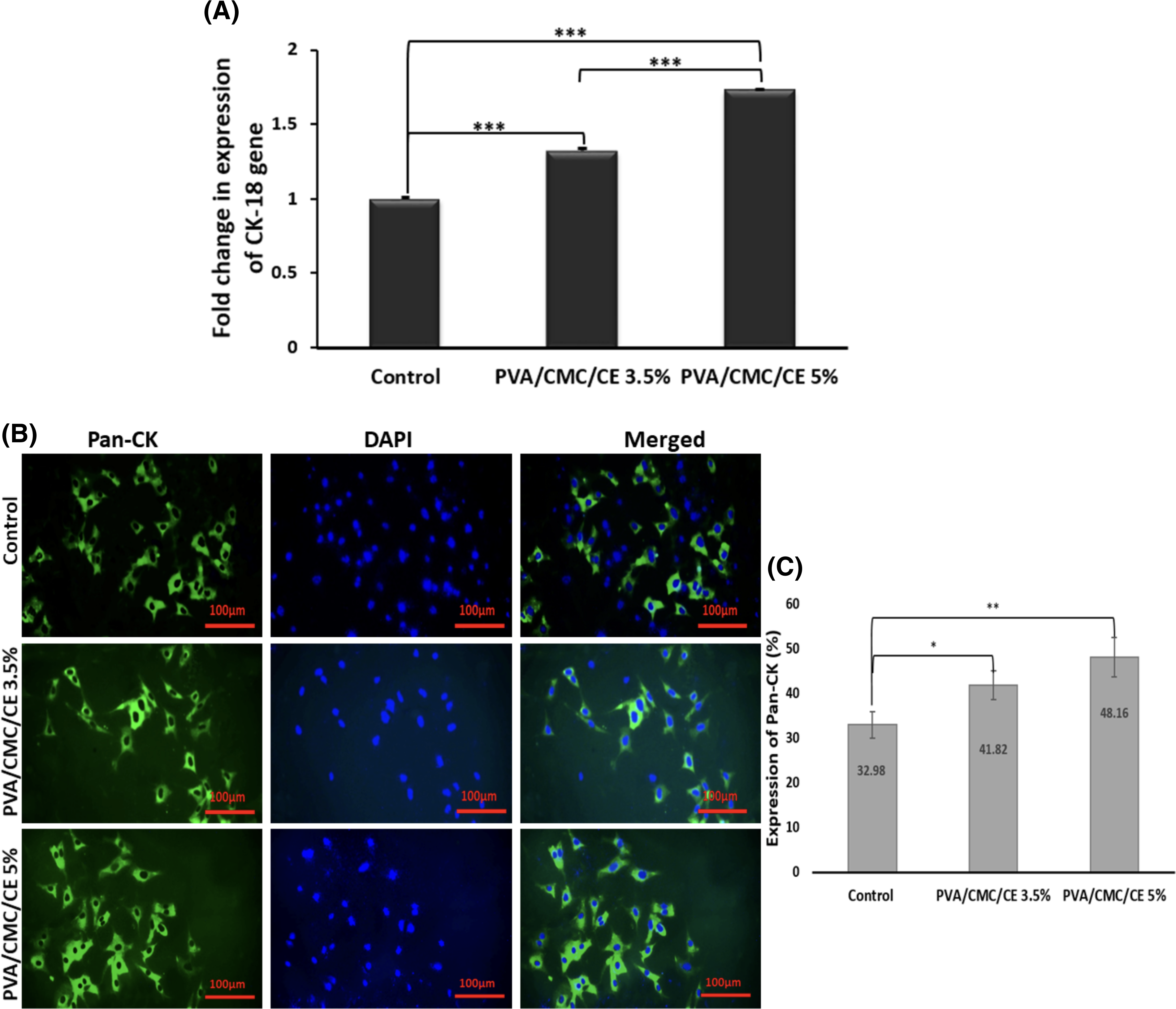
Genes and protein expression analysis.
ICC
The nuclear staining with DAPI displayed a blue color, while the green signal resulting from specific antibody binding indicated the presence of pan-CK (Fig. 4B). Quantitative analysis of the immunostained images revealed that 32.98%, 41.82%, and 48.16% of cells in the control, PVA/CMC/CE 3.5% (p < 0.05), and PVA/CMC/CE 5% (p < 0.01) expressed pan-CK, respectively (Fig. 4C).
Discussion
Recent studies have highlighted the potential of CE to enhance skin regeneration by initiating mechanisms such as antioxidant activity, collagen synthesis, and anti-inflammatory effects.5–7,9 The GC/MS analysis confirmed the diverse lipid composition of CE, which includes saturated and unsaturated fatty acids (ω3, ω6, and ω9) and cholesterol (Table 1). Hydrogels are now regarded as cutting-edge and effective structures for delivering bioactive cosmeceutical ingredients due to their substantial water content, versatility in manufacturing from a wide range of polymers, and gradual release of active compounds.11,12 Specifically, the CMC hydrogel structure is intriguing due to its hydrophilic nature and capability of being broken down by human enzymes, making it both biocompatible and biodegradable.18,19,32 PVA, a nonionic hydrophilic polymer known for its nontoxicity and mechanical strength, when combined with CMC, forms a combination that could serve as a noninvasive carrier for active ingredients.13,33 The chosen method to prepare the functional structure must be safe and simple, generate a practical texture, avoid hazardous components, and be capable of being stored under ambient conditions.34,35 Utilizing the freeze-thaw as a physical method in this study was advantageous, as it prevented the encapsulated biomolecules (CE) from exposure to harsh chemical or thermal conditions and the absence of additional chemical cross-linkers that could impair bioactivity.36,37 The crystallization of PVA during the freezing process enables physical crosslinking with CMC, forming a polymer network upon thawing. 38 It facilitates simultaneous crystallization throughout the entire prepolymer solution through phase transition, leading to synchronized gelation and yielding gels with consistent polymer chain packing. 39 In contrast, sol–gel methods may result in uneven regions that degrade at different rates or swell variably in response to external stimuli over time, compromising the uniform properties of scaffolds such as pore size, diffusion characteristics, and mechanical behavior.39,40 SEM analysis of the prepared hydrogel in this study validated a well-defined porous structure and enlarged pore size with increased CE concentration (Fig. 2A, B), which is crucial for hydrogel materials to retain large amounts of water. ImageJ-based porosity analysis is a valid method for estimating surface porosity, particularly when SEM images are well-calibrated and thresholding is applied carefully. Our SEM-based approach aligns with common practices in hydrogel morphology studies, and the porosity values obtained are consistent with expectations for lyophilized porous matrices.41,42 Studies have demonstrated that hydrogels’ porosity and pore interconnectivity significantly affect the diffusion of nutrients, bioactive ingredients, and fluid. Consequently, present of functional groups (Fig. 2C) effect on the progressive swelling and degradation of designed hydrogels (Fig. 2D, E) led to release of CE from PVA/CMC structure (Fig. 2F). As a result, releasing of bioactive compounds like CE into the skin, supporting processes such as collagen production, cellular renewal, reducing signs of aging, and performance as rejuvenate and antiaging treatments.43–45 It is essential to note that the addition of CE is beneficial for initial swelling; therefore, it is crucial to carefully consider these effects to maintain the hydrogel’s overall structural stability over time (Fig. 2F). 46 One of the functional groups in CE comprises fatty acids, which are rich in C-H bonds (Table 1 and Fig. 2C). 47 These groups can interact with hydrogel polymers such as PVA, which contains numerous oxygen elements in its structure, as well as with CMC.19,48 The increase in such interactions at higher concentrations of CE likely contributes to the observed swelling-degradation behavior. Interestingly, the PVA/CMC/CE 3.5% hydrogel displayed improved mechanical properties, suggesting that the 3.5% CE concentration offers a balance between supporting the hydrogel structure and maintaining its flexibility. Conversely, the PVA/CMC/CE 5% hydrogel, despite having the highest elastic strength, showed reduced maximum elongation, indicating that while the hydrogel becomes stronger, it also becomes less flexible at this concentration. An interesting point to note is that PVA/CMC formulations are usually combined with a plasticizer, such as glycerol, to enhance flexibility. 49 However, in this study, the encapsulated CE removed the need for additional plasticizers. CE effectively acted as a plasticizer, providing the CMC/PVA hydrogel with both flexibility and enhanced porosity (Fig. 2F). These findings suggest that while including CE can enhance certain properties of PVA/CMC hydrogels, the concentration must be carefully optimized to polymeric behavior, particularly for applications in cosmetic and skin repair treatments where these properties are critical (Fig. 2A–F). The CE release study (Fig. 2G) indicated that both PVA/CMC/CE 5% and PVA/CMC/CE 3.5% hydrogels displayed a rapid initial release of CE, with the PVA/CMC/CE 5% hydrogel achieving complete release more quickly than the PVA/CMC/CE 3.5% hydrogel. This swift release from both samples could be particularly advantageous for applications requiring the immediate availability of the bioactive component. 50 For instance, in topical applications such as rejuvenating creams or eye patches, a significant amount of the compound needs to be released rapidly to ensure immediate effects, as users typically do not expect to wait long for benefits.51,52 Hence, rapid release in the early stages is advantageous for effectively delivering rejuvenating compounds. It is essential to note that this differs from pharmaceutical applications, where a gradual, controlled release may be preferred. 50 In vitro evaluation confirmed that PVA/CMC/CE 5% and 3.5% hydrogels are suitable for supporting cellular behavior (Fig. 3). Initial metabolic activity was higher in the 3.5% CE hydrogel, while long-term activity increased in the 5% formulation. This indicates that higher CE concentrations could provide sustained benefits as cells adapt and proliferate over time. The enhanced migration (Fig. 3D) could be attributed to the hydrogels’ bioactive component (CE), which may alter the microenvironment by stabilizing the hydrogel structure (Fig. 2G) and improving cell–hydrogel interaction. 53 Enhanced cell migration is particularly valuable in cosmetic and skin rejuvenation applications where promoting cell renewal and maintaining skin structure is crucial. 54 Gene expression results confirmed the cellular signaling pathways influenced by CE, particularly the PI3K/Akt pathway, which is essential for cell survival, growth, and migration (Fig. 4A).55–57 The interaction of CE’s active components with keratin filaments may accelerate skin healing processes, leading to improved durability and resilience of the skin. 58 This interaction could potentially reduce recovery times and enhance efficient skin repair mechanisms.8,9 The upregulation of CK-18 in both CE-loaded groups indicates a potential role of CE in promoting cellular processes related to skin regeneration and repair. The PI3K/Akt pathway is vital for regulating the survival and growth of keratinocytes and fibroblasts, encouraging cell proliferation, differentiation, and the formation of new tissue, which are essential for sustaining skin integrity and potentially restoring damaged skin.57,59,60 Furthermore, this pathway influences angiogenesis, ensuring an adequate blood supply to meet metabolic needs and nutrients during the healing process. 61 In cosmetic applications, CE targeting the PI3K/Akt pathway may enhance the efficacy of topical formulations aimed at skin rejuvenation and antiaging. For instance, several studies have shown that compounds targeting the PI3K/Akt pathway can improve skin elasticity.62–65 The findings of this study further support this potential, as the PVA/CMC/CE hydrogels demonstrated improved mechanical strength and elasticity, besides rapid release of CE from the structure, which could promote pan-CK expression to skin regeneration.
Conclusion
In the present study, PVA/CMC hydrogels loaded with varying concentrations of CE (3.5–5%) were developed and characterized to optimize their performance. Their potential application as skin therapeutic agents for cosmeceutical use was also evaluated. The results showed that cellular behaviors, such as attachment, proliferation, and differentiation toward epithelial cells, occurred, indicating that PVA/CMC/CE hydrogels could enhance skin repair by activating signaling pathways, including PI3K/Akt. The rapid initial release of CE could provide immediate rejuvenating effects, which are desirable in cosmetic products. These findings suggest that PVA/CMC hydrogels have potential for topical CE delivery to promote skin repair and antiaging effects. Further studies are necessary to evaluate their efficacy and safety in clinical models.
Authors’ Contributions
F.S.S.: Investigation; S.I.: Supervision; S.M.A.: Methodology; S.M.A.: Writing—original draft preparation; F.S.: Writing—review and editing and conceptualization.
Footnotes
Funding Information
No funding was received for this article.
Data Availability
The datasets generated and/or analyzed during the current study are available from the corresponding author upon reasonable request.
Author Confirmation Statement
F.S.S., S.I., and F.S. are from Islamic Azad University (Tehran, Iran), and S.M.A. is from the Pasteur Institute of Iran (Tehran, Iran), both where education and research are the primary functions. S.M.A. Moradi is based outside of Iran.
Ethical Responsibility of Authors
The work presented is submitted to this journal simultaneously. The work is original and has not been published elsewhere in any form or language, unless it involves an extension of previous work and is properly cited in the text. The work has not been divided into multiple parts to increase submissions and is not being submitted to multiple journals or the same journal over time. The results of the work are presented honestly, without fabrication, falsification, or inappropriate data manipulation. The data or theories in the work were not published by others.
Statement of Human and Animal Rights
This article does not contain any studies with human and animal subjects performed by any of the authors.
Disclosure Statement
No competing financial interests exist.
Supplemental Material
References
Supplementary Material
Please find the following supplemental material available below.
For Open Access articles published under a Creative Commons License, all supplemental material carries the same license as the article it is associated with.
For non-Open Access articles published, all supplemental material carries a non-exclusive license, and permission requests for re-use of supplemental material or any part of supplemental material shall be sent directly to the copyright owner as specified in the copyright notice associated with the article.