
Abstract
Pelvic organ prolapse (POP) is an urgent clinical challenge, and traditional surgical treatment is limited by high recurrence rate, erosion, mesh exposure, pain, and other complications. Recently, umbilical cord mesenchymal stem cells (UMSCs) have emerged as a promising modality for tissue regeneration and Pelvic floor repair. This study aimed to assess the effectiveness of polypropylene mesh (GynemeshTM PS) loaded with human UMSCs (HUMSCs) primed with IFN-γ for the treatment of POP. In this study, an ideal IFN-γ concentration was chosen to stimulate HUMSCs and create an enhanced version by incorporating the activated HUMSCs onto the mesh. Balloon distension was used to create a rat POP model, and the mesh was implanted into the vaginal wall of rats. The meshes were removed at week four after implantation in order to assess the treatment effect. The improved mesh displays good biocompatibility. In vitro results indicate that IFN-γ stimulates HUMSC proliferation and enhances paracrine effects. In vitro results also demonstrate that it effectively reduces inflammation and promotes angiogenesis, collagen deposition, and cell proliferation, thereby accelerating tissue repair. Overall, this innovative therapeutic approach offers a new avenue for POP.
Impact Statement
The study primarily examined the effect of IFN-γ pretreated human umbilical cord mesenchymal stem cell meshes on pelvic organ prolapse (POP). Experiments conducted in vitro and in vivo showed that the improved mesh greatly significantly ameliorates POP, promotes tissue repair, and decreases adverse reactions after implantation, demonstrating exceptional biocompatibility.
Introduction
Pelvic organ prolapse (POP) describes the descent and protrusion of the bladder, vagina, and uterus. The main causes are chronic cough, decreased postmenopausal estrogen, and obstetric trauma.1,2 The overall prevalence of POP is high; it has been reported that approximately 30–40% of adult women suffer from POP to varying degrees, which significantly affects their physical and mental health. POP can currently be treated with surgical or nonsurgical methods, and patient preference heavily influences the choice of treatment. Examples of conservative methods include pelvic floor muscle training and pessaries. However, while surgery relieves pelvic discomfort and corrects vaginal prolapse, it can also enhance sexual function. Despite this, high recurrence rates and postoperative problems such as erosion, mesh exposure, and discomfort persist, and surgery is generally considered the ideal treatment option, although it can also lead to complications, as previously noted. 3 Therefore, it is important from a clinical perspective to investigate new therapeutic techniques that could improve the quality of life and treatment outcomes of patients with POP. Rather than using inert implants, the focus of current research is on enhancing the biocompatibility and tissue-interaction properties of mesh, including the use of adult stem cells from various sources.4–6 Nowadays, many researchers are aware that tissue engineering techniques can significantly improve surgical outcomes for POP, and mesenchymal stem cells (MSCs) are attracting increasing attention for their potential use in repairing the pelvic floor.7,8
Stem cell therapy has attracted a lot of attention lately due to its unique regenerative and reparative capacities. Human umbilical cord mesenchymal stem cells (HUMSCs) are abundant, accessible, and have low immunogenicity, making them highly promising for use in regenerative medicine.9,10 With established clinical treatment procedures, HUCMSC therapy has been shown to be safe and effective in treating coagulation problems, 11 metabolic disorders, 12 autoimmune illnesses, 13 tissue damage, 14 inflammation, 14 and POP. 15 Crucially, HUMSC products are currently transitioning from laboratory research to clinical practice, having met the technical conditions for clinical application. By directly injecting HUMSCs into the vaginal wall, Zhang et al. discovered that injected HUMSCs into the vaginal wall encourages the growth of smooth muscle, neovascularization, and extracellular matrix (ECM). 15 Furthermore, Mao et al. observed enhanced angiogenesis and collagen production without MSC differentiation in vivo when they injected hydrogels containing HUMSCs into the vaginal walls of rats. 16
Interferon-gamma (IFN-γ) is a proinflammatory cytokine that plays a role in the transcriptional regulation of immune-related genes. Treating MSCs with IFN-γ enhances the production of transforming growth factor-β (TGF-β), interleukin-10 (IL-10), and indoleamine 2,3-dioxygenase (IDO). These are all essential for immunomodulation, opening up new avenues for stem cell therapy.17,18 In fact, animal models of immunological diseases, such as Alzheimer’s disease, 19 graft-versus-host disease, 20 and experimental autoimmune encephalomyelitis, 21 have shown the improved therapeutic effects of IFN-γ-stimulated MSCs. Wobma found IFN-γ promotes antipathogenic protein expression, inducing MSCs to limit inflammation and fibrosis while enhancing self-survival. 22 Furthermore, Calligaris observed enhanced immunosuppressive activity in IFN-γ-treated human amniotic membrane-derived mesenchymal stem cells (HAMSCs). 23
This project aims to examine the therapeutic effectiveness of HUMSC scaffolds that have been pretreated with IFN-γ in the treatment of POP. We will evaluate their contribution to immunosuppression, vascular regeneration, and pelvic floor muscle regeneration through in vitro testing and animal model validation.
Methods
Culture of human umbilical cord mesenchymal stem cells
The HUMSCs were purchased from Procell and cultivated using 4–6 mL of DMEM/F12 medium supplemented with 100 U/mL penicillin G, 100 U/mL streptomycin sulfate, and 10% fetal bovine serum (FBS). The cells were passaged with trypsin/EDTA until confluence and cultured at 37°C in a humidified incubator with 5% CO2. For subsequent investigations, cells taken from the third to sixth passage were utilized.
Characterization of HUMSCs in vitro
The HUMSC phenotype was verified by examining cell morphology under an optical microscope. For these studies, cells from passage three were selected. Their potential to differentiate into adipocytes and osteoblasts was evaluated using alizarin red and oil red O staining. Cell surface and intracellular markers, such as CD44, CD73, CD90, CD34, HLA-DR, CD45, and CD105, were examined using flow cytometry. The absence of induction was used as a negative control for differentiation assays. For flow cytometry, unstained cells and isotype-matched controls were used.
Alizarin red staining
HUMSCs at the third passage were selected. Once the cells had reached around 80% confluence, they were digested with trypsin replacement and seeded at a density of 2 × 10⁴ cells/cm2 into gelatin-coated 6-well plates; 2 mL of complete culture medium was added to each well. The cells were incubated in a 5% CO2 incubator at 37°C. Once the cells had reached 60–70% confluence, the complete medium was replaced with osteogenic induction differentiation medium (175 mL DMEM low-glucose basal medium + 20 mL premium fetal bovine serum + 5 mL osteogenic induction supplement).
The medium was changed approximately every 3 days. After approximately 3 weeks, the cells were fixed with 4% neutral formaldehyde for 20 min. 1 mL alizarin red stain (Fuheng Biotechnology, China) was added to each well. After 5–10 min, the wells were washed 2–3 times. Then, 1 mL of phosphate-buffered saline (PBS) was added to each well, and the osteogenic staining results were examined under a microscope. All experiments were performed in triplicate wells.
Oil red O staining
HUMSCs at the third passage were selected. Once the cells had reached approximately 80% confluence, they were digested with trypsin-EDTA and seeded at a density of 2 × 10⁴ cells/cm2 into gelatin-coated 6-well plates; 2 mL medium was added per well, and the cells were cultured using all of the medium. The cells were incubated in a 5% CO2 incubator at 37°C. After the cells had reached 100% confluence, adipogenic induction medium A (175 mL DMEM low-glucose basal medium + 20 mL FBS + 5 mL adipogenesis inducer + 200 μL IBMX) was applied for approximately 72 h, followed by adipogenic induction medium B (90 mL DMEM low-glucose basal medium + 10 mL premium fetal bovine serum + 200 μL Insulin) for an additional 72 h. The two medium were alternated for a total of three cycles. After around 3 weeks, the cells were fixed with 4% neutral formaldehyde for 20 min, stained with oil red O (Fuheng Biotechnology, China) for 10 min, and then examined under a microscope for signs of adipogenic differentiation. All experiments were performed in triplicate wells.
Flow cytometry
The phenotypic analysis of HUMSCs was performed using flow cytometry and standard markers, including CD29, CD44, CD90, CD73, CD105, CD34, CD45, and HLA-DR. Third-generation HUMSCs were trypsinized, washed, and resuspended in PBS. The cells were then treated with antibodies at 4°C for 30 min and analyzed using flow cytometry. FlowJo software was used to process the data.
CCK-8 cell proliferation assay
The CCK-8 cell proliferation assay was conducted using a 96-well plate. After the cells had been counted, 200 µL of cell suspension at a density of 4 × 10⁵ cells per well was added. Before each measurement, 10 µL of CCK-8 solution and 100 µL of fresh medium were added to each well, followed by an additional 60-min incubation. Finally, a microplate reader was used to measure the absorbance at 450 nm. Six replicate wells were used for each condition, and the experiment was repeated three times independently.
ELISA
After stimulating HUMSCs with IFN-γ at concentrations of 0, 20, 35, and 50 ng/mL, the cell culture supernatant was collected, and an ELISA kit (Elabscience, China) was used to measure the expression levels of tumor necrosis factor-alpha (TNF-α), IDO, and interleukin-1 beta (IL-1β).
Preparation of matrices loaded with IFN-γ-pretreated HUMSCs
The Gynemesh™ PS (Ethicon) polypropylene (PP) mesh was cut into tiny pieces measuring 0.6 × 0.8 cm. After immersion in 75% ethanol for 10 min, the fragments were disinfected using UV radiation for at least 4 h. The PP mesh pieces were then seeded with HUMSC suspensions containing 20, 35, 50 ng/mL of IFN-γ stimulation at a density of 105 cells/well. Before implantation, the seeded meshes were placed in 24-well culture plates containing media and incubated at 37°C in a humidified incubator with 5% CO2. All experiments were performed in triplicate wells.
Live/dead cell assay
The Live/Dead Viability Assay Kit (Abbkine, China) was used to test the cytotoxicity of the mesh. Mesh samples were placed in a 12-well plate and left in complete medium overnight. HUMSCs were then seeded onto the mesh at a density of 5 × 10⁴ cells/well per milliliter. Cell viability was evaluated using the Live/Dead Assay Kit following a 4- to 5-day incubation period. Acridine orange (green) was used to stain live cells, and propidium iodide (red) was used to label the nuclei of dead cells. Flow cytometry was used for detection. The experiment was performed in triplicate wells and repeated three times.
Establishment of a rat POP model
Thirty female strain of rats (SD rats), aged 8–10 weeks, were selected for the experiment. The rats lived in controlled animal facilities with unrestricted access to food and water. All of the study’s procedures were approved by the Soochow University Animal Care and Use Committee. The study was conducted in accordance with the National Research Council’s recommendations on the care and use of laboratory animals.
There were 15 rats in each of the two groups: the modeling group and the control group. At the start of the investigation, a bilateral ovariectomy was performed via full subcostal excision on the modeling group. The control group underwent the same skin incisions in the same location, but no removal was performed once the ovaries were located using the same method. Two weeks after surgery, rats in the modeling group underwent vaginal balloon dilatation. The vaginal and perineal regions were disinfected with povidone-iodine. The catheter portion at the balloon tip was cut out carefully, taking care not to damage the balloon itself, using a size 12 Foley catheter. Rats were euthanized at 2, 4, and 6 weeks postsurgery (n = 5 per group), and tissue samples were collected to assess histology and collagen expression.
Mesh implantation animal experiment in rats
The POP model was implanted with three mesh groups (n = 5 per group), including: those loaded with HUMSCs that had been pretreated with IFN-γ, those loaded with HUMSCs alone, and those with PP mesh. The rats were placed in a supine position and given 3% isoflurane gas anesthesia for the mesh implantation procedure. The vagina was easily visible. A transverse incision was created at the posterior vaginal wall opening. The submucosal space was separated by blunt dissection. The patch was then inserted into the area effortlessly and fastened without the need for sutures. Afterwards, 4-0 silk sutures (Ethicon, USA) were used to seal the vaginal wall. The animals’ general health was checked 4 weeks following implantation. Rats were euthanized 4 weeks after implantation via cervical dislocation, and the complete mesh-tissue complex, together with the surrounding tissue, was collected.
Protein Western blot assay
The vaginal wall tissue was lysed using RIPA and PMSF (RIPA:PMSF = 1000:1), and the protein content was measured using the BCA assay. The samples were then electrophoresed on polyacrylamide gels containing sodium dodecyl sulfate and transferred to PVDF membranes. Following blocking with 5% skimmed milk, the membranes were incubated with the following primary antibodies: anti-TGF-β1 antibody (ImmunoWay, China), anti-IL-1β antibody (ImmunoWay, China), anti-IL-6 antibody (ImmunoWay, China), anti-IL-10 antibody (ImmunoWay, China), anti-TNF-α antibody (ImmunoWay, China), anti-IDO antibody (ImmunoWay, China), anti-programmed cell death ligand 1(PD-L1) antibody (ImmunoWay, China),anti-alpha-smooth muscle actin (α-SMA) antibody (ImmunoWay, China), anti-collagen type I alpha 1 chain (COL1A1) antibody (ImmunoWay, China), anti-collagen type III alpha 1 chain (COL3A1) antibody (ImmunoWay, China), anti-CD68 (OmniAbs, USA), anti-CD86 (OmniAbs, USA), anti-vascular endothelial growth factor A (VEGF-α) antibody (OmniAbs, USA), anti-matrix metallopeptidase 2 (MMP2) antibody (OmniAbs, USA), anti-MMP9 antibody (OmniAbs, USA), and anti-tissue inhibitor of metalloproteinases 1 (TIMP1) antibody (OmniAbs, USA). The membranes were incubated with the following primary antibodies at 4°C overnight: anti-TIMP2 antibody (ImmunoWay, China), anti-GAPDH antibody (Abways, China). The membranes were then incubated with a secondary antibody (Proteintech, China) for 1 h at room temperature. Using an enhanced chemiluminescence kit (GLPBIO, China), we measured protein expression. ImageJ software was then used to quantify the band intensities of protein expression.
Immunohistochemistry
Paraffin sections were stained using anti-MMP2, anti-MMP9, anti-TIMP1, anti-TIMP2, anti-α-SMA, anti-CD68, anti-CD86 and anti-CD206 antibodies. The primary antibodies were incubated at 4°C overnight. Hematoxylin was used as a counterstain after the addition of secondary antibodies, followed by diaminobenzidine as a chromogen. After sealing with mounting media, the slides were examined under a light microscope and photographed.
Sirius red staining
The vaginal tissue was submerged in 4% paraformaldehyde for 24 h. After dehydration, the samples were embedded, sectioned, baked and stained with Sirius Red (Solis BioDyne, China). A polarized light microscope was then used to take pictures of the vaginal wall tissue from each group in order to assess its collagen state.
Masson trichromatic staining
The vaginal tissue was submerged in 4% paraformaldehyde for 24 h. After dehydration, the samples were embedded, sectioned, baked, and stained using Masson’s Trichrome (Sino Biological Inc., China). To assess the organization of collagen and the integrity of muscle fibers in the vaginal wall tissue of each group, stained tissue samples were examined under an optical microscope.
Hematoxylin and eosin staining
The vaginal tissue was submerged in 4% paraformaldehyde for 24 h. The samples then underwent dehydration, embedding, sectioning, baking, and hematoxylin and eosin (H&E) staining (Solarbio, China). The health of each layer of vaginal wall tissue and any changes to the surrounding mesh tissue were examined under an optical microscope.
Statistical analysis
The data were plotted and analyzed using GraphPad Prism 8.0. ImagePro 6.0 was used to analyze images of immunohistochemistry, Masson trichrome, and Sirius red polarized light. Student’s t-test was used to compare the means of two groups, and one-way ANOVA was used for several groups. All experiments were independently repeated three times (n = 3). Data are presented as mean ± SD from three independent experiments. A statistically significant difference was defined as a p value of less than 0.05.
Result
Identification of HUMSCs
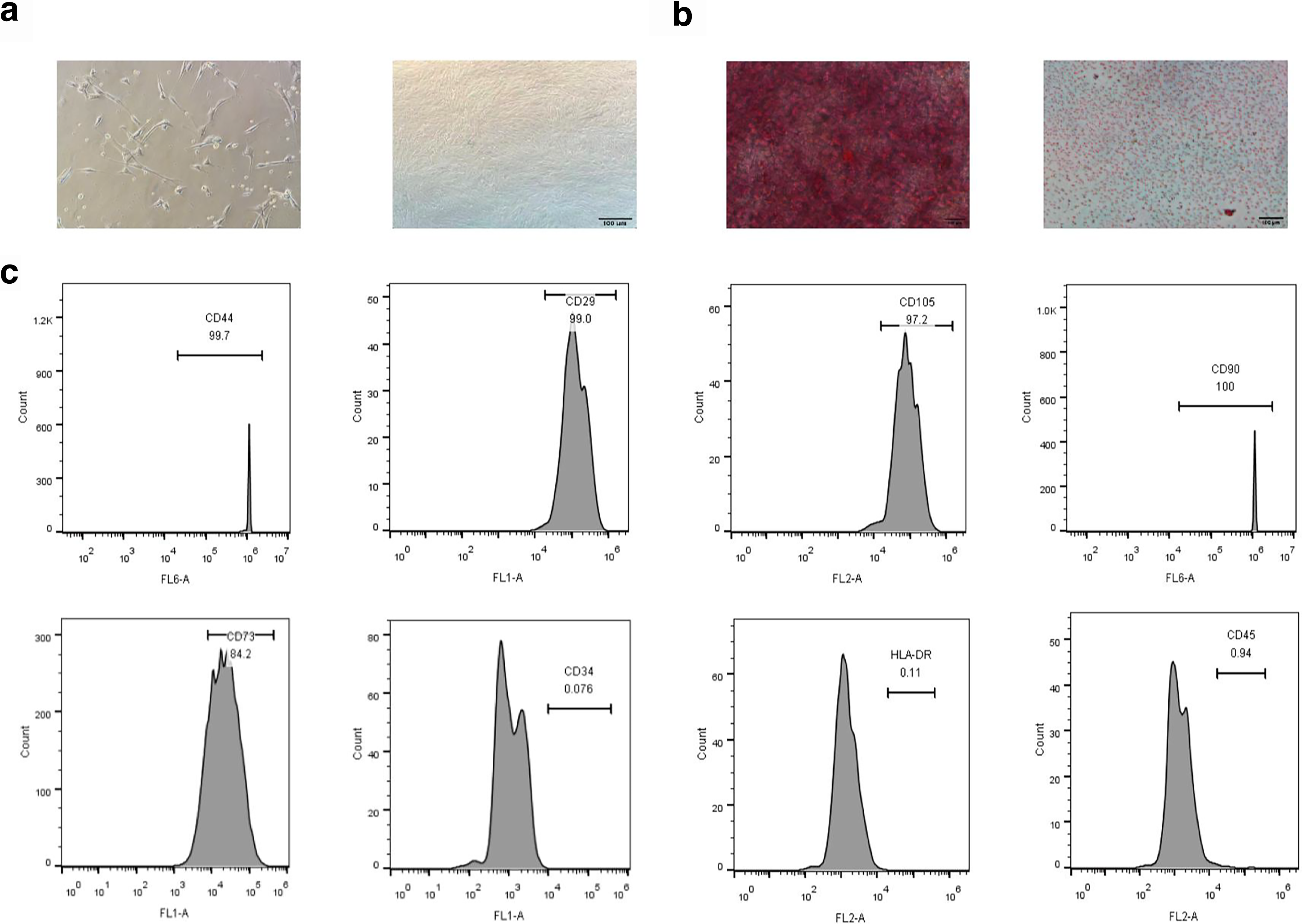
After 7–10 days of cultivation, the HUMSCs began to form vortex-like configurations, resembling fibroblasts with spindle-like morphology (Fig. 1a). Flow cytometry results (Fig. 1e) showed that negative markers such as CD34 (0.076%), CD45 (0.96%) and HLA-DR (0.11%) were sparsely expressed, while positive HUMSC markers including CD29 (99%), CD44 (99.7%), CD73 (84.2%), CD90 (100%), and CD105 (97.2%) were highly expressed. Additionally, after differentiation activation through osteogenesis (Fig. 1b) and lipogenesis (Fig. 1c), cells displayed a large number of red lipid droplets and calcium nodular deposits. These results are consistent with previous publications on the phenotypic traits of HUMSCs.
Isolation and characterization of HUMSCs.
Effects of IFN-γ stimulation on HUMSCs
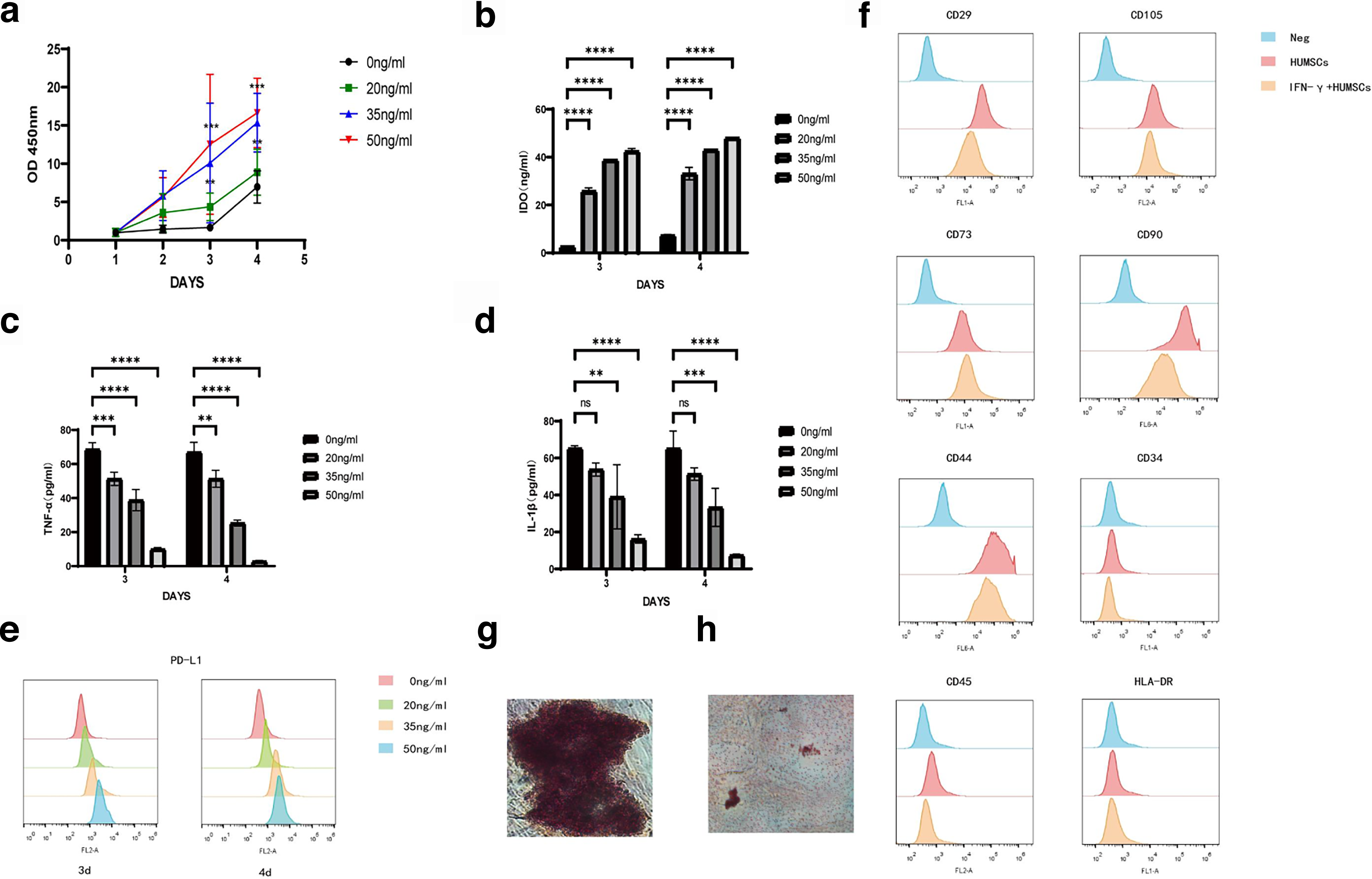
IFN-γ was used to induce HUMSCs at varying concentrations, including 0, 20, 35, and 50 ng/mL. Following stimulation with different doses of IFN-γ, the HUMSCs exhibited significantly higher viability on days 3 and 4. These results suggest a positive correlation between cell proliferation capability and IFN-γ concentration, indicating that optimal levels of IFN-γ can stimulate cell viability and proliferation (Fig. 2a). On days 3 and 4, we examined the expression of paracrine factors in HUMSCs, including IDO, TNF-α, IL-1β, and PD-L1. IFN-γ activation decreased the production of inflammatory factors, including TNF-α and IL-1β (Fig. 2c and d), whereas the production of immune-regulatory proteins such as IDO and PD-L1 increased (Fig. 2a and e). Based on these findings, an IFN-γ concentration of 50 ng/mL was chosen for further research.
Effects of IFN-γ at concentrations of 0, 20, 35, and 50 ng/ml after stimulation of HUMSCs.
Following IFN-γ activation, there was no change in HUMSCs’ surface antigen markers (Fig. 2f), nor in their ability for osteogenic and adipogenic differentiation (Fig. 2g and h). These results suggest that IFN-γ does not affect the characteristics of HUMSCs.
Preparation of matrices loaded with IFN-γ-pretreated HUMSCs and In vitro evaluation of biocompatibility
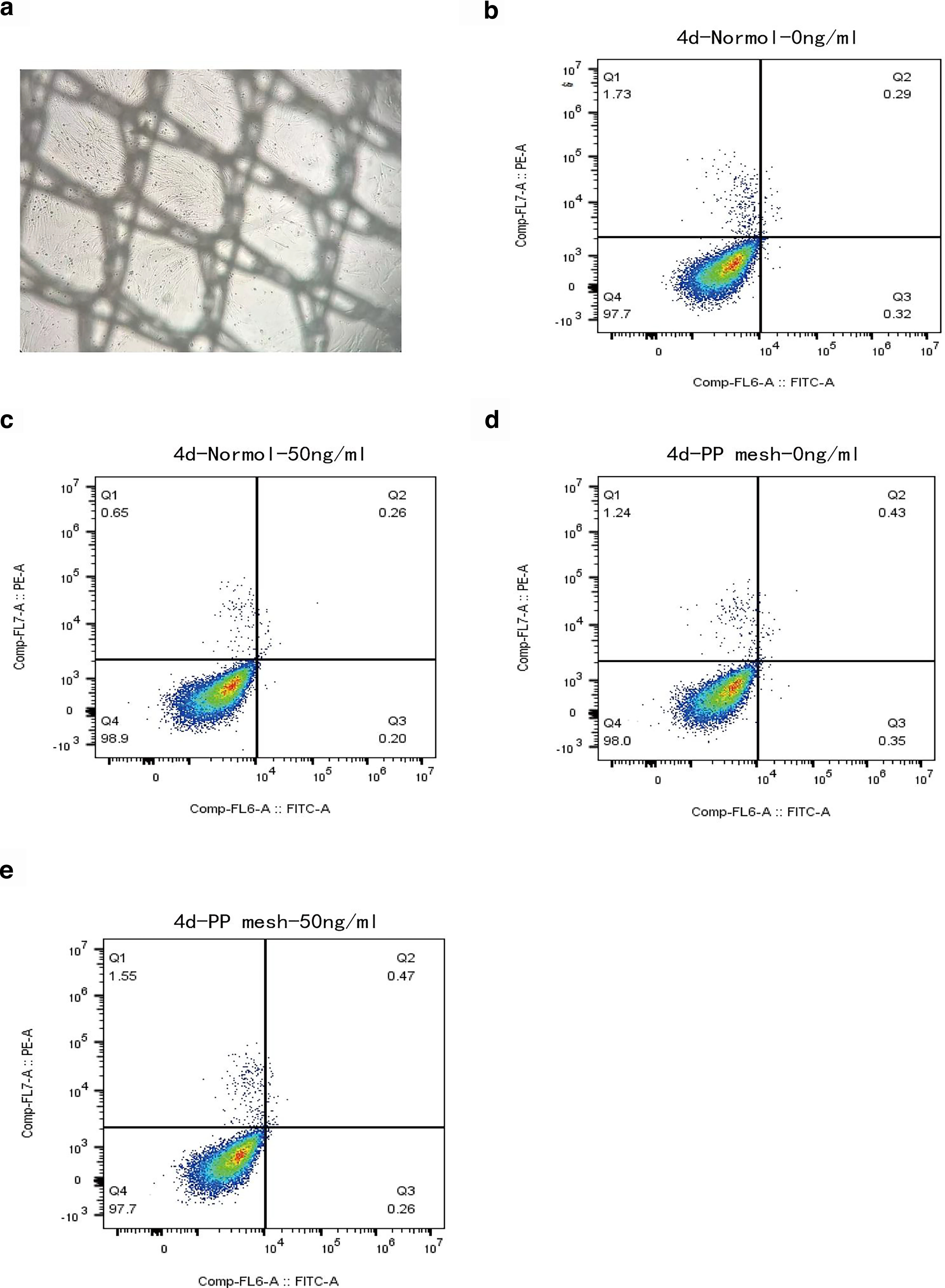
PP mesh that had previously been soaked in a UV-irradiated solution was used to seed the cells. Once the cells had attached, the medium was changed to one containing 50 ng/mL of IFN-γ. Four days later, microscopic examination revealed that the cells were healthy and had attached well (Fig. 3a). Live/dead cell experiments (Figs. 3b–e) showed that cells treated with 50 ng/mL IFN-γ and seeded onto PP mesh for 4 days did not demonstrate substantial cell death, suggesting that the PP mesh is free of apparent cytotoxicity and appropriate for cell loading.
Preparation and biocompatibility of mesh.
Establishment and evaluation of a rat POP model
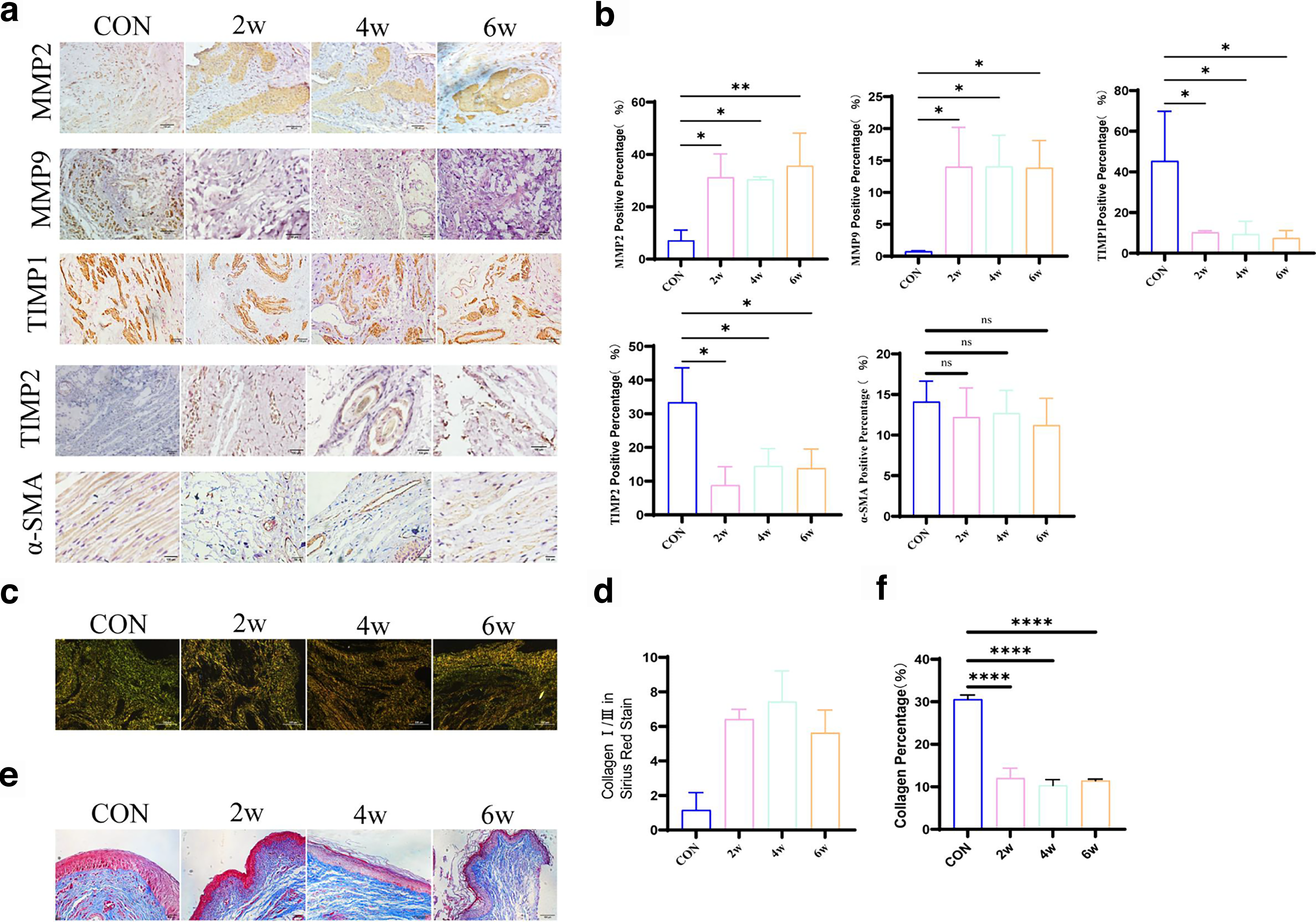
The experiment involved creating a rat POP model by initially performing a bilateral full subcostal ovariectomy, followed by balloon dilatation 1–2 weeks later, as illustrated in Supplementary Figure S2 and in accordance with the literature description. Two, four, and six weeks after dilation, the rats were anesthetized, and tissue samples were taken from their vaginal walls for further analysis. Changes in collagen, α-SMA, and the ECM are associated with the POP model. MMPs are associated with ECM breakdown. MMP-mediated ECM breakdown is controlled in vivo by matrix metalloproteinase inhibitors (TIMPs), which bind to MMPs and reduce their activity. Collagen catabolism is regulated by MMPs and TIMPs in both directions, and the degree of collagen catabolism is determined by dynamic fluctuations in their expression levels. The expression levels of MMP2, MMP9, TIMP1, and TIMP2 were measured at various time points. At various weeks in the modeling group, TIMP1 and TIMP2 expression levels dramatically dropped (Fig. 4a,b), COL I and COL III expression levels significantly decreased (Fig. 4c,d), and MMP2 and MMP9 expression levels significantly increased (Fig. 4a,b). There were no discernible variations within the modeling group. These results were confirmed by Western blot analysis (Fig. 5a and b).
Histopathologic analysis of rats model of pelvic organ prolapses.
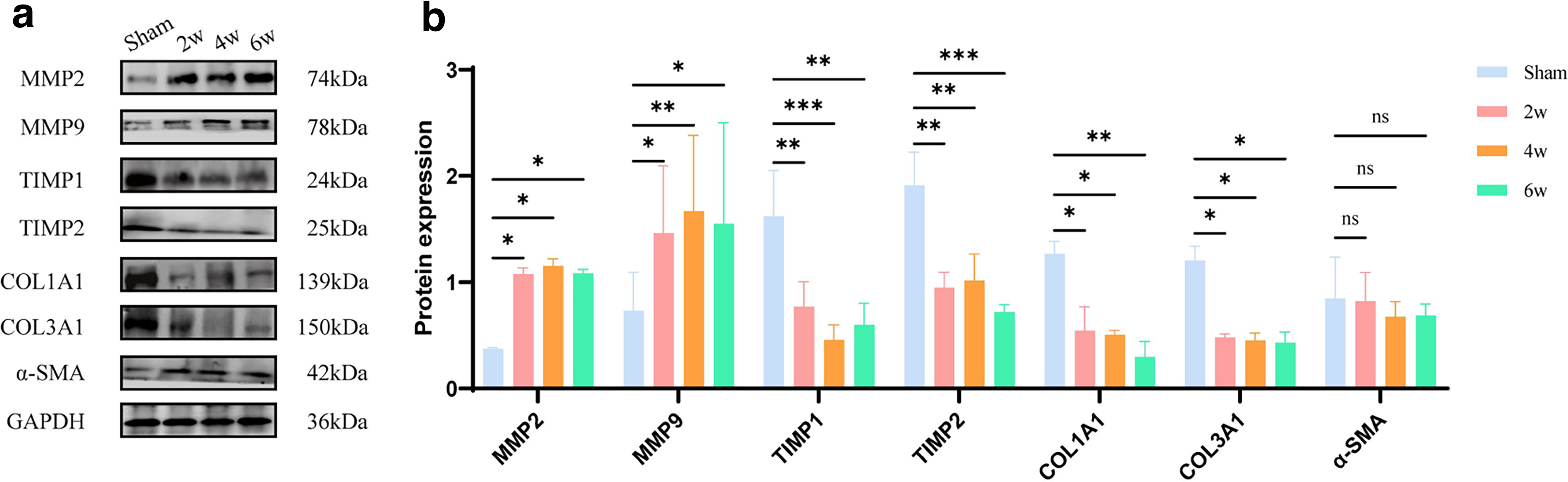
WB detection of changes in POP model-related indicators.
The control group’s α-SMA immunohistochemical results revealed a well-ordered muscle fiber structure, whereas the modeling group’s results indicated a disorganized muscle fiber configuration. However, there was no discernible difference in the proportion of muscle fibers with positive staining areas between the groups (Fig. 4a,b). These results were confirmed by Western blot analysis (Fig. 5a,b).
Comparing the modeling group with the control group, Sirius red staining revealed a significantly higher COLI/COLIII ratio. However, no discernible variations in this ratio were found within the modeling group throughout the weeks (Fig. 5c,d). A higher ratio in the modeling group indicates tissue hardening, elasticity loss, and organ dysfunction.
Masson staining showed substantial atrophy of the vaginal epithelium and notable collagen tissue loosening in the vaginal wall with visible spaces between the collagen fibers during all modeling weeks (Fig. 4e,f).
These tests verified the rat POP model, indicating that it is appropriate for further mesh implantation research.
In vivo biocompatibility evaluation of scaffolds loaded with IFN-γ-pretreated HUMSCs
Three different mesh types were inserted into the aforementioned successful POP model: PP mesh loaded with HUMSCs; PP mesh loaded with IFN-γ-pretreated HUMSCs; and PP mesh (Supplementary Fig. S3). Four weeks after implantation, the IFN-γ group exhibited fewer instances of exposure and wrinkling, as detailed in Supplementary Table S1. This suggests that pretreating HUMSCs with IFN-γ improves the biocompatibility of traditional PP meshes and reduces the risk of adverse effects.
The effect of mesh implantation of IFN-γ-preconditioned HUMSCs on rat POP
After the rats were euthanized, the tissue-mesh grafts were removed approximately 4 weeks after implantation. H&E staining was used to assess the structural integrity of the tissue and the placement of the mesh. Figure 6e shows the significant wrinkling and asymmetrical arrangement of standard PP meshes and meshes loaded exclusively with HUMSCs within the tissue. The typical structure of the surrounding tissue was lost. In exposed cases, the mesh pierced the muscular and epithelial layers of the vaginal wall. In contrast, the altered mesh remained flat within the tissue and retained its natural shape, with stable and visible surrounding tissue structures.

After implanting several mesh types into the POP model, the specimens were collected upon euthanasia and analyzed using staining.
Particular evaluations were carried out on the muscular layer and connective tissue in light of the H&E staining, which indicates disturbed tissue architecture. According to the results, the muscular layer was severely disrupted with a disordered muscle fiber arrangement in tissues implanted with PP mesh and PP mesh loaded solely with HUMSCs. In many instances, the mesh divided the muscle layers after invading them. By contrast, the muscle layer in the modified mesh group had an organized structure and complete integrity, with the mesh situated beneath it. According to immunohistochemical data (Fig. 6a,b) and Western blot (Fig. 7a,b), the IFN-γ group showed a significant increase in α-SMA expression after mesh implantation.
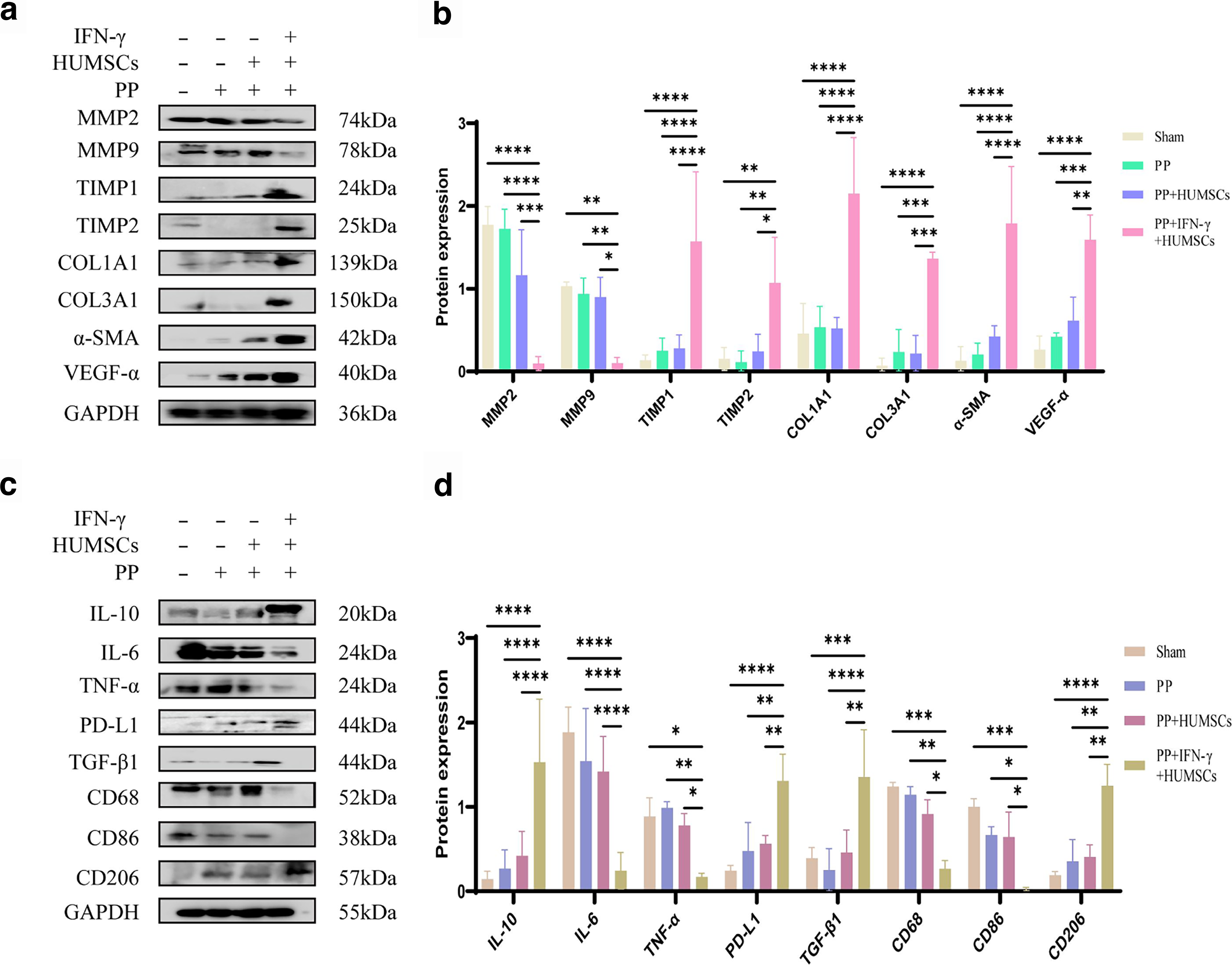
WB detection of proteins associated with tissue repair, inflammation, and immune regulation.
Also, the group treated with IFN-γ showed significantly higher VEGF-α expression than the other groups, suggesting that angiogenesis was boosted in this group (Fig. 7a and b).
IFN-γ upregulates the expression of immunosuppressive molecules such as PD-L1 and TGF-β1 (Fig. 7c and d), while also stimulating MSCs, which exhibit strong immunological activation. By inhibiting the generation of proinflammatory factors by surrounding immune cells, IFN-γ-induced molecules such as PD-L1 have an overall effect of reducing proinflammatory factors while increasing anti-inflammatory factors. The anti-inflammatory factor IL-10 increased significantly, while the proinflammatory factors IL-6 and TNF-α decreased considerably (Fig. 7c,d). Macrophages are impacted by the activation of MSCs by IFN-γ, which causes them to proliferate and polarize from M1 to M2. The main indicators of this are increased expression of the M2 macrophage marker CD206, decreased expression of the M1 macrophage marker CD86, and increased expression of the macrophage surface marker CD68. The results of the Western blot (Fig. 7c and d) and immunohistochemistry (Fig. 6a and b) were consistent.
Discussion
POP is the term used to describe the descent and protrusion of the bladder, vagina, and uterus. 24 Synthetic mesh implants used in transvaginal pelvic floor restoration surgeries carry a comparatively high risk of complications. Furthermore, the limited clinical use of biomaterial meshes is due to their poor mechanical properties and rapid degradation rate, resulting in suboptimal anatomical repair. Consequently, tissue-engineered scaffolds have emerged as a promising therapeutic approach. Tissue regeneration involves repairing and regenerating damaged or injured organs and tissues using scaffolds, cellular integration, and the right biochemical and physicochemical elements to support tissue growth. 25 MSCs appear to be an ideal cell type for clinical implantation and scaffold seeding. MSCs can be induced to differentiate into bone, cartilage, fat, tendon, and muscle. They have been found to exhibit immunomodulatory action and multipotent differentiation capability, as well as the ability to self-renew.26,27 The basic idea is to combine stem cells with existing muscle tissue in order to produce growth factors that encourage muscle and nerve regeneration. MSCs primarily work through paracrine and homing processes.
Skin healing, 28 abdominal wall hernias, 29 and POP 30 have all been successfully treated with tissue engineering based on stimulatory factors and cells. This is mainly accomplished by decreasing proinflammatory factors and raising paracrine factors linked to immunomodulation, 31 angiogenesis, ECM, and anti-inflammation. 32 The mesh’s therapeutic effectiveness for tissue regeneration is influenced by important factors such as cell adhesion, density, and vitality. For this investigation, IFN-γ-stimulated HUMSCs were chosen to boost paracrine factors and improve stem cell survival. Using stimulated stem cells further facilitates HUMSC activity in rat models, as they maintain their natural characteristics, adhere well to the PP mesh, and exhibit exceptional cell proliferation potential.
It is still difficult to establish animal models in POP-related research. Most implants are placed in the back, abdomen, or under the vaginal wall. Commonly used modeling techniques include vaginal dilatation, ovariectomy, or spontaneous prolapse in sheep, 33 rhesus macaque, 34 or rodent. 35 Rats were chosen as the experimental subjects for this study. A bilateral ovariectomy was initially carried out to create a menopausal model. Oophorectomy is a straightforward, controllable, and repeatable modeling technique.16,36 Then, the biomechanical benefits of stretching the vaginal wall following dilatation were combined with the histological benefits of ovariectomy to create a POP model using balloon dilatation after ovariectomy. This model of vaginal wall prolapse in rats was successfully created. Assessments showed that, in human POP patients, modifications to the vaginal wall were correlated with changes in collagen, MMPs, and TIMPs. However, the small size of the rodents and their inability to walk continue to be fundamental limitations of this model. Without clinically useful markers for tissue prolapse assessment, we have only modeled the histological and biomechanical elements and cannot quantify prolapse grading.
Although the precise mechanisms and pathways are unclear, our research shows that the implantation of a mesh containing IFN-γ-pretreated HUMSCs enhances rat POP. Further research is needed.
Conclusion
The results of the study show that meshes loaded with IFN-γ-pretreated HUMSCs significantly improved the condition of rat POP after implantation, demonstrating superior tissue-repair capabilities, improved immunomodulatory functions, and weaker host reactions. Therefore, IFN-γ-stimulated HUMSCs are an excellent option for cell transplantation and regenerative medicine, as they are capable of enhancing the biocompatibility of implanted scaffolds and reducing postoperative complications.
Authors’ Contributions
S.Y.L. drafted the article, designed the experiments, and analyzed the data. F.M., Y.T.S., and Q.Z. contributed to data analysis and edited the article. S.Y.L., F.R.S., J.W., H.M.D., J.H.Z., and Y.G.C. wrote the article, designed the overall testing protocol, and determined the final article version. All the authors have read and approved the article for publication.
Data Availability Statement
The data that support the findings of this study are available from the corresponding author upon reasonable request.
Ethical/Institutional Review Board Approval
All animal experimentation protocols were strictly approved and implemented by the Suzhou University Animal Care and Use Committee (SUDA20251013A13).
Footnotes
Acknowledgments
The authors would like to thank Professor Fu Fengqing, of the Suzhou Institute of Immunology in Jiangsu Province, and his colleagues, for their leadership and assistance in conducting the tests.
Author Disclosure Statement
The authors declare no conflict of interest.
Funding Information
Research Project on health of the elderly in Jiangsu Province (LKM2022020).
Supplemental Material
Supplemental Material
Supplemental Material
Supplemental Material
References
Supplementary Material
Please find the following supplemental material available below.
For Open Access articles published under a Creative Commons License, all supplemental material carries the same license as the article it is associated with.
For non-Open Access articles published, all supplemental material carries a non-exclusive license, and permission requests for re-use of supplemental material or any part of supplemental material shall be sent directly to the copyright owner as specified in the copyright notice associated with the article.