
Abstract
To develop bone grafts with superior osteoconductivity, we have fabricated unidirectionally connected porous carbonate apatite (CAp) via gelatin gelation combined with freeze-drying. A composite of calcium carbonate (CaCO3) and gelatin was first gelled at low temperature and then subjected to directional freeze-drying to create well-aligned macroporous channels. The samples were then sintered to remove the organic components and subsequently phosphatized to convert the composition into CAp. The resulting material exhibited a porosity exceeding 80% with fully interconnected unidirectional pores favorable for cell migration and vascular infiltration. Bone regeneration was evaluated by implanting the material into rabbit femoral bone defects; a comparison was also made with nonoriented porous controls. Histological and radiographical analyses demonstrated that the unidirectional porous structure significantly enhanced directional ingrowth of new bone tissue and accelerated early-stage bone formation relative to the control. This study demonstrates that unidirectional porous CAp fabricated via a gelatin-based freeze-casting method is promising for bone regeneration, particularly in clinical applications requiring guided bone formation.
Impact Statement
The architecture of bone grafts plays a critical role in guiding cell migration, vascularization, and subsequent bone regeneration. While carbonate apatite (CAp) is widely recognized for its excellent osteoconductivity, strategies to precisely control its three-dimensional pore orientation remain limited. In this study, we demonstrated that a gelatin-based freeze-casting approach enables the fabrication of unidirectionally connected porous CAp with high porosity. This porous architecture significantly enhances directional bone ingrowth and early-stage bone formation in vivo. Our findings highlight the importance of pore orientation, beyond porosity alone, in optimizing osteoconductive performance and suggest that unidirectional porous CAp is a promising bone graft material for clinical applications requiring guided bone regeneration.
Introduction
Maintaining the structural and functional integrity of bone, a key component of the locomotor system, is essential for preserving mobility, quality of life, and healthy life expectancy.1–3 Large bone defects resulting from trauma, tumor resection, or degenerative diseases are commonly treated using autologous bone grafts, allografts, and/or xenografts.4,5 However, each approach has inherent limitations. For example, autologous grafting requires additional surgery and is associated with donor site morbidity; whereas allografts and xenografts carry risks of immunogenicity and disease transmission.4–6
To overcome these drawbacks, synthetic bone grafts have been developed as alternatives.7–9 They can be designed to mimic both the chemical composition and the structural characteristics of natural bone, thereby providing osteoconductivity and controlled resorption without the disadvantages of biological grafts. Among the parameters governing scaffold performance, composition and architecture are particularly critical. Regarding compositional performance, carbonate apatite (CAp) is attractive because it closely resembles bone mineral and undergoes bioresorption via osteoclastic activity that promotes subsequent osteoblast recruitment and bone remodeling.10,11 Structurally, porous scaffolds enable fluid transport, cell migration, and vascularization; the osteoconductivity is strongly influenced by pore size, interconnectivity, and orientation.12–15
Recent studies have highlighted the unique advantages of unidirectional porous architectures, which provide long-range guidance for tissue ingrowth.16,17 Conventional pore-forming methods such as gas foaming and porogen leaching often produce closed or poorly connected pores.18–20 By contrast, directional freezing can yield open, aligned channels by controlling thermal conduction during cooling to orient ice columns and the resulting pores. Gelatin-mediated directional freezing has been widely used to prepare aligned porous bioceramics such as β-tricalcium phosphate and hydroxyapatite. 17 However, the effect of pore orientation on bone formation in CAp scaffolds has not been investigated.
Here, gelatin-mediated directional freezing was employed to fabricate unidirectional porous CAp scaffolds. The in vivo efficacy was evaluated in a rabbit femoral defect model, with randomly porous CAp (rad-CAp) used as a control.
Materials and Methods
Fabrication of unidirectional porous CAp blocks
Calcium carbonate (CaCO3) powders (Sakai Chemical Industry Co., Ltd., Osaka, Japan) were dispersed in a 3-wt% gelatin solution (Sigma-Aldrich, Co., USA). The suspension was maintained at 5°C overnight to induce gelation. Subsequently, the CaCO3 gels were subjected to unidirectional freezing at −10°C in a cooling device (MyBL-100CS; AS ONE Corporation, Osaka, Japan). After complete freezing, the samples were placed in a freeze dryer (FDU-1200; Tokyo Rikakikai Co., Ltd., Tokyo, Japan) under low pressure (<15 Pa). The resulting dried samples were sintered at 700°C for 24 h to remove the gelatin component. The sintered CaCO3 blocks were immersed in a 0.1 mol/L Na2HPO4 aqueous solution at 80°C for 7 days to induce conversion from CaCO3 to CAp. Finally, the unidirectional CAp blocks (uni-CAp) were shaped into cylinders (6 mm diameters, 4 mm lengths) using a computer-controlled milling machine (SRM-20; Roland DG Corp., Hamamatsu, Japan).
Fabrication of randomly porous CAp blocks
Randomly porous CAp blocks (rad-CAp) were fabricated using a procedure similar to that described above in Section 2.1, except that the −10-°C freezing was performed by placing the CaCO3 gels directly in a conventional freezer, without unidirectional freezing. All other steps, including freeze-drying, sintering, and chemical conversion, were performed under the same conditions described above.
Characterization of physicochemical, structural, and mechanical properties
The microstructure morphologies of uni-CAp and rad-CAp longitudinal cross-sections were imaged via scanning electron microscopy (S-3400N, Hitachi, Tokyo, Japan). Structural analyses were performed via microcomputed tomography (ScanXmate-L090T, Comscan, Kanagawa, Japan).
Phase compositions of uni-CAp and rad-CAp were analyzed with X-ray diffraction (Rigaku, Tokyo, Japan) at 40 kV and 50 mA and a scanning speed of 20°/min. Functional groups were analyzed using Fourier-transform infrared spectroscopy (FT/IR-6200, JASCO, Tokyo, Japan). The carbonate content was measured using an MT-6 carbon-hydrogen-nitrogen elemental analyzer (Yanako Analytical Instruments, Kyoto, Japan).
Mercury intrusion porosimetry (AutoPore 9420, Shimadzu Corporation, Kyoto, Japan) was used to determine sample pore size distributions and total porosities.
The compressive strengths of uni-CAp and rad-CAp were measured via a universal testing machine (Autograph AGS-J, Shimadzu Corp., Kyoto, Japan). Compression was applied along the longitudinal axis of the cylindrical specimens; for uni-CAp, this direction was parallel to the freezing direction. The crosshead speed was 1 mm/min, and five samples were used for each group.
Calcium ion release from uni-CAp and rad-CAp was measured by immersing each sample (50 mg) in 20 mL of an isotonic 4-(2-hydroxyethyl)-1-piperazineethanesulfonic acid–sodium chloride (HEPES–NaCl) buffer solution (Fujifilm Wako Pure Chemicals, Osaka, Japan) at 37°C. The solution consisted of 10 mmol/L HEPES and 140 mmol/L NaCl, adjusted to pH 7.4 with 1 mol/L NaOH. The buffer was replaced every 7 days with an equal volume of fresh solution to prevent ion oversaturation and precipitation. The calcium ions (Ca2+) released into the buffer were analyzed after 7, 14, and 21 days using inductively coupled plasma optical emission spectroscopy (Optima 7300DV, PerkinElmer, Waltham, MA, USA). To further evaluate the degradation behavior of the scaffolds, an in vitro weight loss test was performed. Each sample was weighted before immersion to determine the initial dry weight. The samples were then immersed in 20 mL of isotonic HEPES–NaCl buffer solution at 37°C for up to 21 days. At each predetermined time point, the samples were collected, gently rinsed with distilled water, dried until a constant weight was obtained, and weighed again. The weight loss percentage was calculated from the decrease in dry weight after immersion relative to the initial dry weight and expressed as a percentage.
Sterilization procedures
Before in vitro cell experiments and surgical implantation, the samples were sterilized via dry heat at 170°C for 3 h. Surgical instruments were autoclaved at 121°C for 20 min before use.
In vitro cell adhesion and proliferation assays
To evaluate the early cellular response to the different pore architectures, in vitro cell adhesion and proliferation assays were performed using MC3T3-E1 cells. The cells were cultured in culture medium supplemented with 10% fetal bovine serum and 1% penicillin-streptomycin at 37°C in a humidified atmosphere containing 5% CO2.
For the cell adhesion assay, cells were directly seeded onto sterilized uni-CAp and rad-CAp scaffolds at a density of 40,000 cells/sample and cultured for 24 h. After incubation, the samples were gently washed with phosphate-buffered saline to remove nonadherent cells and fixed with 10% formalin. The fixed cells were then permeabilized with 1% polyethylene glycol mono-4-octylphenyl ether (Triton X-100, Nacalai Tesque, Inc., Kyoto, Japan). The actin cytoskeleton and nuclei were stained with phalloidin (Acti-stain 555 phalloidin, Cytoskeleton, Inc., Denver, CO, USA) and Hoechst 33258 (Dojindo), respectively. Cell attachment, distribution, and morphology on the scaffold surfaces were observed using a fluorescence microscope (BZ-X 700, Keyence, Osaka, Japan).
For the proliferation assay, scaffold extracts were prepared by immersing sterilized uni-CAp and rad-CAp samples in culture medium at 200 mg/mL and incubating them at 37°C for 24 h. Cells were seeded in a 48-well plate at a density of 8,000 cells/well and cultured with uni-CAp or rad-CAp extract medium for 1, 3, and 7 days. At each time point, cell proliferation was evaluated using a Cell Counting Kit-8 assay (CCK-8; Dojindo). CCK-8 regent was added to each well and incubated at 37°C for 2 h. The absorbance at 450 nm, used as an indicator of cell proliferation, was measured using a microplate reader. Blank wells containing extract medium and CCK-8 reagent without cells were used for background correction.
Animals
Japanese white rabbits (Japan SLC Inc., Shizuoka, Japan) were individually housed in cages under standard laboratory conditions, with free access to standard feed and water, at the Center of Biomedical Research in the Research Center for Human Disease Modeling at the Graduate School of Medical Sciences, Kyushu University.
Surgical procedures
All animal experiments were conducted according to the ethical policies and procedures approved by the Animal Care and Use Committee of Kyushu University, Japan (Approval No. A24-478-0; Issued August 1, 2018).
Rabbits were sedated by intramuscular injection of ketamine (30 mg/kg; Ketalar, Daiichi Sankyo Propharma, Tokyo, Japan) and xylazine (5.0 mg/kg; Selactar, Elanco, Indianapolis, IN, USA). The medial femoral region was shaved, and anesthesia was maintained throughout surgery via intravenous injection of ketamine (10 mg/kg) and xylazine (3 mg/kg) into the marginal ear vein. The surgical site was disinfected with 10% povidone-iodine solution, and local anesthesia was applied using 2% lidocaine (Nipro, Osaka, Japan).
The skin, fascia, and muscle were sequentially incised, and the periosteum was carefully separated to expose the femoral condyle. A cylindrical bone defect (6 mm diameter, 4 mm depth) was created using a surgical drill under continuous saline irrigation to prevent thermal damage. Subsequently, either uni-CAp or rad-CAp samples were randomly implanted into the defects. The surgical wounds were then closed sequentially from the periosteum to the skin. Postoperatively, rabbits received an intraperitoneal injection of gentamicin (4 mg/kg; Takata Pharmaceutical, Saitama, Japan) to prevent infection.
The rabbits were euthanized at 4 weeks or 8 weeks postimplantation. Tissue specimens from the surgical sites, including implanted samples and surrounding bone tissues, were harvested and fixed in 10% neutral buffered formalin solution.
Microcomputed tomography and histological analysis
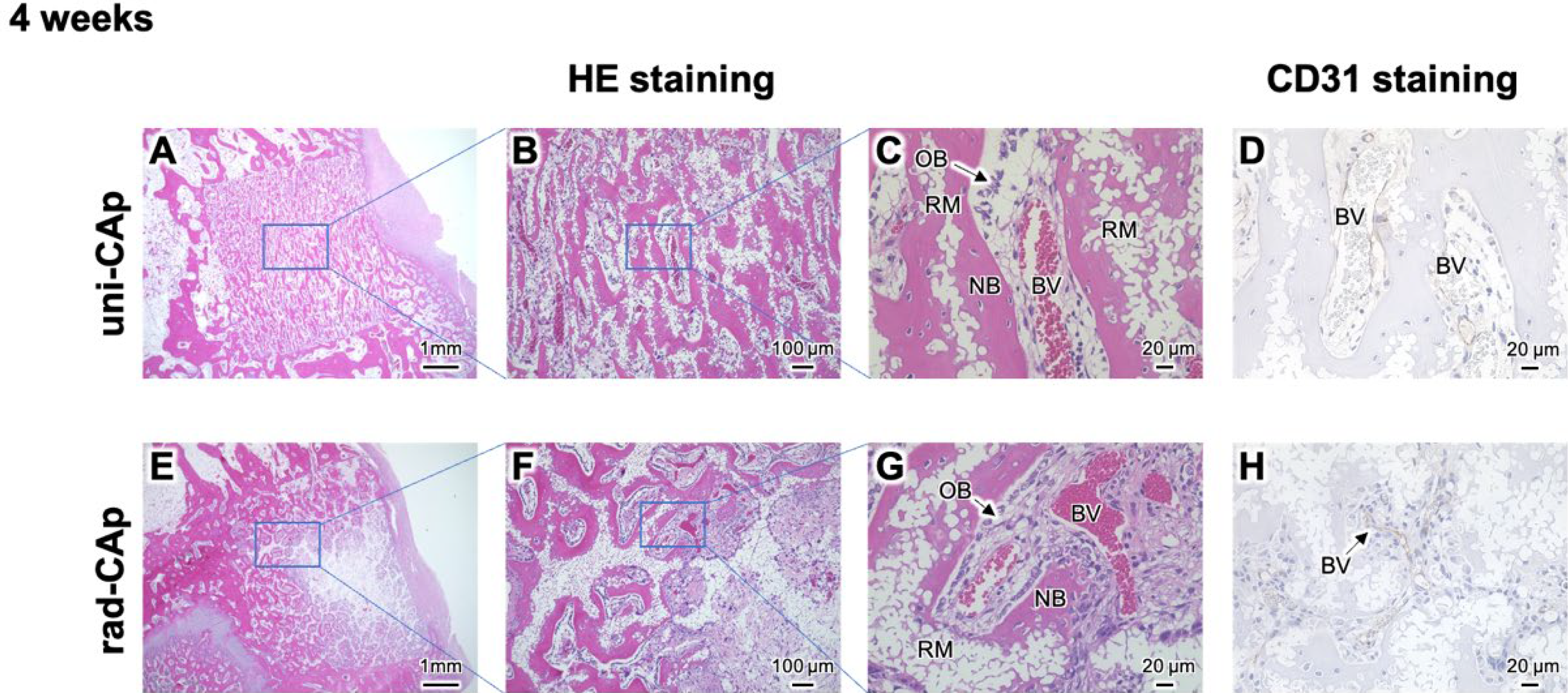
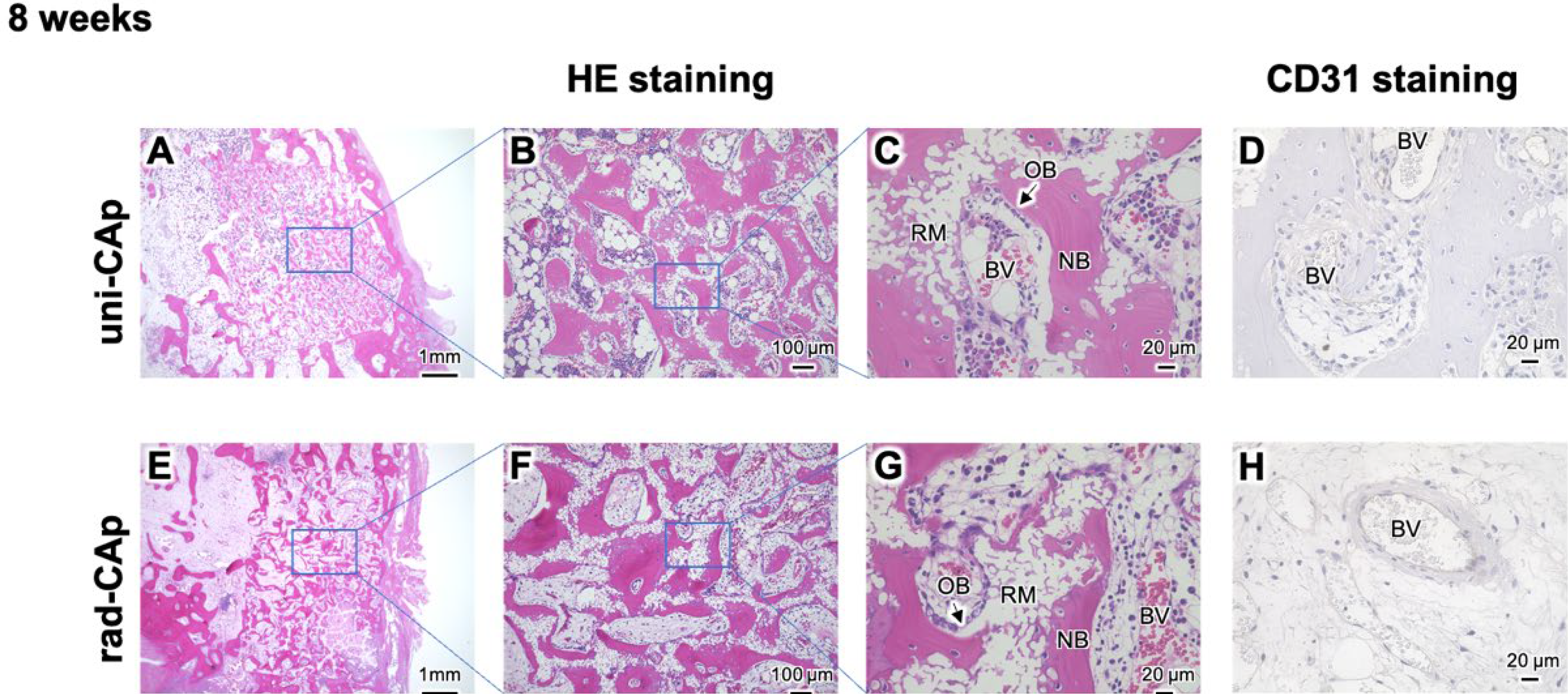
Collected samples were scanned using microcomputed tomography (ScanXmate-L090T, Comscan, Kanagawa, Japan) and analyzed using commercial software (CTAn; Bruker, MA, USA). Subsequently, the specimens were decalcified, embedded in paraffin, and sectioned. Following deparaffinization, sections were stained with hematoxylin and eosin (HE) and subjected to CD31 (platelet endothelial cell adhesion molecule-1) immunohistochemistry. For CD31 staining, monoclonal mouse antihuman CD31 (C31.7; Abcam, Cambridge, UK) and polyclonal goat anti-mouse immunoglobulins/biotinylated (ab6788; Abcam) were used as the primary and secondary antibodies, respectively. An avidin-biotin complex (ABC) detection system (Ultra-Sensitive ABC peroxidase Standard Staining Kit, #32050; Thermo Fisher Scientific, MA, USA) was used to detect immunoactivity. The sections were stained with the chromogen 3,3′-diaminobenzidine (D12384; Sigma-Aldrich). Histological images of HE- and CD31-stained sections were obtained using an optical microscope (BZ-X700; Keyence, Osaka, Japan). For histomorphometric analysis, the region of interest (ROI) was defined as the entire original defect area in each histological section. Within this ROI, the areas of new bone (NB), remaining material (RM), and blood vessel (BV) were quantitatively analyzed using digital image analysis software.
Statistical analysis
All data are presented here as the mean ± standard deviation. For the statistical analysis of differences between the two groups, a Student’s t-test was performed. For comparisons involving multiple groups, a Tukey-Kramer test was conducted. Statistical analyses were performed using R software [R Core Team (2022), version 4.2.2]. Statistical significance is indicated by *p < 0.05.
Results and Discussion
Fabrication and characterization of uni-CAp and rad-CAp scaffolds
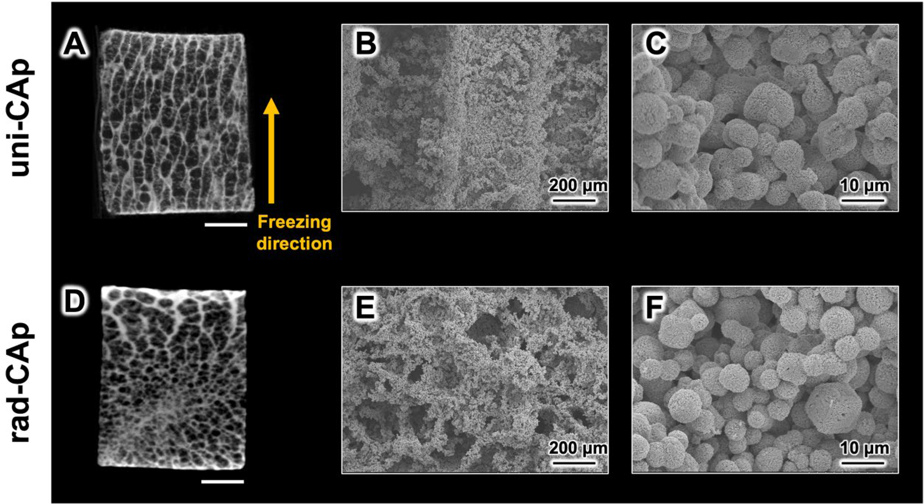
In this gelatin-based system, the imposed thermal gradient (directionally facilitated by a cold surface) governed ice-crystal growth and the porogenic architecture.21–23 Blocks obtained by this unidirectional freezing exhibited long-range channel alignment (uni-CAp) that reflected the direction of the advancing freezing front. Meanwhile, nondirected freezing resulted in a porous network without a preferential orientation (rad-CAp).22,24 Microcomputed tomography and scanning electron microscopy confirmed continuous, axially aligned macropores in uni-CAp and more tortuous, isotropic pore networks in rad-CAp (Fig. 1). Typical macroscopic features in uni-CAp had ∼300-μm channel widths and ∼50-μm strut (wall) thicknesses; while rad-CAp displayed ∼200–300-μm pores with shorter, discontinuous pathways.
Microcomputed tomography
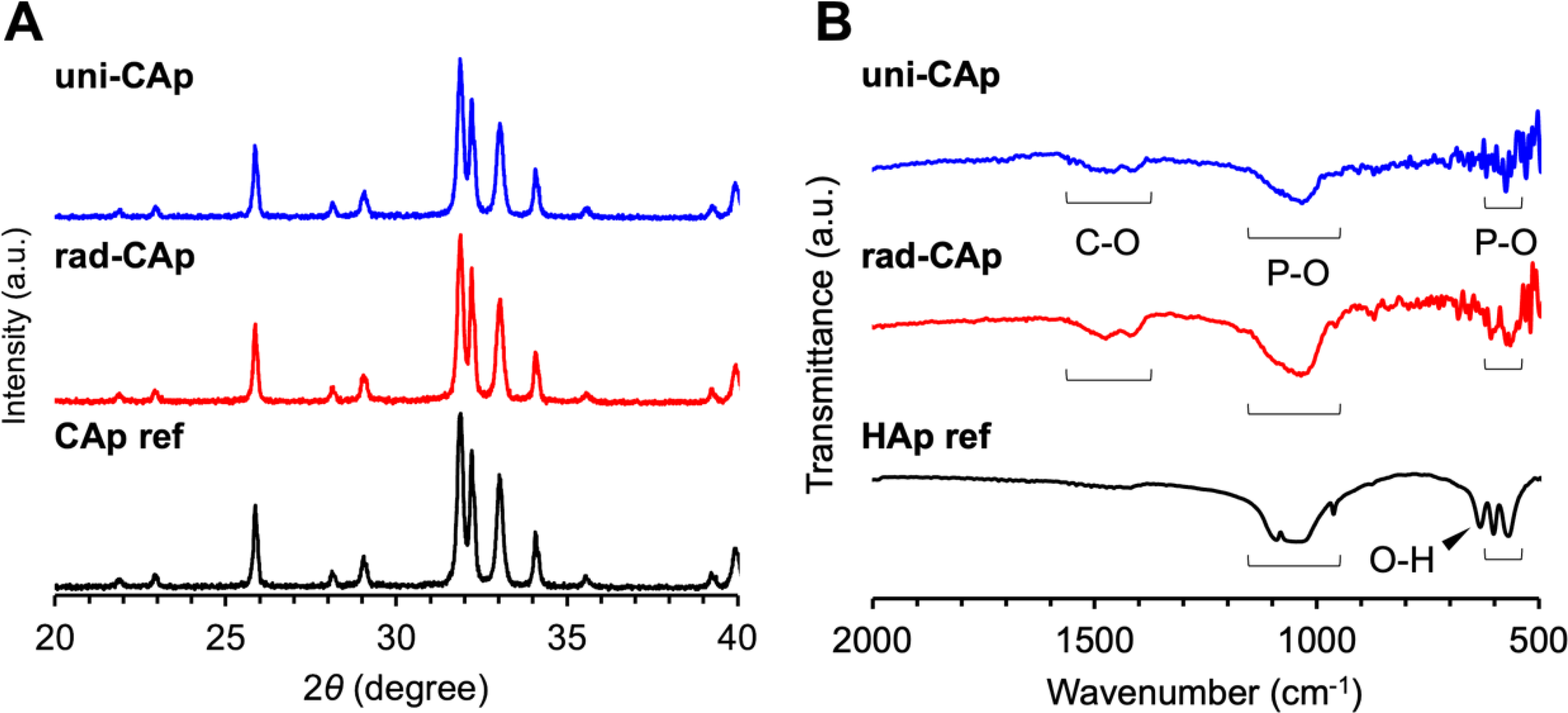
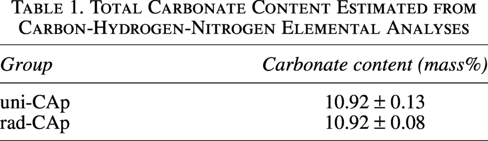
X-ray diffraction patterns of both groups revealed apatite crystalline phases, and Fourier-transform infrared spectra exhibited phosphate vibrational bands (υ3 1100–1000 cm−1; υ1 960 cm−1; υ4 600–560 cm−1) and carbonate vibrational bands (υ3 ∼1470–1415 cm−1; υ2 875 cm−1) consistent with carbonate-substituted apatite (Fig. 2). 25 The hydroxyl signature typical of stoichiometric HAp (O-H libration at ∼630 cm−1) was absent, consistent with bone-like, carbonate-rich apatite.25,26 Furthermore, no gelatin remnants were detected. Specifically, amide I (1700–1600 cm−1), amide II (1560–1500 cm−1), and amide III (1300–1200 cm−1) vibrational bands were absent above the noise, consistent with effective pyrolysis and unobstructed CAp formation.27,28 Total carbonate, estimated by carbon-hydrogen-nitrogen (CHN) analysis, was ∼11% (Table 1), which was suitably close to human bone. 10 It should be noted that the CHN analysis does not directly distinguish inorganic carbonate from residual carbon. However, the obtained samples were white, without any visual indication of residual carbon. In addition, the nitrogen content was 0%, indicating that no gelatin-derived residue remained after sintering, in agreement with the absence of amide bands in the FTIR spectra. The measured carbon was therefore predominantly derived from carbonate in the apatite phase. Thus, both conditions can be described conservatively as carbonate-substituted apatite, without assigning quantified A- vs B-site substitution states.
X-ray diffraction (XRD) patterns
Total Carbonate Content Estimated from Carbon-Hydrogen-Nitrogen Elemental Analyses
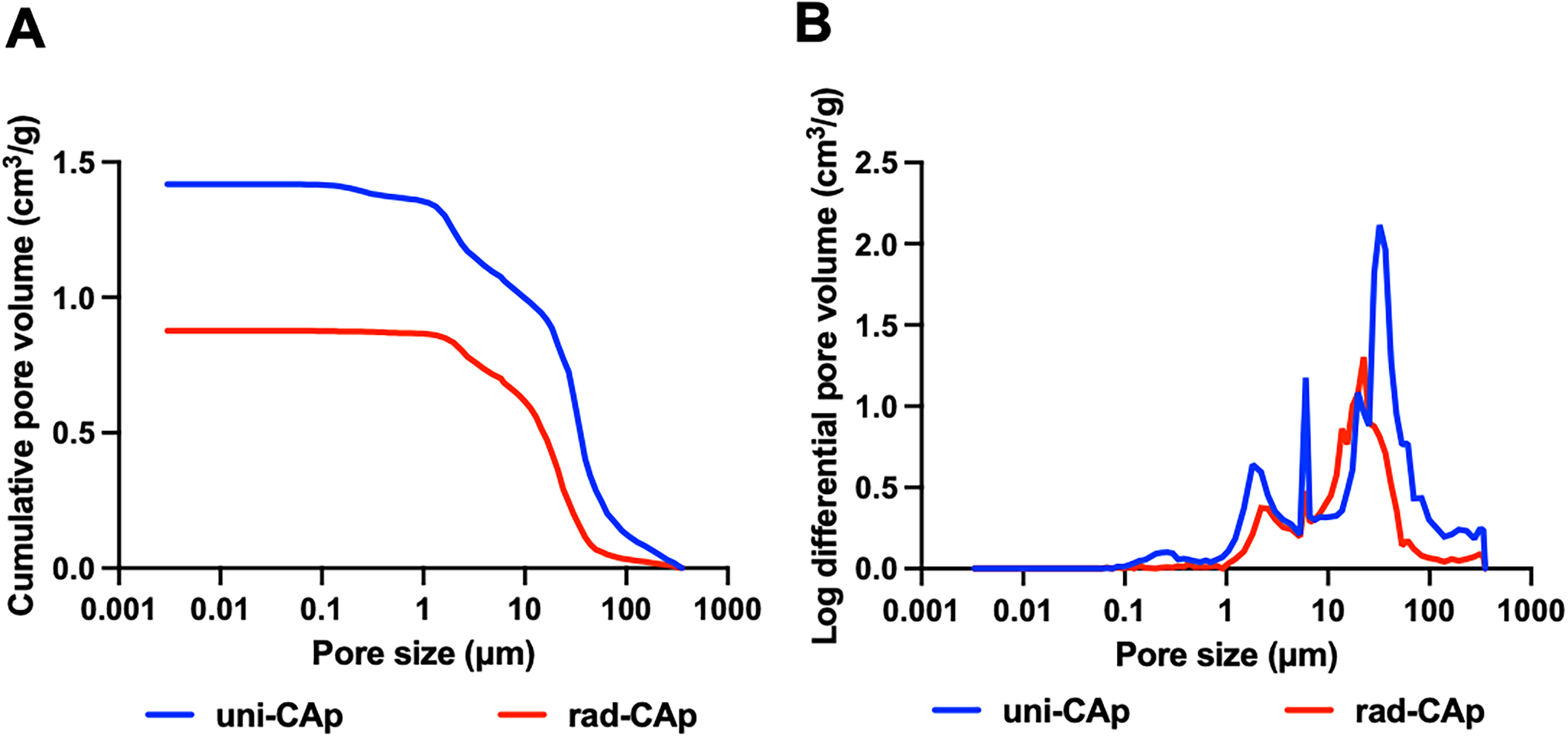
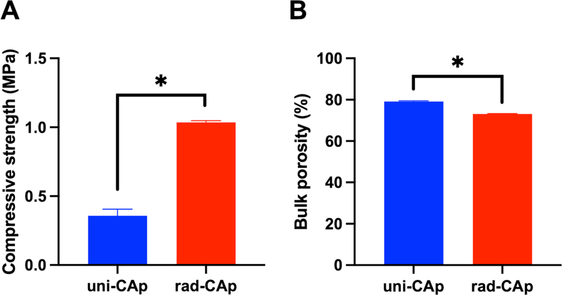
Mercury intrusion porosimetry indicated high porosity in both architectures, where uni-CAp generally exhibited a right-shifted intrusion profile and a higher cumulative porosity across most pore-size ranges (Fig. 3). The bulk porosity followed the same trend (Fig. 4B), where uni-CAp had 79.1 ± 0.4% and rad-CAp had 73.1 ± 0.2%, indicating that alignment did not compromise the total void fraction. Compressive strength values (Fig. 4A), measured with the load applied the longitudinal axis of the cylindrical specimens, parallel to the freezing direction in uni-CAp, showed that uni-CAp exhibited a lower strength (0.4 ± 0.05 MPa) than rad-CAp (1.0 ± 0.01 MPa). This difference was consistent with differences in load-path continuity associated with the unidirectional channel architecture. 29
Cumulative pore volume
Compressive strength
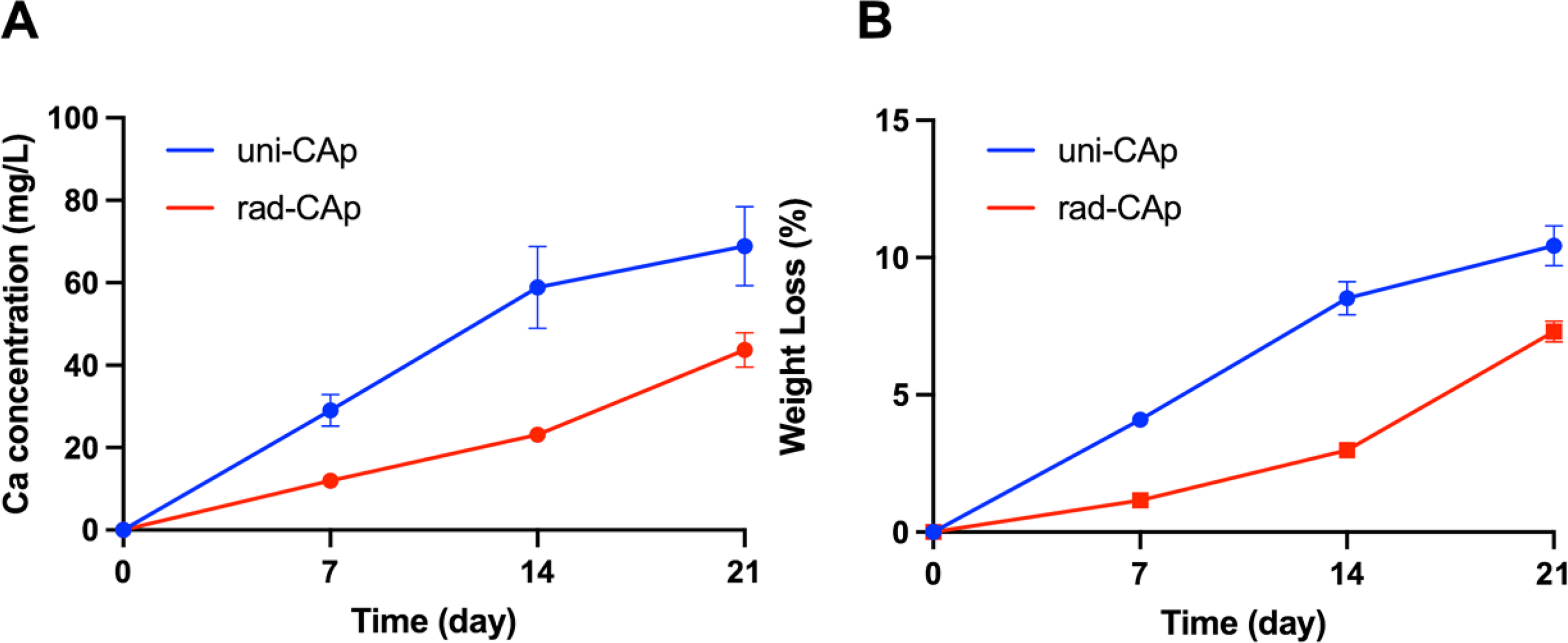
In HEPES–NaCl (pH 7.4, 37°C) over 3 weeks with weekly renewal, uni-CAp showed faster early Ca2+ release during the first 14 days, followed by a slower increase from 14 to 21 days; whereas rad-CAp showed a slower release initially with a later increase (Fig. 5A). The Ca2+ concentrations released from uni-CAp at 7, 14, and 21 days were 29.0 ± 3.9, 58.9 ± 9.9, and 68.8 ± 9.6 mg/L, respectively; the corresponding values for rad-CAp were 12.0 ± 0.8, 23.1 ± 0.7, and 43.7 ± 4.2 mg/L, respectively. To further evaluate material degradation, an in vitro weight loss test was performed under the same immersion conditions (Fig. 5B). The weight loss increased over time in both groups, but uni-CAp showed greater values than rad-CAp throughout the 21-day period. The weight loss values of uni-CAp at 7, 14, and 21 days were 4.1 ± 0.1%, 8.5 ± 0.1%, and 10.4 ± 0.7%, respectively; the corresponding values for rad-CAp were 1.2 ± 0.1%, 3.0 ± 0.2%, and 7.3 ± 0.4%, respectively. The higher early Ca2+ release and greater weight loss in uni-CAp were attributed to its high interconnected unidirectional channels, which reduced stagnant zones and shortened diffusion paths, allowing the immersion medium to penetrate more efficiently into the internal regions of the scaffold. Therefore, the Ca2+ release and weight loss results together indicate that pore orientation and a percolated channel network enhanced fluid accessibility and material degradation.
In vitro Ca2+ release
In vitro evaluations
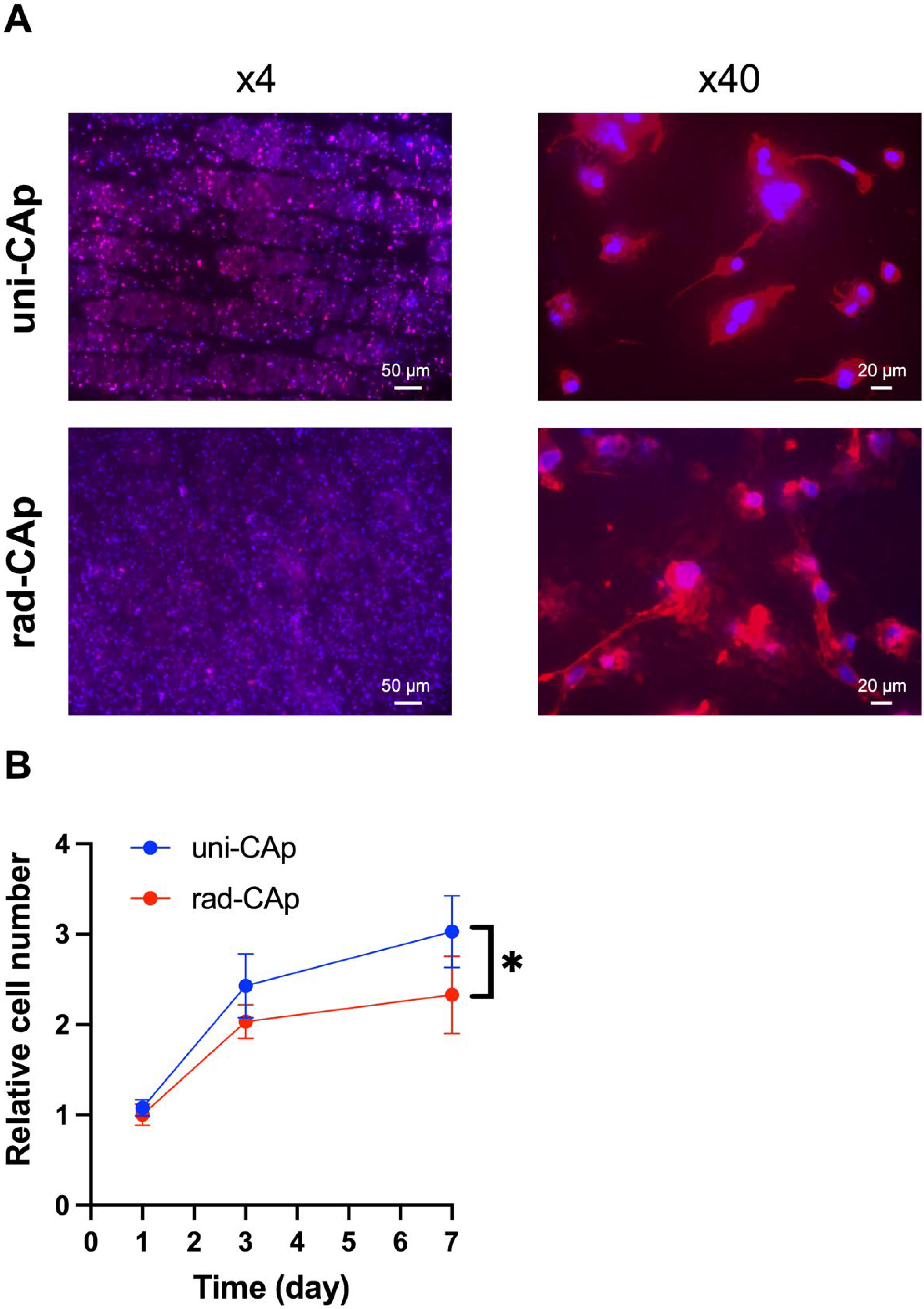
To further evaluate early cellular responses, cell adhesion and proliferation assays were performed. For the adhesion assay, cells were directly seeded onto uni-CAp and rad-CAp scaffolds and cultured for 24 h. Fluorescence staining showed that cells adhered to both scaffold surfaces, indicating that both materials supported early cell attachment (Fig. 6A). At low magnification, cells on uni-CAp appeared to be distributed along the aligned channel direction, suggesting that the unidirectional pore structure may have influenced early cell distribution. In contrast, cells on rad-CAp were randomly distributed over the scaffold surface, reflecting the nonoriented pore architecture. At high magnification, cells on uni-CAp tended to exhibit an angular or polygonal morphology with cytoskeletal spreading, whereas cells on rad-CAp showed relatively elongated actin filaments. These morphological differences suggest that the distinct pore architectures and local surface microenvironments influenced early cell attachment and spreading behavior.
Fluorescence images
Cell proliferation was further evaluated using scaffold extracts. Cell numbers increased from day 1 to day 7 in both groups (Fig. 6B). At 1 and 3 days, cell proliferation was comparable between the uni-CAp and rad-CAp groups. At 7 days, the uni-CAp group showed significantly higher cell proliferation than the rad-CAp group. These results suggest that the higher early Ca2+ release from uni-CAp scaffolds provided a more favorable ionic environment for cell proliferation during the later culture period.
In vivo evaluations
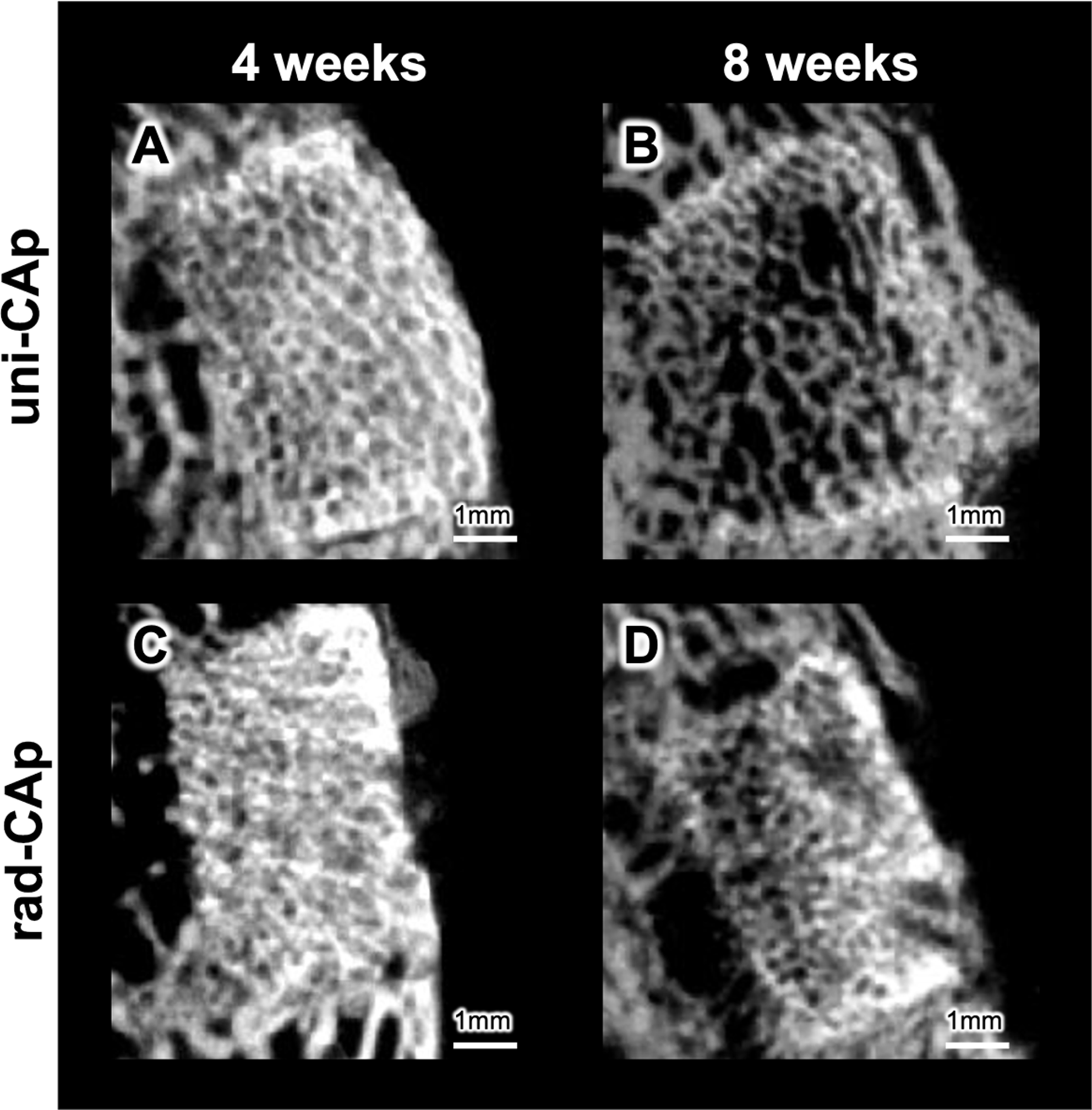
After 4 weeks and 8 weeks, microcomputed tomography showed extensive inward bone formation within implanted uni-CAp, with trabecular-like columns tracking a channel axis that were particularly evident at 8 weeks (Fig. 7A,B). In contrast, implanted rad-CAp exhibited slower, less directed ingrowth (Fig. 7C,D). This was corroborated with HE staining, where uni-CAp had larger and more contiguous regions of NB and BV along channels, abundant osteoblasts at interfaces, and a lower fraction of RM at matched timepoints (Figs. 8 and 9). To further confirm vascular formation within the regenerated tissue, CD31 immunohistochemical staining was performed (Figs. 8D,H and 9D, H). At 4 weeks, the uni-CAp group showed more abundant CD31-positive vascular structures than the rad-CAp group, particularly along the unidirectional channels. At 8 weeks, CD31-positive vessels were observed in both groups, with a higher signal in the rad-CAp group. Quantification (Fig. 10) revealed higher NB percentages and lower RM percentages for uni-CAp than for rad-CAp at both timepoints, as well as a significantly higher BV area for uni-CAp at 4 weeks. In uni-CAp, NB, RM, and BV were 30.0 ± 5.7%, 13.3 ± 2.4%, and 2.5 ± 0.1% at 4 weeks and 26.6 ± 4.5%, 9.6 ± 1.4%, and 1.6 ± 0.4% at 8 weeks, respectively. In rad-CAp, the corresponding values were 21.0 ± 3.5%, 23.6 ± 4.0%, and 1.1 ± 0.3% at 4 weeks and 18.3 ± 6.0%, 14.3 ± 1.7%, and 2.1 ± 0.5% at 8 weeks, respectively.
Microcomputed tomography images of implanted uni-carbonate apatite (CAp) blocks after 4 weeks
Histological hematoxylin and eosin staining images of uni-carbonate apatite (CAp) blocks
Histological hematoxylin and eosin staining images of uni-CAp blocks
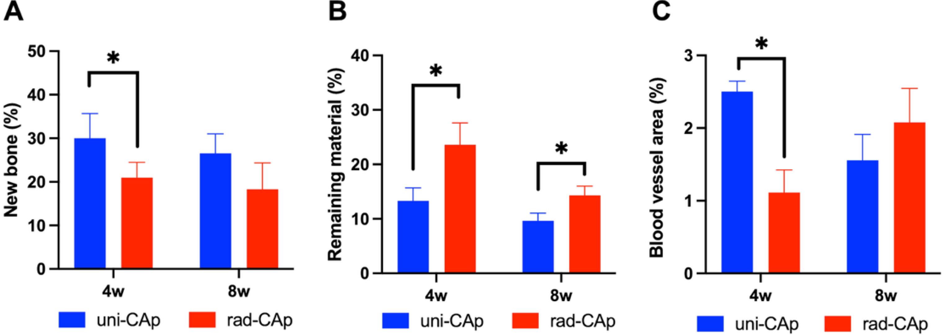
Quantified new bone formation
Collectively, these findings are consistent with previous reports on unidirectional freeze-cast scaffolds, which also demonstrated guided bone ingrowth. Our results extend this concept by establishing a gelatin-to-CAp fabrication route. Moreover, the unidirectional scaffolds exhibited enhanced in vivo bone regeneration relative to the randomly porous scaffolds. The aligned and highly interconnected channels appeared to facilitate early fluid transport and cellular access, thereby enabling resorption from both external and internal surfaces. This interpretation was supported by the higher in vitro Ca2+ release and greater weight loss profiles of uni-CAp than of rad-CAp. Similarly, cells on uni-CAp distributed along the aligned channels, and uni-CAp supported higher cell proliferation. Overall, the unidirectional, highly interconnected pore architecture of uni-CAp drove faster material resorption and enhanced osteogenesis, supported by faster ion release and enhanced cellular response.
The present architectural features highly benefited from the chemical composition of CAp. The carbonate content facilitates osteoclast-mediated remodeling, whereby the rate and spatial distribution of scaffold resorption and new bone formation are governed by the channel architecture. Clinically, the unidirectional pore architecture of uni-CAp may be advantageous when directional guidance of bone ingrowth is desired. However, the relatively low compressive strength of uni-CAp in the present study indicates that its current application may be more appropriate for nonload-bearing or low-load-bearing defects, whereas its use in load-bearing defects requiring substantial mechanical support, such as segmental bone defects, remains limited by insufficient mechanical strength. Therefore, improving the mechanical strength while preserving the beneficial unidirectional pore architecture will be an important objective of future studies through optimization of the precursor composition and processing conditions.
Conclusions
In this study, CAp scaffolds with a unidirectional porous structure were fabricated via gelatin-based freeze-casting. The aligned channel architecture facilitated early fluid transport and cellular infiltration that enabled resorption to progress from both external and internal surfaces of the scaffold. This accelerated bone regeneration. Compared with randomly oriented porous structures (rad-CAp), the uni-CAp samples exhibited increased new bone formation and lower remaining material fractions at both 4 weeks and 8 weeks. Hence, these findings demonstrate that pore orientation, rather than overall porosity alone, plays a critical role in governing bone ingrowth and remodeling. Such architecture-function synergy has a strong potential as a bone graft substitute, particularly for clinical applications requiring directional bone regeneration.
Authors’ Contributions
Z.L.: Writing—original draft, investigation, validation, methodology, and visualization. R.K.: Writing—review and editing, visualization, supervision, methodology, and funding acquisition. P.F.: Writing—review and editing and methodology. K.I.: Supervision, conceptualization, and funding acquisition.
Ethical Consideration
All animal experiments were conducted according to the ethical policies and procedures approved by the Animal Care and Use Committee of Kyushu University, Japan (Approval No. A24-478-0; Issued August 1, 2018).
Footnotes
Author Disclosure Statement
No competing financial interests exist.
Funding Information
This research was supported, in part, by Japan Agency for Medical Research and Development (Grant No. JP25ym0126161h0002), Japan Society for the Promotion of Science KAKENHI (Grant No. JP23K09233).