






Abstract
Open fractures have been associated with high rates of morbidity and mortality. Morbidity and mortality rates have improved with the advancements in infectious disease, plastic and reconstructive surgery, as well as damage control orthopaedics. Despite these advancements, few reports have focused on management of open foot and ankle fractures. This review outlines evidence-based principles for open fracture management and how these principles are applied to the foot and ankle.
Keywords
“Historically, open fractures have been associated with high rates of morbidity and mortality secondary to infection and sepsis.”
Open fractures are those in which there exists a soft tissue deficit that communicates directly or indirectly with a fracture site. Historically, open fractures have been associated with high rates of morbidity and mortality secondary to infection and sepsis. Prior to the development of antisepsis and antibiotic therapy, open fractures were viewed as a life-threatening injury. Subsequently, many were treated with emergency amputation. 1 This was especially true of war-time injuries well into the 20th century. During the Franco-Prussian war, French military casualties included 10 006 deaths and 13 173 amputations directly attributable to open fractures. 2 Similarly, the mortality rate of individuals suffering open fractures throughout the American Civil war have been estimated to be 26%. 3
Today, open fractures remain serious and sometimes life-threatening injuries. However, with advancements in the fields of infectious disease, plastic/reconstructive surgery, and damage control orthopaedics, morbidity and mortality rates have dramatically decreased. In an epidemiologic study of 2386 open fractures, Court-Brown et al found that 2.3% of all fractures studied were considered open. 4 Furthermore, they found that 80% of open fractures were the result of low-energy mechanisms while 17% of all open fractures affected the foot or ankle. 4 Despite the historically documented considerable incidence of foot and ankle open fractures, there is scant literature guiding surgeons in the management of these fractures. The purpose of this review is to outline the advancements in the management of open fractures and explain how these principles can be applied to the foot and ankle.
Evaluation
The goals when managing any open fractures are to preserve life, limb, and function. This begins with thorough evaluation and documentation of the environment in which an injury occurred, contaminants present at the injury site, neurovascular status of the affected anatomy, quality of the surrounding integument, and other associated injuries. The first step in the evaluation of any fracture is the determination of whether the fracture has communication with the outside environment. Traumatic wounds with persistent oozing, return of injected fluid after diagnostic injection, extruding fat, and/or subcutaneous gas evident on plain radiographs are suggestive of open fractures. Furthermore, fractures where skin tenting/mottling over the injury site exists should be treated as impending open fractures and reduced and stabilized in a timely fashion to prevent the development of an open skin portal. Gross debris within open fractures should be removed immediately, followed by proper reduction and provisional stabilization. When possible, a “one look” approach should be utilized, minimizing exposure of the open wound to the outside environment and further minimizing iatrogenic tissue trauma.
Prior to splinting, a thorough vascular, neurologic, and musculoskeletal examination should be performed. Halvorson et al 5 reported a series of risk factors for arterial injury with 9% of all open fractures having concomitant arterial injury. In the absence of palpable pedal pulses and absent Doppler signals, other noninvasive vascular studies such as duplex ultrasound or computed tomography angiography may be sought. 5 Alternatively, immediate vascular consultation should be sought and a traditional angiogram should be obtained in conjunction with orthopaedic management. Neurological status to the lower extremity should also be thoroughly documented. Traditionally, an insensate limb was presumed to portend poor prognosis; however, more recent studies have suggested this may not be true. In a prospective multicenter trial, Bosse et al 6 analyzed severe extremity trauma distal to the femur including open fractures, crush injuries, and foot trauma. Plantar foot sensation was evaluated with pin-prick examination and documented as present or absent. Three treatment groups were established including amputation for the insensate limb, salvage of the insensate limb, and salvage of the sensate limb. The researchers found no difference in rate of late amputation and insensate limb salvage did not correlate with poorer overall outcomes. Interestingly, a fraction of patients initially perceived to have an insensate limb had significantly improved sensation at later evaluations. Bosse et al concluded that lack of plantar foot sensation cannot be assumed to be resultant of nerve transaction. 6
Patients with open fractures are traumatized, in pain, and often unable to participate in a thorough musculoskeletal and neurologic evaluation; however, certain injures are vital to be ruled out. Despite open injury, compartment syndrome is one such entity. In a laboratory study using rabbit models, researchers found no difference in compartment pressures after iatrogenic open and closed tibia fractures, with both groups exhibiting statistically significant rises in compartment pressures. This data led the researchers to conclude that open tibia fractures should be monitored for compartment syndrome similarly to closed tibia fractures. 7 A French study of open tibia fractures in adults found a 10% incidence of compartment syndrome 8 ; however, no similar laboratory or clinical study has been applied to open foot or ankle fractures. Other often overlooked injuries associated with open fractures include tendon injuries. Patillo and Rayan, 9 found that in the setting of open tendon injuries to the hand, 46.7% of patients also suffered open osseous injuries. Acute tendon injury may also occur with open foot and ankle fractures despite being underreported in the literature. 10
Debridement Principles
Early and adequate debridement of traumatic wounds associated with open fractures has been a topic of discussion for decades. Cleveland and Grove 11 attributed prompt and adequate debridement of soft tissues among other treatment principles to a 93% limb salvage rate of open fractures sustained during World War II. What constitutes “adequate debridement” is poorly defined. In the leg, inspection of muscle for qualities such as contractility, color, capacity to bleed, and consistency all are evaluated to determine tissue viability. With open foot and ankle fractures, this recommendation is problematic given the lack of large skeletal muscle within the foot and distal leg. Conversely, appearance of other surrounding soft tissues including skin may be used to guide the surgeon. Adequate debridement of open fractures should (Figure 1) also include exposure of free bone ends of the fracture followed by curettage and debridement of any grossly necrotic bone. Ultimately, all necrotic or questionable tissue should be removed and traumatic wounds should be extended to ensure adequate debridement of the zone of injury is performed.

Open fractures of the talus and ankle treated provisionally with spanning external fixation.
Timing to initial debridement has been a long debated topic throughout the literature. The golden period of 6 hours to debridement has been considered the gold standard for timing to operative intervention. The golden period appears to be based on a laboratory study using a guinea pig model in 1898, where animals debrided after 6 hours had a higher incidence of infection. 12 Robson et al lent further support to this concept, showing that the time to grow 105 bacteria took an average of 5.17 hours. 13 More recently, there has been increasingly abundant evidence discrediting the 6-hour rule. Skaggs et al 14 reported on 554 pediatric open fractures where they found a 3% infection rate when fractures were debrided within 6 hours and a 2% infection rate when debridement occurred more than 7 hours after injury. Al-Arabi et al 15 reported similar results without statistical benefit for debridment within 6 hours in a prospective study of 248 long bone fractures. Thus, while prudence dictates that open fractures should be managed promptly, the dogma that open fractures must be emergently debrided within 6 hours is largely unsupported. In a systematic review, Schenker et al 16 reviewed 3539 open fractures, all of which had adequate and early antibiotic therapy. The authors concluded, with regard to deep infections, that the 6-hour rule has little support in the literature; however, the golden period may be influenced by optimal antibiotic therapy. 16
Antibiotic Therapy
Optimal antibiotic therapy has been the topic of multiple studies. Using a rat femur model, Penn-Barwell et al 17 induced iatrogenic fractures and surgical contamination with Staphylococcus, then treated specimens with antibiotics and surgery at less than 2 hours, between 2 and 6 hours, and finally between 6 and 24 hours, with reverse testing on the 2 variables. The authors concluded that delaying antibiotics 6 or 24 hours significantly increased infection rates regardless of surgical timing. 17 Patzakis and Wilkins 18 revealed lower infection rates when antibiotics were given less than 3 hours from the time of injury compared with more than 3 hours, 4.7% versus 7.4%, respectively.
Surgical site infection (SSI) appears to be predominately caused by gram-positive species, while varying prevalence of gram-negative species has been reported. To a lesser extent, reports of gas-forming species exist as well. 19 In 1974, Patzakis et al 20 provided the basis for selecting first-generation cephalosporin therapy for antibiotic prophylaxis in the management of open fractures. Gram-positive coverage for prophylactic antibiotic therapy has subsequently been supported by multiple other studies.21-24
The majority of recommendations regarding antibiotic prophylaxis were made prior to the emergence of methicillin-resistant Staphylococcus aureus (MRSA). Approximately 5% of general orthopaedic patients and roughly 10% of trauma patients will culture MRSA from their anterior nares and skin, which translates to a 2.5-time higher likelihood of an MRSA SSI. 25 Carsenti-Etesse et al 24 reported a rate of 20.8% of MRSA SSI secondary to open fractures. In another study, Chen et al 26 demonstrated an infection rate of 2.5% in their open fracture population. The authors noted that lower extremity open fractures were more likely than upper extremity open fractures to become infected. Of the fractures that developed infection, 25% of infections were caused by MRSA. Additionally, 55% infections included at least one gram negative organism. This suggests it may be prudent to revise antibiotic selection to target MRSA and gram-negative organisms in the setting of open fractures. Randomized trials are needed to determine whether revised antibiotic selection positively affects open fracture infection rates.
Term of antibiotic administration has also been debated. In a double-blinded, randomized trial, Dellinger et al 23 revealed no additional benefit of prophylactic antibiotic administration after 24 hours postoperatively. In a meta-analysis, the Surgical Infection Society Guideline 19 showed insufficient data to support prolongation of any prophylactic antibiotic beyond 48 hours of the perioperative period. Additionally, prolonged prophylactic antibiotics may increase the probability of resistant organisms; however, this has not been confirmed specifically in regard to open fracture antibiotic prophylaxis. 27
Timing to Permanent Fixation
In their landmark classification system for open tibia fractures, Gustilo and Anderson 28 recommended no primary internal fixation for open fractures secondary to the risk of hardware infection. They advocated for traction pins incorporated into a plaster cast for temporary fixation. For many years, no specific guidelines regarding internal fixation existed with specific interest to the foot and ankle. In 1984, Franklin et al 29 reported on 38 open ankle fractures, all of which were treated with immediate debridement and internal fixation. The authors reported no infections and hypothesized that bony stabilization protected the soft tissue envelope, thereby decreasing the risk of infection. Bray et al 30 performed a comparative study of 31 open ankle fractures, of which 16 were treated with immediate internal fixation and 15 with delayed internal fixation. One infection was reported in each group; however, the authors noted a trend for decreased hospital stay for the patients treated with immediate internal fixation. 30 With the emergence and access to external fixation, the philosophy of damage control orthopaedics has rapidly gained popularity. 31 The principles of damage control may be applied to open foot and ankle fractures. These principles involve placement of an external fixator as temporizing fixation, which may be placed promptly, even in hemodynamically unstable and polytrauma patients. External fixation allows increased fracture stability, decreased soft tissue and bone damage, and allows easy access to the soft tissues until permanent fixation can be safely obtained (Figure 1). The principles of damage control orthopaedics should be applied to high-grade open fractures and open fracture that occur in polytraumatized and hemodynamically unstable patients with external fixation being placed outside of the zone of injury if possible. There are cases in which primary internal fixation is indicated and can be safely utilized but extreme care and caution should be taken (Figure 3).
There is scant literature regarding the use of bone grafting with open fractures. Blick et al 32 performed a retrospective review of 53 open tibia fractures. Autogenonous canellous grafting was performed at a mean of 10 weeks postinjury or 8 weeks after coverage. The authors noted a mean time to union of 45 weeks, which was considerably lower than historic controls. 32 Trabulsy et al 33 performed a prospective comparative study involving 45 type III-B open tibia fractures with 19 of those being treated with bone grafting between 8 and 12 weeks postinjury. The authors noted a mean time to union of 41 weeks in the bone grafting group compared with 52 weeks when no bone grafting was utilized. 33 Okike et al 34 provided a grade C recommendation for use of bone grafting in open fractures in a systematic review. In the same study they made a grade B recommendation for the use of bone morphogenic protein-2 for open fracture management. To date there is no conclusive data supporting or refuting bone grafting or bone substitutes for foot and ankle fractures. 34
Timing to Closure
Historically, fear of gas gangrene with Clostridial species led surgeons to abstain from closing open fracture wounds primarily. 35 In fact, Gustilo et al 36 advocated against primary closure of all type III open tibia fractures. In 1986, Godina 37 published a landmark study of 532 patients with open extremity fractures that were treated with flap coverage within 72 hours, between 72 hours and 3 months, or after 3 months. In that study, early (less than 72 hours) flap coverage was associated with significantly fewer microsurgical procedures, fewer postoperative infections, shorter time to bony union, and shorter hospital stays. 37 The “fix and flap” protocol was termed by Gopal et al, 38 based on many of Godina’s principles. Gopal et al initiated an aggressive protocol with debridement outside of the zone of injury, immediate fracture stabilization, and flap coverage immediately (less than 24 hours), early (less than 72 hours), or late (greater than 72 hours). Of the 84 type IIIB or IIIC open tibia fractures, better outcomes and lower infection rates were obtained with the immediate and early flap coverage groups (Figure 2). 38
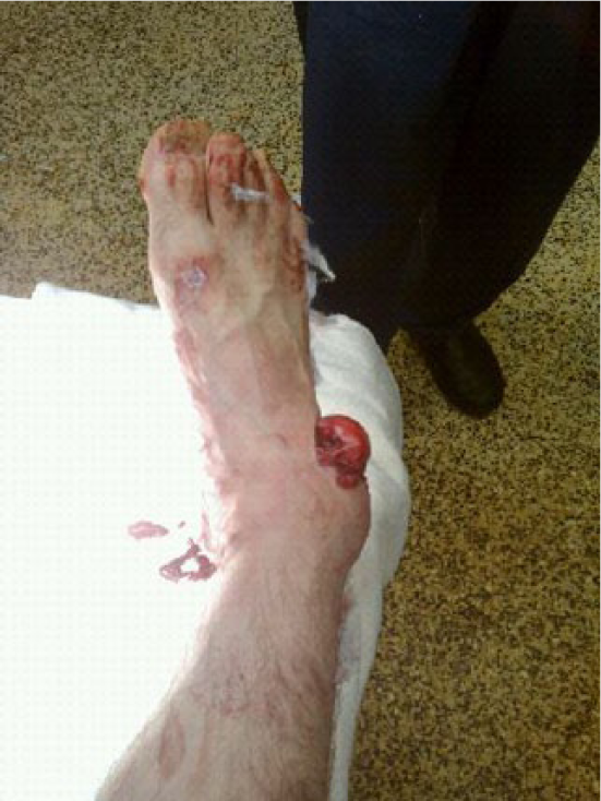
Open fracture of the talus with talar extrusion treated with primary internal and external fixation and subsequent delayed primary closure within 72 hours.

Primary internal fixation of the extruded talus fracture.
Until flap coverage can be safely obtained, traumatic wounds should be temporarily covered. Blum et al 39 performed a retrospective comparative cohort study involving 229 open tibial fractures with 166 of the 229 patients having negative pressure wound therapy (NPWT) during the interim to permanent coverage while 63 had conventional dressings. On multivariate analysis, the NPWT group had an 80% risk reduction for deep infection over the conventional dressing group. Patients in the NPWT were also less likely to have polymicrobial infections. 39 Liu et al 40 reported retrospectively on 105 free flap reconstructions for lower extremity trauma. The patients who received soft tissue coverage within 3 days had significantly fewer operations and flap revisions, preflap infections, deep infections, cases of osteomyelitis, and shorter hospital stays compared with those patients who were reconstructed beyond 7 days. Furthermore, the patients who received NPWT had significantly fewer flap revisions and VTE compared with wet-to-dry dressings; however, the benefits for NPWT were not noted if flap reconstruction was performed beyond 7 days. 40 Likewise, Bhattacharyya et al 41 retrospectively compared infection rates for coverage obtained less than 7 days versus more than 7 days after injury and debridement. All 38 patients with type III-B open tibia fractures had NPWT during the interim to closure. The authors noted a 12.5% infection rate when coverage was obtained in less than 7 days compared with 57% if coverage was obtained after 7 days. The authors concluded that NPWT can reduce complications for up to 7 days, however after 7 days the benefit of NPWT diminishes. 41
Amputation
Despite the existence of multiple injury scoring systems, the authors are unaware of any studies specifically addressing primary or secondary amputation for open fractures of the foot and ankle. The Mangled Extremity Severity Score has been shown to be prognostic of secondary amputation in high-grade tibia diaphyseal open fractures 42 ; however, it is not reasonable to apply these results to the foot and ankle. Rather, the best measures of when to amputate continue to be tissue viability and coverage, presence of infection, and non-reconstructable vascular injury or inadequate vascularity (Figure 4).

Open fracture of the hallux treated with primary amputation.
Grades of Recommendations (as used by the National Guideline Clearinghouse).

(from Shekelle PG, Woolf SH, Eccles M, Grimshaw J. Developing clinical guidelines. West J Med. 170(6):348-51, 1999 June)
Conclusion
While significant advances have been made in the management of open fractures, literature guiding the treatment of open fractures of the foot and ankle is lacking. There is low level of evidence to support the use of primary fixation and closure in select cases. Also, there is emerging evidence to support the timely use of cephalosporin antibiotics in the management of all open fractures. While the majority of open fracture literature is based on long bone studies, it remains to be determined whether these studies may be applicable to the management of foot and ankle fractures. Prospective, randomized trials should be undertaken to determine the best management for open fractures of the foot and ankle.