
Abstract
Chondroid syringoma is a cutaneous sweat gland tumor. Despite its relative rarity, a benign and malignant variant have been described. We present a case report of chondroid syringoma of the foot in a healthy patient. Definitive diagnosis required histopathologic examination, while treatment included wide resection. Surgeons who are presented with a painless, solid nodule in the lower extremities should be cognizant of this neoplasm.
“. . . one must consider chondroid syringoma a possibility when faced with a soft tissue mass involving the foot or ankle.”
Chondroid syringoma, also known as a “mixed tumor of the skin,” is a cutaneous sweat gland tumor that shares epithelial and mesenchymal structure and function. 1 The incidence of chondroid syringoma is reported to be less than 0.01% of all primary skin tumors. 2 Although rare, benign and malignant variants have been described. In the 2 largest case series reported in the literature, the extremities are involved 15% and 10% of the time.1,3 A review of malignant chondroid syringoma observed that the extremities were affected in 82% of cases. 4 Therefore, one must consider chondroid syringoma a possibility when faced with a soft tissue mass involving the foot or ankle. We describe a case of chondroid syringoma in the forefoot, with histological analysis, magnetic resonance imaging, and review of the literature.
Case Study
A 43-year-old Caucasian male presented with a firm mass on the plantar aspect of the right forefoot over 6 months. The patient described an achy pain, which was exacerbated with shoe wear. There was no history of local trauma, other tumors, or infection to this area. A magnetic resonance image was obtained, which displayed a mass isointense to skeletal muscle on T1 and hyperintense to skeletal muscle on T2, suggestive of a fibrous-type tumor (Figures 1 and 2). At this time, it was decided to take the patient to the operating room for excision of the mass.
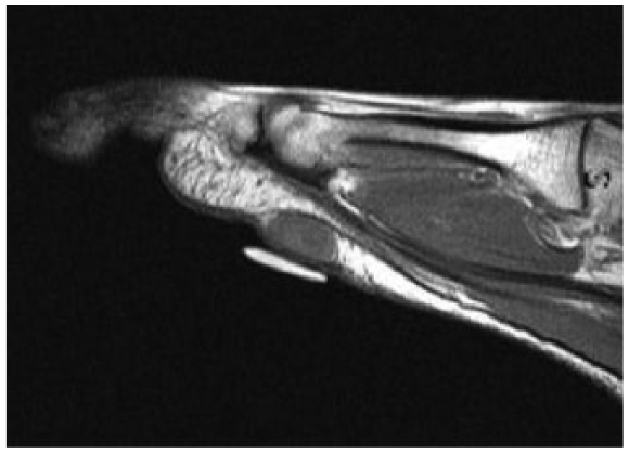
Sagittal plane T1-weighted magnetic resonance image. Homogenous isointensity to skeletal muscle.
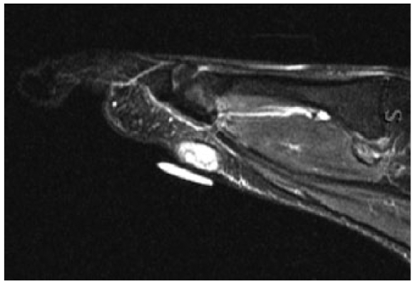
Sagittal plane T2-weighted image. There is homogenous hyperintensity in the subcutaneous tissue.
The patient was placed in a supine position and a lazy-S type incision was created over the plantar aspect of the second metatarsal head. Dissection was carried down through subcutaneous tissue where the mass was identified. The mass was well circumscribed with no discrete stalk or invasive structures, measuring 2.0 × 1.3 × 1.2 cm with a gray hue. This was then sent to pathology in formalin. The incision was then closed in standard fashion and a sterile dressing applied. The patient began weightbearing 2 weeks postoperatively. The patient followed an unremarkable postoperative course at 18 months’ follow-up and returned to all activities of daily living without restriction.
Pathological Findings
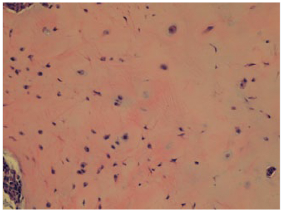
Histological analysis of the tumor revealed findings consistent with a chondroid syringoma. Features of the specimen included epithelial-myoepithelial islands in a tubulogranular pattern within a chondromyxoid stroma (Figures 3-5).
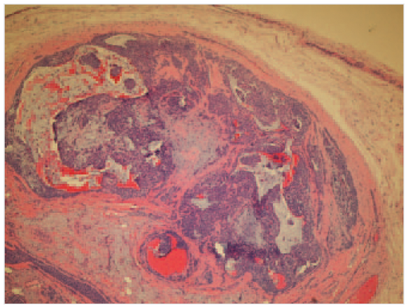
Chondromyxoid stroma (hematoxylin and eosin, 100×).
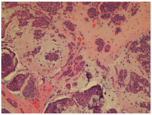
Tubulogranular pattern surround the epithelial-myoepithelial islands.
Myoepithelial islands within a chondromyxoid stroma.
Discussion
In 1961, Hirsch and Helwig 1 first introduced the term chondroid syringoma. Its nomenclature derives from its histologic appearance of sweat gland features in a cartilage-like stroma. 1 Clinical diagnosis is often difficult because prevalence is rare and have silent clinical presentations, and thus are often overlooked or confused with other types of skin lesions, such as sebaceous cysts, dermatofibromas, basal cell carcinomas, histiocytomas, and seborrheic keratosis.5-7 Though benign and malignant variants may be indistinguishable until late in the disease process, there are clinical differences that assist in diagnosis.
The most common site for the benign tumor is in the head and neck and, as seen in our patient, is most often observed in middle-aged males with a male-to-female ratio of 3:1.1,8,9 The gross appearance is typically a slow-growing, solitary, nonulcerating mass ranging from 0.5 to 3.0 cm. 6 Malignant variants tend to affect the lower extremities and trunk. Although there is a predilection toward females, however, there does not seem to be a correlation with age. 5 Tumors greater than 3.0 cm are associated with malignancy. 6
Magnetic resonance imaging with and without contrast is able to provide characterization of a soft tissue mass. While able to provide a diagnosis for fatty tumors or cysts, in most cases a diagnosis must be confirmed with histopathological examination. 10 When the diagnosis is unclear from magnetic resonance imaging, a fine-needle aspiration biopsy (FNAB) may be performed prior to definitive surgical resection. FNAB is most useful for diagnosis of tumor recurrences and differentiation between benign and malignant lesions, and therefore aiding in consultation of an oncology specialist. Specificity of histological diagnosis through FNAB varies from 36% to 96%, 10 and therefore definitive diagnosis is best confirmed by an excisional biopsy.11-14
Expected features with benign chondroid syringoma are a combination of epithelial and myoepithelial structures within a chondromyxoid and fibrous stroma. 15 Histologic features that suggest malignancy include cytologic atypia, infiltrative margins, tumor necrosis, numerous mitoses, excessive mucoid matrix, and poorly differentiated chondroid components.16,18 Malignant chondroid syringomas typically arise de novo and not from a preexisting benign tumor. 6 In 1983, Ishimura et al, 19 stated that the presence of a large amount of mucoid matrix and poor chondroid differentiation were the true determinants of metastatic potential.
In the series reported by Hirsch and Helwig, 1 recurrence was only seen if there was incomplete excision or if electrodessication was used. Because of its lobulated nature, removal is often inadequate, which leads to higher recurrence rates. 6 Excisional biopsy remains the most reliable method prevent recurrence. Excision techniques that do not allow pathologic evaluation of the entire specimen should be avoided. 8
Indications for excision include moderate discomfort with shoe wear. Total excision should be considered when presented with a painless nonilluminating mass in the foot and ankle. Chondroid syringoma in our case was small with no histological findings suggestive of malignancy. This case is unique because the location of the benign mass on the plantar aspect of the forefoot. Although we do report an atypical case of the benign chondroid syringoma, malignancy should remain in the differential diagnosis.
Conclusion
Despite the rarity of a chondroid syringoma, it must be remembered when presented with an insidious, painless, solid nodule in the soft tissues. The relative rarity of this neoplasm, and its greater incidence and potential for malignancy in the lower extremity, make it worthy of acknowledgment.