
Abstract
Context:
Running is one of the most popular sports worldwide. However, controversies exist regarding how running affects runner’s intervertebral discs (IVD).
Objective:
The purpose of this study was to systematically review studies that evaluated IVD morphology or composition changes in response to running exercise, to determine the impact of running exercise on IVD.
Data Sources:
A systematic literature search was performed for 4 major databases: PubMed, Cochrane, Embase, and Web of Science.
Study Selection:
Inclusion criteria were as follows: (1) healthy people without known IVD disease or major complications such as tuberculosis (IVD degeneration or low back pain are considered as minor complications); (2) subjects performed 1-time or regular running exercises; (3) pre and post comparison of runners or comparison between runners and healthy control subjects; (4) direct or indirect IVD morphology or composition measured; (5) IVD assessed before and after either acute or chronic running exercise, or compared cross-sectionally between runners and controls. Exclusion criteria were as follows: (1) reviews, editorials, letters or abstracts only; (2) animal studies; (3) subjects performed exercise other than running.
Study Design:
Systematic review.
Level of Evidence:
Level 3.
Data Extraction:
The extracted data included study design and primary outcomes of the included studies. The Newcastle-Ottawa scale (NOS) was used to evaluate study quality and risk of bias.
Results:
A total of 13 studies with 632 participants were included in the final analysis; 4 studies measured IVD changes using stature or spinal height, and the other 9 measured IVD changes using magnetic resonance imaging; 6 studies found that running acutely and negatively impacts IVD; 3 out of 5 cross-sectional studies found that IVD parameters are better for runners than controls; 1 longitudinal study found no significant difference in IVD before and after training for marathon in runners; 1 longitudinal study found no significant difference in changes of IVD between runners and controls after 15 years of follow-up.
Conclusion:
Negative changes in IVD exist for a short period of time after running, which may be due to the temporary compression pushing water content out of the disc. Cross-sectional studies suggest that long-term running exerts a mild positive effect on IVD; however, this inference has not been confirmed by high-quality longitudinal studies.
Running is a very popular sport that is potentially accompanied by a series of both physical benefits and injuries. Current research regarding the benefit effects of running focuses mainly on muscle, bone, and cardiovascular systems.25,42 Studies evaluating how running may affect other connective tissues, such as intervertebral discs (IVD), have been scarce in general. From an anatomic perspective, IVD can be divided into 2 parts: the nucleus pulposus and the annulus fibrosus. 37 Healthy nucleus pulposus is a gelatinous tissue rich in water and glycosaminoglycans, 46 the content of which decreases linearly from the center to the outer ring of the nucleus pulposus. 58 The annulus fibrosus is composed of about 15 to 25 concentric sheets of inclined collagen fibers, and surrounds the gelatinous nucleus pulposus. 34 When the axial pressure generated by running is applied to the IVD, pressure acting on the nucleus may be dispersed to the annulus fibrosus.28,58 Ohshima et al 39 found that, when the IVD of pig tail was compressed, the swelling pressure of nucleus pulposus and inner layer of annulus fibrosus increased proportionally, and water diffusion in IVD was inhibited. In another animal experiment, Zhu et al 61 found that long-term static load will lead to progressive IVD degeneration, while short-term static load may stimulate the generation of collagen type II alpha 1. These studies indicate that loading conditions may significantly influence IVD.
Studies that evaluated how running may affect human IVD date back to as early as the 1990s. In the early days, studies tended to evaluate changes in IVD by measuring the stature and length of the spine as surrogates.2,12,57 These studies generally found that the stature or vertebral column height of runners decreased after acute bouts of running. With advancements in imaging technology, IVD conditions can now be detected noninvasively using magnetic resonance imaging (MRI) techniques. 38 Regarding the concept of “disc health,” Belavý et al 7 suggested that the best possible definition might be “absence of disc degeneration.” The characteristics of IVD degeneration include reduction in IVD height, loss of IVD signal intensity, uneven structure, and other functional MRI-detected changes in the IVD component.1,7 For example, MRI T2 mapping can evaluate the water content and proteoglycan content of IVD quantitatively using T2 values 38 ; diffusion-weighted imaging (DWI) can be used to calculate the apparent diffusion coefficient (ADC) to reflect the average diffusion rate of water molecules. 6 Using MRI, Horga et al 23 found no significant changes in IVD height or width over a 16-week period of chronic running totaling approximately 500 miles during a marathon race. However, Dimitriadis et al 16 and Kingsley et al 24 found that IVD height decreased after 1 bout of acute running (running for 1 hour and 30 minutes, respectively) compared with pre-run through MRI scan examination. On the other hand, the cross-sectional study performed by Belavý et al, 9 which compared runners (≥20 km/week for the preceding 5 years) with nonsporting controls indicated that runners had higher T2-times values and IVD height. Another study that compared runners (≥20 km/week for 5 years) vs sedentary controls indicated that runners had higher IVD water content from a q-Dixon sequence. 8 Such cross-sectional studies suggest that runners may have heathier IVD compared with their counterparts who do not run.
Summing the above perspectives and evidence together, it is clear that controversies exist regarding how running may affect IVD in humans. The nature of the studies, the evaluation methods employed, as well as the characteristics of participants may all potentially impact the conclusions drawn. Hence, a study summarizing current knowledge regarding this topic is needed. Therefore, the purpose of this study was to systematically review literature that measured IVD changes associated with running to answer the following question: what is the impact of running exercise on IVD?
Methods
Systematic Review Scales
This systematic review followed the Preferred Reporting Items for Systematic Reviews and Meta-analyses (PRISMA) statement as well as the Cochrane Handbook for Systematic Review.31,36
Literature Search
A systematic literature search was performed for 4 major electronic databases (PubMed, Cochrane, Embase, and Web of Science) using keywords (“lumbar disc herniation” OR “LDH” OR “intervertebral disc” OR “IVD” OR “annulus fibrosus” OR “nucleus pulposus”) AND (“jogging” OR “jogger” OR “runner” OR “long-distance runners” OR “run” OR “runs” OR “running” OR “marathon” OR “marathon runner” OR “half-marathon” OR “half-marathon runner”). References of the retrieved articles were further checked to identify additional studies. Related articles were identified up to April 18, 2023 (search date).
Study Selection
After eliminating duplicates, articles were screened according to abstract and title to find all relevant literature for this review according to the inclusion and exclusion criteria described below. All studies were assessed for eligibility by applying the PICOS framework (population, intervention, comparison, outcomes, and setting). 45 Inclusion criteria were as follows: (1) healthy people without known IVD disease or severe complications such as tuberculosis (IVD degeneration or low back pain are considered minor complications); (2) subjects performed 1-time or regular running exercises; (3) pre and post comparison of runners or comparison between runners and healthy persons; (4) direct or indirect IVD morphology or composition measured (any measurement that reflects IVD conditions, eg, MRI scans and surrogate measures via body surface); (5) IVD assessed before and after either acute or chronic running exercise, or compared cross-sectionally between runners and controls. The acute running includes 1 bout of running, and chronic running includes multiple runs over a longer time span. 33 Exclusion criteria were as follows: (1) reviews, editorials, letters or abstracts only; (2) animal studies; (3) subjects performed other exercise with running (except warm-up exercise). Two reviewers independently screened the title and abstract of the selected papers, and retrieved the full text when necessary. Discussions were carried out with a third researcher to make a definitive decision where unanimous agreement could not be reached.
Data Extraction
Data extracted for the current review including study design and primary outcomes of the included studies, specifically (1) authors; (2) study design; (3) participant characteristics including age, sex, and anthropometrics if available; (4) outcome measurement methods; (5) running regime or history; and (6) primary study outcomes.
Quality Assessment/Risk of Bias Assessment
The Newcastle-Ottawa scale (NOS) was used to evaluate study quality and risk of bias. 47 This tool is used widely to evaluate methodological quality in nonrandomized studies.15,60 According to the summary score, the studies were divided into very good (9 points), good (7-8 points), satisfactory (5-6 points), and unsatisfactory (0-4 points). 22 One reviewer performed quality assessment for included studies, the results of which were cross-checked by a second reviewer. Disagreement was resolved by consensus. When no consensus could be reached, a third reviewer made the final decision. Because of the heterogeneity of the studied populations and research designs among studies, it was impossible to perform a formal meta-analysis. Thus, a comprehensive qualitative review of the existing literature was performed.
Results
The keyword search identified 2019 articles. Among them, 1085 articles remained after removing 934 duplicates. After reading the titles and abstracts, 1055 articles were excluded for the following reasons: participants did not run (n = 595); IVD morphology or composition was not measured (n = 343); other acute or chronic exercise involved (n = 64); animal studies (n = 40); review studies, editorials, letters or abstracts only (n = 13). Among the 30 studies assessed in full text, 18 studies were further excluded for the following reasons: did not perform IVD assessment before and after either acute or chronic running exercise, and did not compare runners versus control subjects (n = 17); 2 studies using the same cohort (n = 1). Studies performed by Dimitriadis et al16,17 utilized data from the same group of subjects. Therefore, we included only 1 study for the purpose of this review. 17 An additional 1 article was identified by checking references of the retrieved articles. Therefore, a total of 13 studies were included in this systematic review: 6 studies performed IVD assessment before and after 1 acute bout of running exercise, 2 were longitudinal cohort studies, and 5 were cross-sectional studies. A flow diagram of the study selection process is shown in Figure 1.
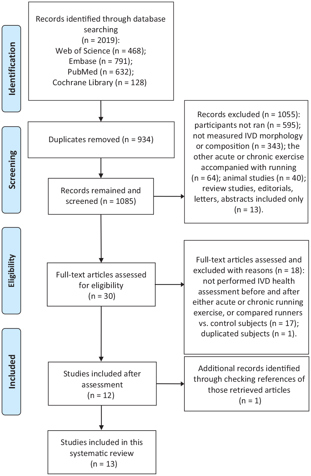
Flow diagram of study selection. IVD, intervertebral discs.
Risk of Bias in Studies
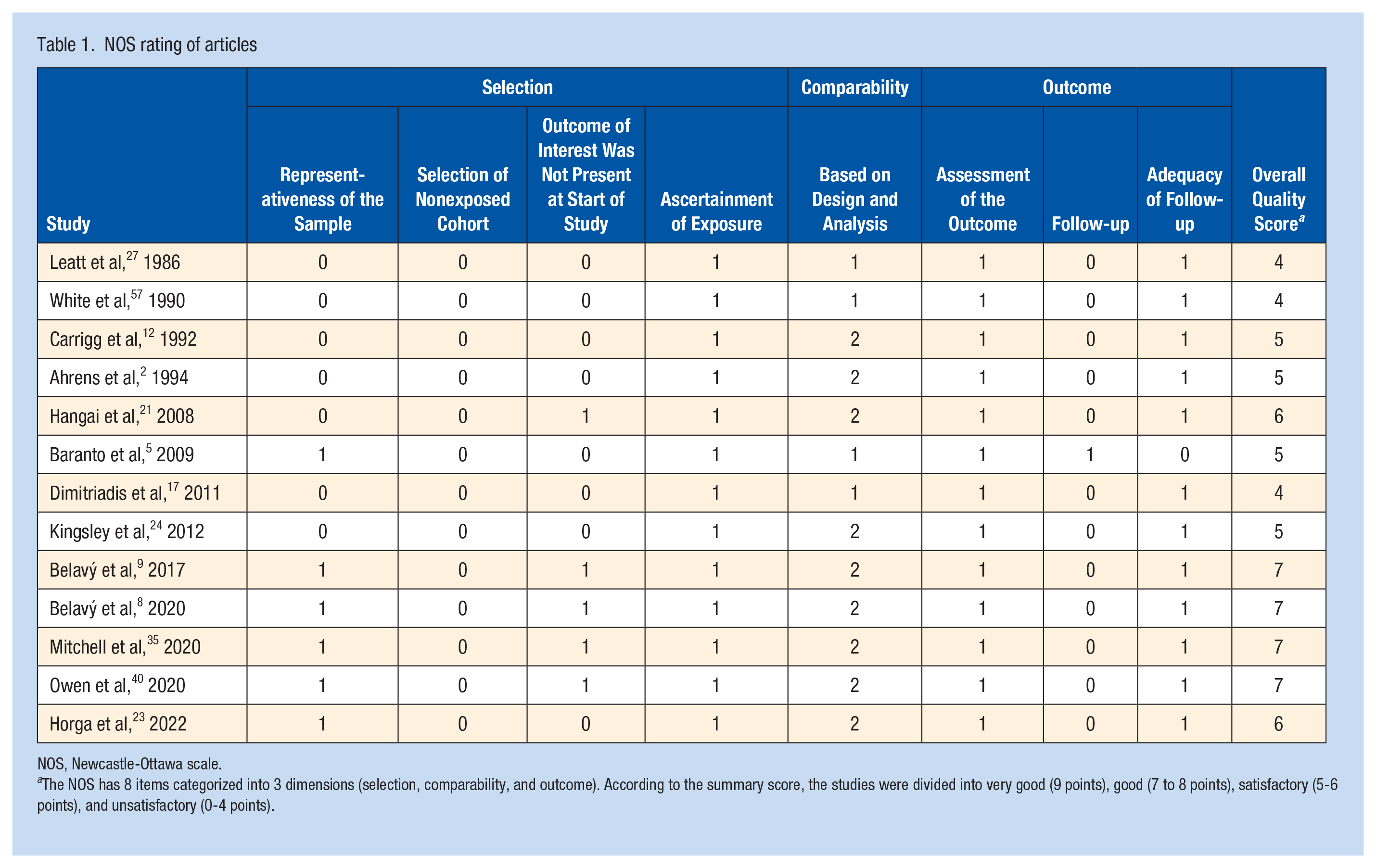
NOS has 8 items, which are categorized into 3 dimensions (selection, comparability, and outcome). The results are listed in Table 1. The study ratings ranged from 4 to 7, with a median of 5. The majority of studies (10 of 13) were rated ≥5: 4 studies were rated 7, 2 studies were rated 6, 4 studies were rated 5, and 3 studies were rated 4. Among them, the 5 cross-sectional studies were rated ≥5.
NOS rating of articles
NOS, Newcastle-Ottawa scale.
The NOS has 8 items categorized into 3 dimensions (selection, comparability, and outcome). According to the summary score, the studies were divided into very good (9 points), good (7 to 8 points), satisfactory (5-6 points), and unsatisfactory (0-4 points).
Main Study Results
The collections of the study design and primary outcomes of included studies are presented in Tables 2 and 3.
Demographic characteristics of study participants
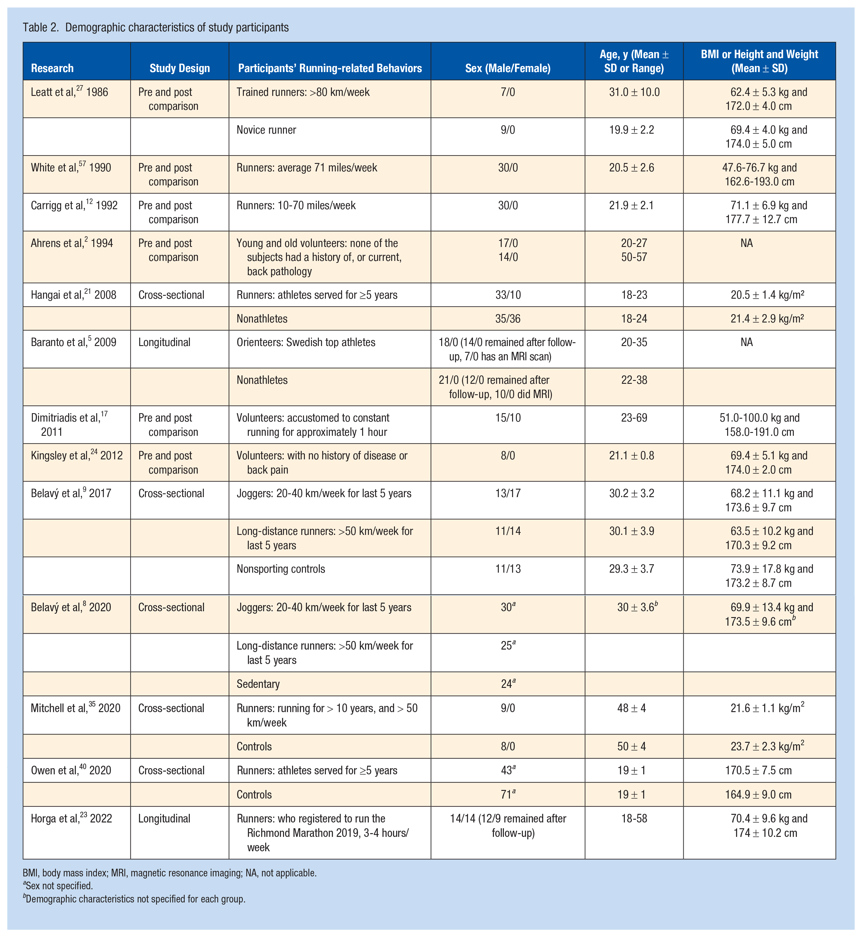
BMI, body mass index; MRI, magnetic resonance imaging; NA, not applicable.
Sex not specified.
Demographic characteristics not specified for each group.
Study design and main results of included studies
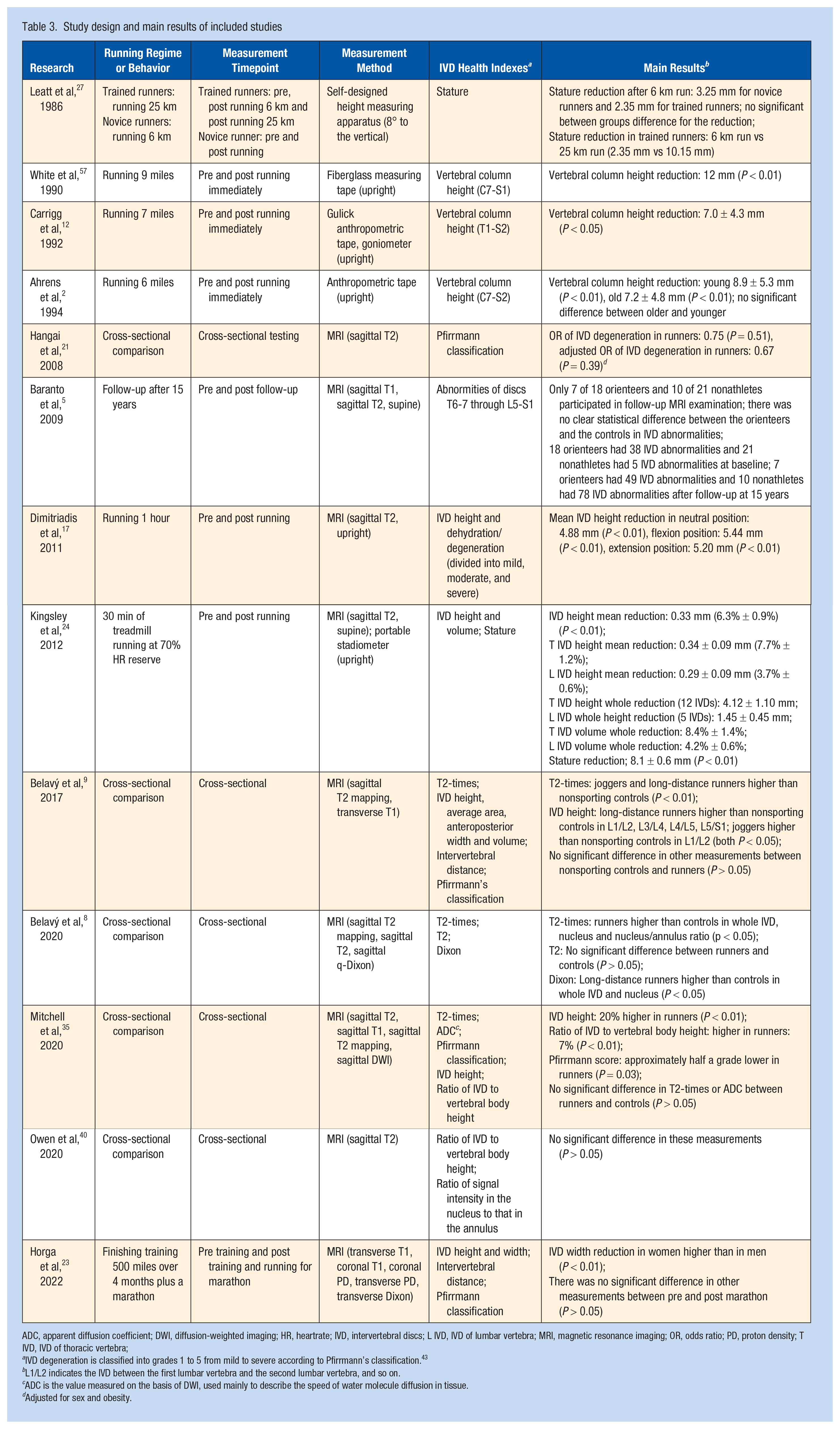
ADC, apparent diffusion coefficient; DWI, diffusion-weighted imaging; HR, heartrate; IVD, intervertebral discs; L IVD, IVD of lumbar vertebra; MRI, magnetic resonance imaging; OR, odds ratio; PD, proton density; T IVD, IVD of thoracic vertebra;
IVD degeneration is classified into grades 1 to 5 from mild to severe according to Pfirrmann’s classification. 43
L1/L2 indicates the IVD between the first lumbar vertebra and the second lumbar vertebra, and so on.
ADC is the value measured on the basis of DWI, used mainly to describe the speed of water molecule diffusion in tissue.
Adjusted for sex and obesity.
Five studies evaluated IVD changes by measuring stature and vertebral column height as surrogates.2,12,24,27,57 Of these 5 studies, 4 found that the stature or vertebral column height of the subjects decreased after running2,12,24,57; Leatt et al 27 found that the stature of the subjects decreased after running, which was more obvious in athletes after running 25 km than 6 km .
Nine studies measured IVD changes by different MRI sequences.5,8,9,17,21,23,24,35,40 Of the 9 studies, 2 evaluated the acute effects of running on IVD and found that IVD height decreased after running.17,24 Of the 9 studies, 5 conducted cross-sectional assessments of the IVD on runners and controls.8,9,21,35,40 Among them, 2 studies found no significant difference in IVD abnormalities between runners and controls21,40; 3 studies found that at least some benefit-related IVD parameters were better for runners than controls.8,9,35 Of the 9 studies, 2 conducted longitudinal experiments and found no significant difference in IVD morphology or degeneration between baseline and follow-up.5,23
Classification According to Study Type
Studies on the Acute Effects of Running
Six studies evaluated the acute effects of running on IVD.2,12,17,24,27,57 Out of the 6 studies, 3 measured the stature or height of the spine using anthropometric tape after running for 6 to 9 miles,2,12,57 and 1 of the 6 studies also evaluated trained runners after running for 6 and 25 km. 27 All 4 studies found that the stature or vertebral column height decreased after running compared with pre-run.2,12,27,57 For the remaining 2 studies, 1 performed measurements after running for 30 minutes and the other performed evaluation after running for 1 hour.17,24 These 2 studies found that IVD height or volume decreased after running compared with pre-run.17,24 In general, studies that evaluated the acute effects of running on IVD generally indicate that running negatively impacts IVD when evaluated within a short period after running.
Cross-Sectional Studies
Cross-sectional evaluation was conducted in 5 studies to compare IVD status between runners and controls, and MRI was used to examine IVD parameters in all 5 studies.8,9,21,35,40 Runners in 4 of the 5 studies had >5 years running experience, and in 1 of the 5 studies had >10 years running experience. For quantitative assessments, 1 of the 5 studies found higher IVD height and higher ratio of IVD to vertebral body height in runners than controls, while no significant difference in T2-times or ADC values between runners and controls was detected. 35 One study found higher IVD height and higher T2 values in runners than controls. 9 Another study found that the water content of q-Dixon increased only in runners compared with controls and the T2 values were higher in runners than in controls. 8 No significant between-group differences in IVD abnormalities were detected in the other 2 studies.21,40
The Pfirrmann grading system, which has a scoring between 1 to 5, with lower score representing healthier IVD, is commonly used for qualitative assessment for IVD.43,51 This system conducts qualitative observation of a series of changes in IVD. 43 Two studies employed the Pfirrmann grading system with the aim to compare IVD qualitatively between runners and controls. One study found lower Pfirrmann scores of IVD for runners compared with controls, 35 and the other found no significant difference in Pfirrmann scores of IVD between groups. 9 In general, most studies found that runners possess better IVD health-related parameters compared with their counterparts; however, exceptions do occur, and more high-quality studies are needed to draw definitive conclusion on this matter.
Longitudinal Cohort Studies
Two cohort studies were identified.5,23 MRI measurements were carried out in marathon runners (3-4 hours of running/week) for 16 weeks before and 2 weeks after a marathon in 1 study, and it was found that IVD height in women decreased significantly compared with that of men after marathon exercise. 23 The participants ran about 500 miles during the 16-week training period leading to the marathon race in this study, which equals approximately 30 miles per week. In another study, 18 orienteers (Swedish top athletes with the highest possible international or national ranking at the time of inclusion) and 21 nonathletes received the baseline MRI evaluation for IVD, and only 7 orienteers and 10 nonathletes received the follow-up MRI scan. 5 Describing the abnormalities, 18 orienteers had 38 IVD abnormalities and 21 nonathletes had 5 IVD abnormalities at baseline and 7 orienteers had 49 IVD abnormalities and 10 nonathletes had 78 IVD abnormalities after 15 years’ follow-up. However, the small number of participants, the low retention rate at follow-up, as well as the lack of clear statistical analysis makes it difficult to interpret the results correctly.
Discussion
The NOS rating ranged from 4 to 7 for included studies, with a median of 5. The relatively low study quality might be attributed to selection bias (from NOS), which is an inherent limitation of nonrandomized studies. For studies that examined the acute effects of running on IVD, a downward trend in height or IVD volume or height of the spine was generally found within a short time after running compared with pre-run, which indicates a negative impact on IVD. Whether including all acute running studies or excluding low-quality studies, the direction of the results was consistent. In cross-sectional studies where runners and controls were compared, the Pfirrmann scores (with lower score indicating better outcome) of IVD in habitual runners were generally lower compared with controls in 1 study, 35 while other parameters such as the height or volume of the IVD, 9 the T2-times value or water content of q-Dixon of IVD,8,9 and the ratio of IVD to vertebral body height, 35 showed an upward trend compared with controls, although exceptions did occur. The 2 cohort studies did not indicate that running exercise was harmful to the IVD specifically.5,23 Together, these results indicate that runners might have healthier IVD compared with those who do not run. Although cross-sectional studies and cohort studies had satisfactory study quality (≥5 points) and consistently positive results, the results still need to be interpreted with caution due to the nature of these studies.
Methods and Parameters for Evaluating IVD
In 5 studies, the stature or vertebral column height was measured by body surface measurement tools in upright or near-upright position such as measuring tapes and self-made height measuring apparatus,2,12,24,27,57 while 9 studies examined the spine using MRI in supine position,5,8,9,17,21,23,24,35,40 except for the study by Dimitriadis et al 16 (some studies did not report the subject’s examination position, but it can be inferred from the MRI machine model provided by the authors). This raises the question as to whether the measurement position might influence the study results. However, it was pointed out that, although gravity can have an effect on IVD height, the few minutes duration of the examination would have little effect on the overall results. 11 In addition, Kingsley et al 24 observed a consistent trend of decreased IVD height in supine MRI examinations and stature examined in upright position. Therefore, the measurement position is unlikely to be a significant influential factor in the study results.
In the 4 early studies that compared spine height before and after 1 bout of acute running using body surface measurement tools, it was suggested that changes in stature or spine height reflected changes in IVD height.2,12,27,57 Lewis et al 29 demonstrated that stadiometry can be used as a reliable indirect measure of IVD height using MRI scans as a reference. They found statistically significant correlations between height loss as measured by stadiometer and reduction in posterior spine length as measured by MRI scan (r = 0.61), which represented changes in IVD height. 29 For studies that employed MRI functional sequence, Belavý et al8,9 and Mitchell et al 35 used T2 mapping sequence to study the IVD. The IVD might have subtle physiological changes before the T2 signal intensity was significantly lost, while the T2 mapping sequence could reflect the water content, collagen orientation, or structure in IVD, allowing early observation of IVD degeneration.32,56 Although generally used to evaluate fat fraction, Belavý et al 8 employed the q-Dixon technique on IVD with the purpose of assessing water content, and found that it could reflect the change in pure water content of IVD, although the q-Dixon technique is more commonly used to evaluate fat fraction. 30 The DWI technique used by Mitchell et al 35 was able to assess the diffusive movement of water molecules throughout the IVD, providing an earlier indication of disc tissue changes than conventional MRI T2 sequence. In addition, studies using MRI T1rho sequence to observe IVD found that it was more suitable than T2 mapping sequence to evaluate the degeneration of annulus fibrosus. 55 More advanced functional MRI sequence examination can evaluate different physiological aspects of IVD and detect very subtle changes in IVD composition, which may provide further insights into how running may affect IVD in future studies.
Running Volume of Subjects
In studies that evaluated the acute effects of running on IVD, most of the running distance was set between 6 and 9 miles, or running time of between 30 minutes and 1 hour.2,12,17,24,57 In 1 study, trained runners ran 6 and 25 km (resumed running for a further 19 km) and found a significant decrease in height after running 25 km compared with at 6 km. 27 This suggests that running distance is an important mediator of the acute IVD response after running. One cohort study conducted MRI examination of marathon runners before and after 16 weeks of training and 2 weeks after a marathon. The participants ran about 500 miles in total during the 16 weeks training in this study, which equals approximately 30 miles per week. 23 This study demonstrated that IVD in women might be more vulnerable to load impact induced by large volume of running, although more studies are needed to confirm this notion.53,54 The other cohort study and 5 cross-sectional studies included habitual runners and controls for comparison.5,8,9,21,35,40 Among them, habitual runners had >5 years of running experience or ran >10 miles per week in running volume. One Swedish study included orienteers who were top athletes. 23 This study found a very high IVD abnormality rate for runners group at baseline, which might be due to the inclusion of Swedish top athletes. However, it should be noted the small number of participants, the high decline rate of MRI scan evaluation for orienteers, as well as the low retention rate for controls at follow-up potentially significant undermines the validity of this study. 5 Although not included in this study as it did not fit the inclusion criteria, the epidemiology study done by Takatalo et al 41 suggested that running at least twice a week might be potentially associated with lumbar IVD degeneration in early adulthood. Combined with the abovementioned information, it is possible that there is an optimal running volume range within which subjects may gain health benefits for IVD from running, while running too much could adversely impact IVD. However, such inference remains to be confirmed.
Potential Mechanisms for Running to Impact IVD
It is generally believed that the rapid deceleration of the body when the feet contact the ground during running could cause shock loading, and the shockwave will pass through the body from the ground to the spine. 19 The impact loading generated by the ground-reaction force could be as high as 3 times the body weight. 13 The resulting load might lead to inflammation around the vertebrae and back muscles, which may eventually cause IVD damage.48,59 Using MRI, Arun et al 3 showed that sustained mechanical loading may impair the diffusion of small solutes entering the IVD. However, an animal study showed that 11 weeks of treadmill exercise increased the concentration of glycosaminoglycans in rat IVD, in direct proportion to training load. 49 Runners also generally had better IVD parameters than controls in the 5 cross-sectional studies included.8,9,21,35,40
Six studies found that IVD reduced in height and volume after 1 bout of acute running.2,12,17,24,27,57 It can be inferred that the load sustained during running has the potential to cause the water in the nucleus to extrude. This extrusion occurs through the vascular pores located in the cartilaginous endplate and moves into the vertebral body.26,41,57 While the outer ring of the IVD receives nutrients from the surrounding vascular system, the inner ring and nucleus pulposus acquire nutrients by moving large amounts of fluid through the IVD tissue and diffusing through the vertebral endplate. 20 The pressure on the IVD needs to be within an appropriate range to promote IVD health. Nucleus pulposus cells showed an anabolic response to light-to-medium intensity static compression, osmotic pressure, or hydrostatic pressure, while higher intensity static compression promoted a catabolic response. 18 Appropriate load, frequency, and duration could promote stem cells to differentiate into “discogenic” cells. 14 Belavý et al7-9 also believed that light to moderate dynamic pressure was beneficial to the IVD, while too high and too fast or extreme range of activity loading was harmful to IVD. Therefore, fluid exchange in the short period of time after running might alleviate the degeneration of IVD. This indicates that IVD has a possible anabolic “loading window,” ie, a certain appropriate pressure might be beneficial for IVD.4,10,50 The results seen in the acute effect studies included in this review might be due to the time window at which measurements were taken. It is possible that, after a period of recovery, these changes might recover and reach a supercompensation state, which may lead to the better IVD parameters observed in habitual runners compared with the control group of cross-sectional studies. Future studies that evaluate participants over a prolonged period of time after running should be carried out to test this hypothesis.
In addition, nutrition, environment, and genetic factors have been shown to relate to IVD degeneration.44,52 Sakai et al 44 believed that the event initiating IVD degeneration is a complex interplay between genetic predisposition and accompanying conditions such as inappropriate loading, nutrient deficiency, and other illnesses. Future relevant studies should consider these factors.
Limitations
There are limitations to this systematic review. First, the quality of the literature included was relatively limited based on NOS, and more high-quality studies are needed before definitive conclusions can be drawn. Second, most of the studies were either cross-sectional comparisons or 1-time follow-ups in nature, and the lack of high-quality longitudinal studies makes it difficult to draw definitive conclusions regarding this topic. Third, the surface on which running occurs may have an impact on load, and only 1 of the studies we included described the running surface. This is an important aspect that should be examined carefully in future studies. A fourth limitation of our study involves the diversity in study designs. This diversity includes various factors like the nature of each study, the methods used for measurement, and the running protocols implemented. A notable difference is in the measurement tools used, particularly between stadiometer and MRI measurements. These variations make it challenging to determine the effective running volume that may be beneficial for IVD, based on the available studies.
Conclusion
Negative changes in IVD exist for a short period of time after running, which may be due to temporary compression pushing water content out of the disc. Cross-sectional studies suggest that long-term running exerts a mild positive effect on IVD; however, this inference has not been confirmed by high-quality longitudinal studies.
Footnotes
The authors report no potential conflicts of interest in the development and publication of this article.
The authors disclosed receipt of the following financial support for the research, authorship, and/or publication of this article: this study was funded by the Key Laboratory of Functional Molecular Imaging of Tumor and Interventional Diagnosis and Treatment of Shaoxing City, the Central China Normal University Faculty Startup Grant (31101222041), the Zhejiang Province Public Welfare Technology Application Social Development Field Project (LGF20H180008), and the Shaoxing Health Science and Technology Project (2022SY009).