






Abstract
Lateral medullary syndrome is a common presentation of posterior circulation ischemia that presents with ipsilateral Horner syndrome, ipsilateral facial numbness, contralateral body numbness, vestibular symptoms, ataxia, dysphagia, and dysarthria. Here, we describe an 84-year-old who presented to the hospital with right upper motor neuron facial weakness and gait abnormality found to have a right lateral medullary ischemic stroke. Multiple MRI’s, including with thin brainstem slices, were without evidence of pontine, midbrain or cerebral ischemia outside the medulla. We postulate that the patient’s ipsilateral upper motor neuron facial weakness was caused by involvement of aberrant corticobulbar fibers in the medulla ascending to the facial nucleus.
Case Report
A man in his eighties with past medical history of hypertension, prediabetes, obstructive sleep apnea, hyperlipidemia, and chronic kidney disease presented to the emergency department with concern for right facial droop and imbalance. In the emergency department, his examination was notable for a right facial droop sparing the forehead and a rightward leaning, imbalanced gait (Figure 1). His blood pressure was 186/99 upon presentation. He was not on anticoagulation. Noncontrast computed tomography (CT) of his brain was negative for hemorrhage or acute ischemic changes. CT angiography of the head and neck was negative for flow limiting stenosis of the head or neck. National Institutes of Health Stroke Scale (NIHSS) was one. The patient did not receive alteplase due to the symptoms being thought to be nondisabling overnight and was admitted to the inpatient service. He was started on aspirin and clopidogrel for low NIHSS based on POINT
1
and CHANCE
2
criteria overnight for secondary stroke prevention. Right sided upper motor neuron facial droop is noted at rest (A) with flattened nasolabial fold and with smile (B) Right sided ptosis is seen (A,B) secondary to right sided incomplete Horner’s Syndrome. Diffusion weighted imaging shows an area of restricted diffusion in the right lateral medulla (C) with a correlate on the apparent diffusion coefficient (D) consistent with an acute infarct of the right lateral medulla.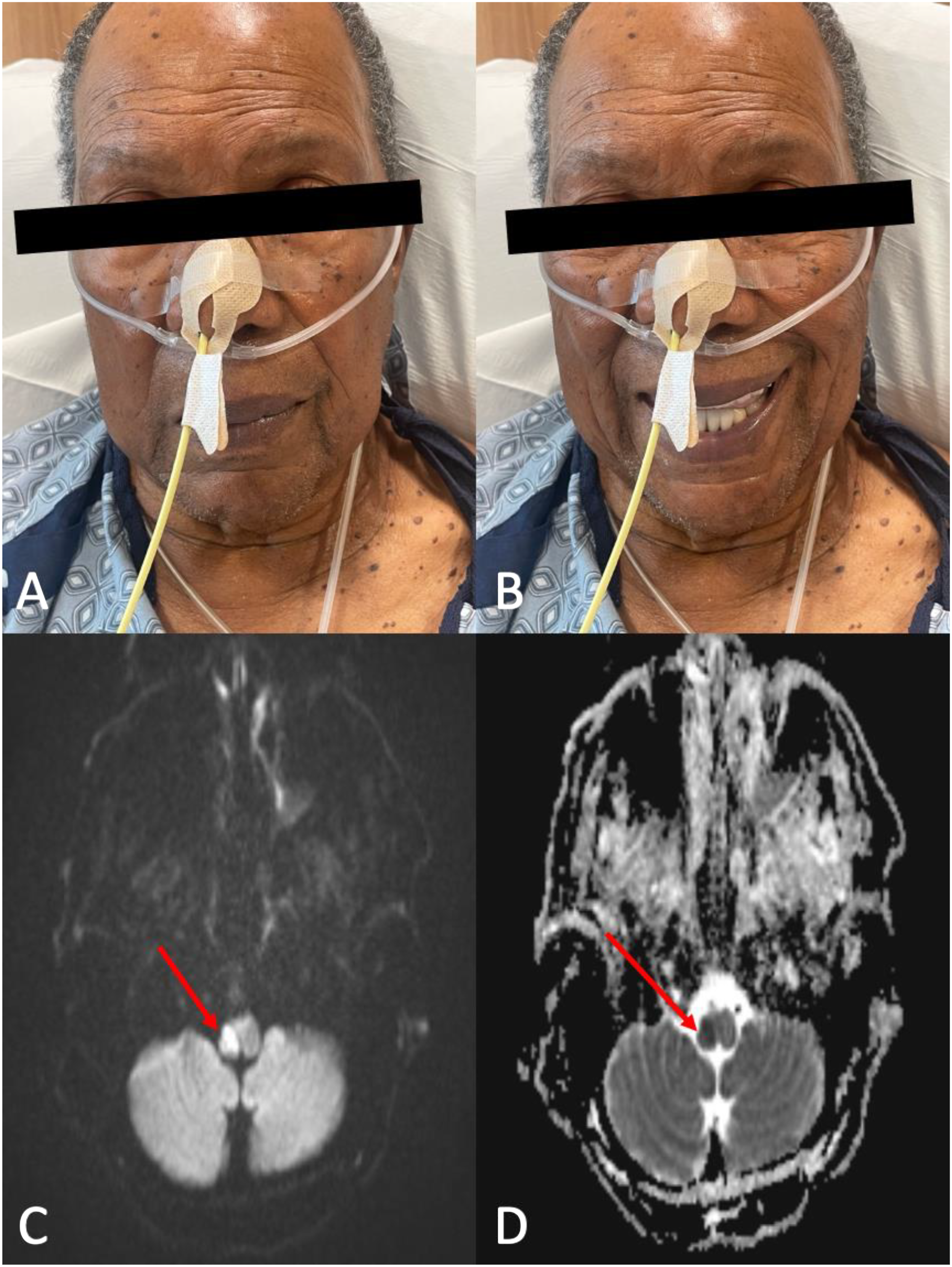
Upon re-evaluation the next morning with the entire stroke service, the patient was noted to have mild right sided ptosis, anisocoria with a constricted but reactive right pupil, right facial droop with sparing of the forehead, hoarseness, dysarthria, dysphagia with inability to pass bedside swallow tests, decreased pinprick sensation in the left upper and left lower extremity, and right sided dysmetria in the upper extremity. NIHSS was 4. Magnetic resonance imaging (MRI) of the brain revealed an acute infarct in the right lateral medulla (Figure 1). Considering the upper motor neuron pattern of facial droop and concern for pontine ischemia in the setting of facial weakness, a repeat MRI was performed with thin cuts through the brainstem. Once again, an acute infarct of the right lateral medulla was demonstrated without evidence of infarct elsewhere. The patient was continued on aspirin and clopidogrel for 21 days with recommendations for aspirin monotherapy thereafter. He was evaluated for a percutaneous endoscopic gastrostomy (PEG) tube for feeding due to persistent dysphagia but was discharged on a full liquid diet. The patient was eventually discharged to acute rehab with continued workup of stroke etiology.
Discussion
Posterior circulation strokes account for approximately 20% of all ischemic strokes 3 and often present with more nonspecific, non-quantified symptoms as compared to anterior circulation strokes. Due to the proximity of nuclei and tracts within the brainstem, small posterior circulation strokes can create significant disability and rarely present with isolated symptoms. 4 Most commonly, patients present with dizziness or vertigo, dysarthria, nausea, ataxia, and limb weakness. 5 Presenting symptoms differ across patients given anatomic variation within the vascular distribution of the posterior circulation. Posterior circulation strokes are five times more likely to have diffusion weighted imaging (DWI) negative strokes as compared to patients with anterior circulation ischemia, 6 which creates diagnostic pitfalls. Therefore, diagnosing cerebral ischemia based on clinical examination findings rather than relying solely on imaging is critical.
Lateral medullary syndrome, first described by Adolf Wallenberg in 1895 and further pathologically confirmed in 1901, 7 is caused by occlusive disease of the posterior inferior cerebellar artery or vertebral artery. Clinical manifestations include ipsilateral Horner syndrome, ipsilateral facial numbness, contralateral body numbness, ipsilateral ataxia, dysphagia, vertigo, nystagmus, and hoarseness. These symptoms are due to the involvement of structures within the lateral medulla such as the descending sympathetic fibers, spinothalamic tract, spinal trigeminal tract, spinal trigeminal nucleus, inferior cerebellar peduncle, vestibular nuclei, and nucleus ambiguus. The pyramidal tracts are often spared, and patients commonly do not present with weakness. 8
In the case described here, the presenting symptom was facial weakness of the lower face in an upper motor neuron pattern. This presentation has been clinically described previously,9,10 but appears to be a rare finding on examination. The facial nucleus resides in the dorsolateral pons, and thus ischemic lesions of the caudal pons can lead to lower motor neuron facial weakness. More commonly, lesions in the cerebrum or ventral brainstem rostral to the facial nucleus can cause upper motor neuron facial weakness.
Dejerine 11 first detailed the corticobulbar projections in human beings in 1914 11 and described fibers that descended alongside the corticospinal fibers. Amongst these fibers, he described five aberrant pyramidal bundles of corticobulbar fibers, the fourth of which was a ponto-medullary bundle leaving the main tract near the ponto-medullary sulcus supplying the motor nucleus of the facial nerve, as well as fibers to the nucleus ambiguus and hypoglossal nucleus. This aberrant anatomy would suggest that a variety of craniobulbar presentations could be created through injury to these pathways within the medulla, which could create diagnostic challenges for neurologists. This fourth aberrant pathway has been postulated to be involved in cases of hypoglossal injury with ipsilateral upper motor neuron facial weakness in patients with medullary strokes. 12
This pathway has been further illustrated in the clinical setting. In a series of 53 patients using transcranial magnetic stimulation (TMS), electromyography (EMG), and MRI, two patients with lateral medullary injury and ipsilateral upper motor neuron facial weakness had absent contralateral cortical TMS responses to the buccinator, though normal EMG responses peripherally. Such findings suggested a corticobulbar loop of fibers descending into the medulla that loop prior to ascending to the facial nucleus. 13
In the patient described here, two MRI’s were performed, one with thin cuts through the brainstem, without evidence of pontine, midbrain, or cerebral infarction that would explain the upper motor neuron facial weakness. Given this patient’s history, examination, and imaging, his presentation is suggestive of involvement of this aberrant corticobulbar loop of fibers in the medulla after these fibers cross midline, leading to an ipsilateral upper motor neuron facial droop (Figure 2). First and foremost, neurologists in training must be aware of the common presentations of posterior circulation ischemia in order to provide appropriate care. However, they must also be aware of anatomic variation and challenges with imaging and diagnostics in patients with such symptoms. Sagittal and coronal planes showing of the descending corticobulbar tract with decussation in the pons to the contralateral facial nucleus. A lesion involving the aberrant tract in the rostrolateral medulla if the tract decussates and ascends could lead to upper motor neuron facial weakness ipsilateral to a medullary lesion.
Footnotes
Declaration of conflicting interests
The author(s) declared no potential conflicts of interest with respect to the research, authorship, and/or publication of this article.
Funding
The author(s) received no financial support for the research, authorship, and/or publication of this article.
