
Abstract
Background
Chronic rhinosinusitis with nasal polyps (CRSwNP) is characterized by significant tissue remodeling, in which Epithelial–mesenchymal transition (EMT) plays a pivotal role in this process. Autophagy, a cellular degradation mechanism crucial for maintaining cellular homeostasis, may also contribute to EMT in CRSwNP.
Objectives
To investigate the role of autophagy in EMT during CRSwNP.
Methods
Tissue specimens were obtained from control subjects and patients with CRSwNP. The expression levels of human neutrophil elastase (HNE), Transforming growth factor-β1 (TGF-β1), the autophagic protein LC3, and EMT-related markers (E-cadherin, N-cadherin, and vimentin) were detected using immunohistochemistry and Western blotting. The expression characteristics of TGF-β1 in CRSwNP and their relationship with autophagy caused by HNE was assessed by in-vitro culture of the human nasal epithelial cell line RPMI-2650 treated with human recombinant HNE.
Results
Patients with CRSwNP had more protein expression of TGF-β1, LC3, N-cadherin, and vimentin, whereas the expression of E-cadherin was significantly decreased (P < .05). Treatment of nasal epithelial cells with recombinant HNE also induced the upregulation of TGF-β1, LC3, N-cadherin and Vimentin, while reducing E-cadherin levels (P < .05). Alternatively, treatment of nasal epithelial cells with an autophagy inhibitor or a TGF-β1 inhibitor mitigated EMT.
Conclusion
HNE induced EMT by activating the autophagy in CRSwNP and TGF-β1 also played an important role in this process.
Keywords
Introduction
Chronic rhinosinusitis with nasal polyps (CRSwNP) is a common inflammatory disease of the nasal and sinus mucosa, severely impacting patients’ quality of life. 1 Histologically, CRSwNP is characterized by significant tissue remodeling, including alterations in epithelial cells, hyperplasia and hypertrophy of mucous glands, increased goblet cells, thickening of the basement membrane, and fibrosis or edema. 2 Epithelial–mesenchymal transition (EMT) is a significant contributor to this tissue remodeling process. 3 EMT disrupts the epithelial barrier and during the repair following epithelial injury, leads to thickening of the basement membrane and tissue fibrosis. Human neutrophil elastase (HNE) plays a key role in regulating the immune response of the mucosa in the nasal cavity and sinuses. Our previous studies demonstrated that HNE promoted mucin secretion from goblet cells, a process implicated in chronic rhinosinusitis (CRS) tissue remodeling.4–6 Understanding the role of various inflammatory factors in EMT is therefore crucial for elucidating the regulatory mechanisms underlying tissue remodeling in CRSwNP.
Transforming growth factor-β1 (TGF-β1), as a critical regulator in respiratory diseases, has accumulated evidence in recent years and demonstrated its ability to promote EMT in CRS, where epithelial cells lose characteristics such as tight junctions and transform into loosely structured mesenchymal cells. 7 Studies indicated that the TGF-β1 signaling pathway could promote collagen deposition in CRS and played a significant role as a major pathway driving EMT in this condition.8,9 Furthermore, research suggested that neutrophil elastase could enhance TGF-β1 production in cystic fibrosis airway inflammation. 10 Consistent with these findings, our preliminary research detected elevated TGF-β1 expression in CRSwNP compared to normal tissues. However, the mechanism of the elevated TGF-β1 expression and subsequently inducing EMT in CRSwNP are not fully understood.
Autophagy is an intracellular catabolic pathway responsible for degrading dysfunctional proteins as well as damaged or superfluous organelles, playing a pivotal role in maintaining cellular homeostasis.11,12 Previous studies indicated that autophagy could regulate the occurrence of EMT.13,14 A growing body of evidence demonstrated that TGF-β1 could promote EMT induction through autophagy. For instance, Huang et al 15 found that TGF-β1-activated cancer-associated fibroblasts promote tumor invasion, lung metastasis, and EMT. Furthermore, in CRS, MUC5AC upregulation was mediated by autophagy and TGF-β3 induced MUC5AC overexpression via autophagy activation. 13
Currently, the role of autophagy in EMT in CRSwNP has not yet been reported. Whether HNE activates EMT in CRSwNP via autophagy or not needs further investigation. This study was designed to elucidate TGF-β1expression characteristics in CRSwNP and their relationship with autophagy caused by HNE. We identified that HNE induced EMT by activating the autophagy in CRSwNP and TGF-β1 also played an important role in this process.
Materials and Method
Subjects
This study was approved by the medical ethics committee of The First Affiliated Hospital of Nanchang University (Nanchang, China), and written informed consent was obtained from each participant prior to the study. In this study, a total of 30 patients were enrolled, comprising 15 with CRSwNP and 15 controls. The sample size was determined with reference to previous relevant literature.5,16 The diagnosis of CRSwNP was based on computed tomography scans, nasal endoscopy findings, and postoperative histopathological examination. Patients with CRSwNP were excluded if they had a history of asthma, allergic rhinitis, vasomotor rhinitis, autoimmune diseases, fungal sinusitis, aspirin-exacerbated respiratory disease (AERD), cystic fibrosis, primary ciliary dyskinesia, gastroesophageal reflux disease or sinonasal tumors. In addition, Oral glucocorticoids and intranasal steroid sprays were discontinued at least 3 months and 1 month prior to surgery, respectively. The control group consisted of 15 patients with simple nasal septum deviation or maxillary sinus mucosal cysts who underwent septoplasty or endoscopic sinus surgery. These control subjects had no history of atopic disease, CRS, vasomotor rhinitis, autoimmune diseases, fungal sinusitis, AERD, cystic fibrosis, primary ciliary dyskinesia, gastroesophageal reflux disease or sinonasal tumors.
During surgery, nasal polyp tissues were collected from the CRSwNP group, while hypertrophic inferior turbinate tissues were obtained from patients with deviated nasal septum, or uncinate process tissues were collected from those with sinus cysts. Subject characteristics are detailed in Table 1.
Clinical Characteristics of the Control Group and Chronic Rhinosinusitis with Nasal Polyp (CRSwNP).
Immunohistochemistry
Immunohistochemistry was performed by the method described by Ye et al 5 Briefly, tissue samples underwent dehydration, embedding, and sectioning. Sections were stained using rabbit monoclonal antibodies against the following proteins: HNE (1:150, Cat No: 27642-1-AP, Proteintech, USA), LC3 (1:250, Cat No: 14600-1-AP, Proteintech), TGF-β1(1:200, Cat No: AF-1027, Proteintech), and E-cadherin (1:2000, Cat No: 20874-1-AP, Proteintech),N-cadherin(1:150, Cat No: SY02-46, Immunoway, USA), Vimentin (1:2000, Cat No:10366-1-AP, Proteinch,USA). Subsequently, slides were counterstained with hematoxylin, dehydrated through a graded ethanol series, and cleared in xylene.
Western Blotting
Western blot analysis was performed to determine the expression levels of the following proteins in tissue and cell samples: HNE (1:3000, Cat No: 27642-1-AP, Proteintech), LC3 (1:3000, Cat No: 14600-1-AP, Proteintech), TGF-β1(1:3000, Cat No: AF-1027, Proteintech), and E-cadherin (1:2000, Cat No: 20874-1-AP, Proteintech),N-cadherin(1:3000, Cat No: SY02-46, Immunoway), Vimentin (1:4000, Cat No:10366-1-AP, Proteinch). Total protein extracts were resolved by 10% sodium dodecyl sulfate-polyacrylamide gel electrophoresis and subsequently electrophoretically transferred onto polyvinylidene difluoride membranes. The membranes were then incubated overnight at 4 °C with primary antibodies and incubated for 1 h at temperature with goat anti-rabbit IgG (H + L) secondary antibody (1:4000, Cat No: RGAR001, Proteintech).
Transmission Electron Microscopy
For transmission electron microscopy (TEM), tissue samples were fixed with 2.5% glutaraldehyde (Cat No: G1102, Servicebio, China) in PBS for 24 h at 4 °C following experimental treatments. After fixation, tissues were rinsed with PBS, postfixed in 1% osmium tetroxide, and were then dehydrated through a graded ethanol series and embedded in epoxy resin. Images were acquired at an accelerating voltage of 80 kv using an HT7700 transmission electron microscope (Hitachi, Tokyo, Japan).
Culture of Nasal Epithelial Cells and Treatments
The human nasal epithelial cell line RPMI-2650 (Wuhan Pricella Biotechnology Co., Ltd,China) was cultured in MEM complete medium (Booster, Wuhan, China).
Cells were divided into seven treatment groups: (1) Normal Control Group. (2) HNE Concentration Gradient Groups: Cells were treated with HNE at final concentrations of 0, 10, 25, 50, or 100 ng/ml. After 24 h, cells were harvested for western blotting to determine the optimal stimulation concentration. (3) HNE Stimulation Time-Course Groups: cells were treated with the optimal HNE concentration determined in Group 2 and harvested after 0, 6, 12, 24, or 48 h to determine the optimal stimulation time. (4) Optimal HNE Stimulation Group: cells were treated with the optimal HNE concentration for the optimal duration as determined in Groups 2 and 3. (5) 3-Methyladenine (3-MA) Groups: cells were treated with the autophagy inhibitor 3-MA (Cat No: T1879, Targetmol, USA) at 100, 300, or 500 μM for 24 h. (6) HY-10431 Groups: cells were treated with the TGF-β1 inhibitor HY-10431 (Cat No: HY-10431, MedChemExpress, USA) at 50 μM for 24 h. (7) HNE + 3-MA Group: cells were pretreated with 300 μM 3-MA for 6 h. The medium was then replaced, and cells were stimulated with 50 ng/ml HNE for 24 h before collection.
The concentrations of inhibitors used in the experiments were selected as recommended in the manufacturer's instructions for the antibody reagents. Following treatments, cells were collected for detection of TGF-β1, LC3, E-cadherin, N-cadherin, and Vimentin expression.
Quantitative Real-Time Polymerase Chain Reaction
Total RNA was isolated from cells using Buffer QLS reagent (Cat No: AG21023, Rccurate Biology, China). cDNA was then synthesized from the extracted RNA using a reverse transcription kit (Cat No: AG11728, Hunan Aikui Biotechnology Co., Ltd), following the manufacturer's protocol. RT-qPCR was performed on an ABI PRISM 7600 instrument (Applied Biosystems, Thermo Fisher Scientific, USA) using SYBR Green solution (Cat No: AG11701, Hunan Aikui Biotechnology Co., Ltd). Relative mRNA levels were quantified using the 2−ΔΔct method, with β-actin serving as the endogenous control gene. The primer sequences are listed in Table 2.
Primer Sequence Used for RT-qPCR Analysis.
BECN1 and ATG5, the autophagy-related gene.
Statistical Analysis
Statistical analyses were performed using GraphPad Prism software (ver.10.0; GraphPad Software, Inc., San Diego, CA, USA). Data are expressed as mean ± standard deviation (SD). Differences between two groups were analyzed using the Student's t-test, while comparisons among multiple groups were assessed by analysis of variance (ANOVA). A P value less than .05 (P < .05) was considered statistically significant.
Results
Upregulation of HNE, TGF-β1, and EMT-Related Proteins and the Activation of Autophagy in Nasal Epithelial Cells from Patients with CRSwNP
Compared to the control group, HNE, TGF-β1, LC3, N-cadherin, and Vimentin were highly expressed in the CRSwNP group. Conversely, E-cadherin expression was significantly reduced in the CRSwNP group (p < .05) (Figure 1). Consistent with these findings, western blot analysis confirmed that protein levels of HNE, TGF-β1, LC3, N-cadherin, and Vimentin were significantly higher in nasal polyps than in control tissues (p < .05). In contrast, E-cadherin protein expression was significantly higher in the control nasal mucosa compared to the nasal polyp group (p < .05) (Figure 2). Moreover, TEM images showed that nasal polyp tissue contained more autophagosomes than healthy tissue. This observation indicated enhanced autophagy in CRSwNP (Figure 3).

Immunohistochemical expression of HNE, TGF-β1, LC3, and EMT-related proteins in CRSwNP and control tissues. Representative images (left panel) are shown at 400× magnification. A-F were the quantitative results. Immunohistochemical staining revealed significantly elevated protein levels of HNE, TGF-β1, LC3, N-cadherin, and Vimentin in nasal polyps compared to control tissues (p < .05). Conversely, E-cadherin protein expression was significantly higher in the control group (p < .05). The arrow denotes cells with positive HNE, TGF-β1, LC3, and EMT-related proteins protein expression. ****P<.0001. HNE, human neutrophil elastase; CRSwNP, chronic rhinosinusitis with nasal polyp; TGF-β1, transforming growth factor-β1; EMT, epithelial–mesenchymal transition.
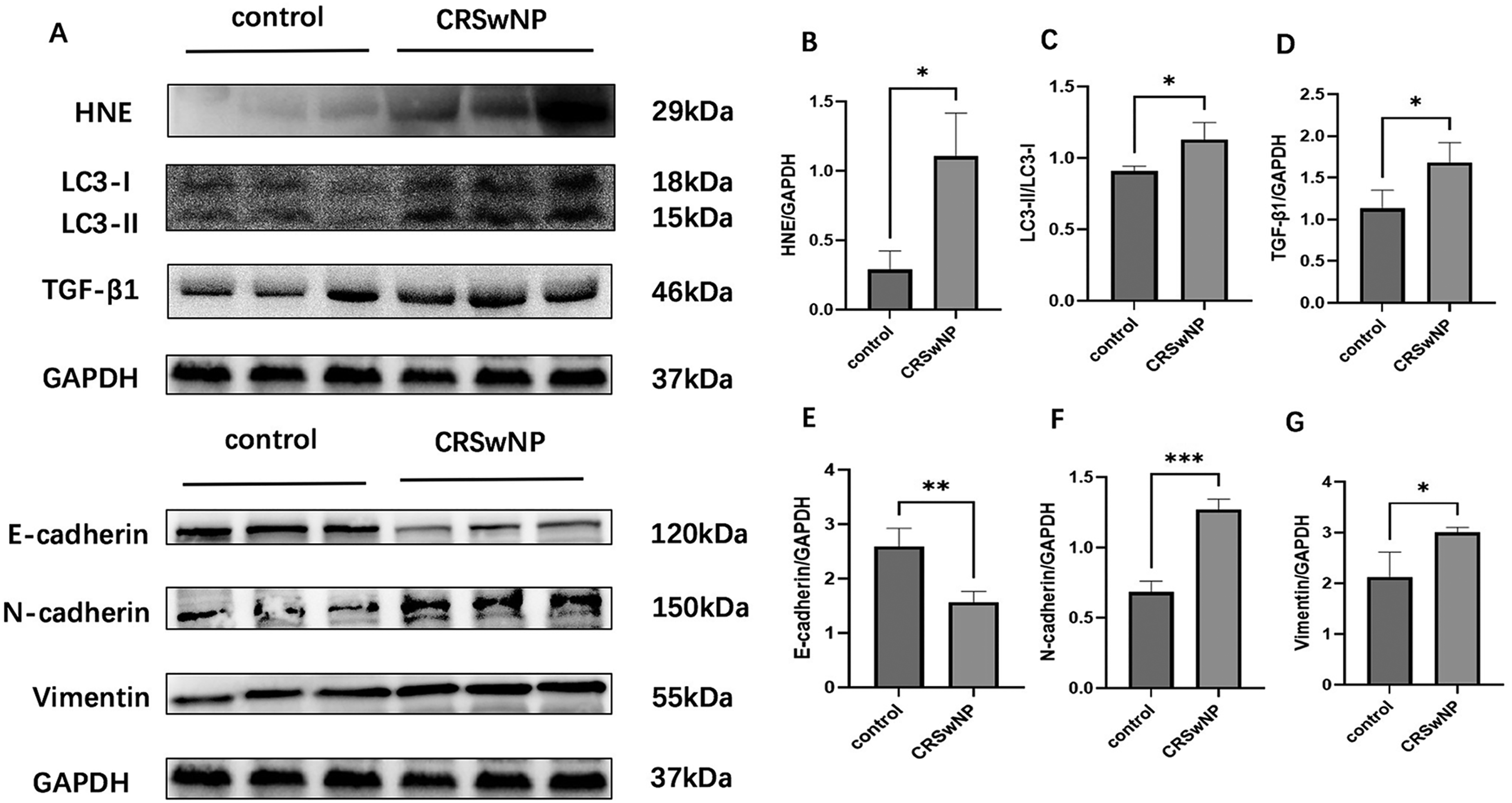
The protein expression levels of HNE, TGF-β1, LC3, and EMT-related proteins in CRSwNP and control tissues. (A) shows the WB bands for the two groups. (B)-(G) are bar graphs presenting the data analyzed using ImageJ and GraphPad Prism software. Western blot analysis revealed that the expression levels of HNE, TGF-β1, LC3, N-cadherin, and Vimentin were significantly upregulated, whereas E-cadherin expression was significantly downregulated in the CRSwNP group compared with the control group (p < .05). *P<0.05; **P<0.01; ***P<.001. HNE, human neutrophil elastase; CRSwNP, chronic rhinosinusitis with nasal polyp; EMT, epithelial–mesenchymal transition; TGF-β1, transforming growth factor-β1.
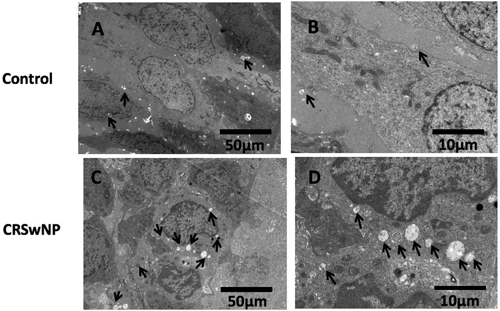
The TEM images of healthy tissue from controls and nasal polyp tissue from CRSwNP patients. (A) and (B) show different magnifications: 50 μm and 10 μm, respectively. C-D show different magnifications: 50 and 10 μm, respectively. An increased number of autophagosomes were observed via TEM in nasal polyp tissue relative to healthy controls. The black arrows indicate autophagosomes. CRSwNP, chronic rhinosinusitis with nasal polyp; TEM, transmission electron microscopy.
The Expression Levels of LC3, TGF-β1, and EMT-Related Proteins in Nasal Epithelial Cells After Stimulation with HNE at Different Concentrations and Time Points
Following HNE stimulation, expression of the epithelial marker E-cadherin decreased significantly, whereas expression of mesenchymal markers N-cadherin and Vimentin increased (P < 0.05, Figure 4A-D). Concurrently, elevated expression of the autophagy-related protein LC3 and TGF-β1 was observed (P < 0.05, Figure 4E-G). These results suggested that 50 ng/mL HNE was a suitable concentration for subsequent experiments.
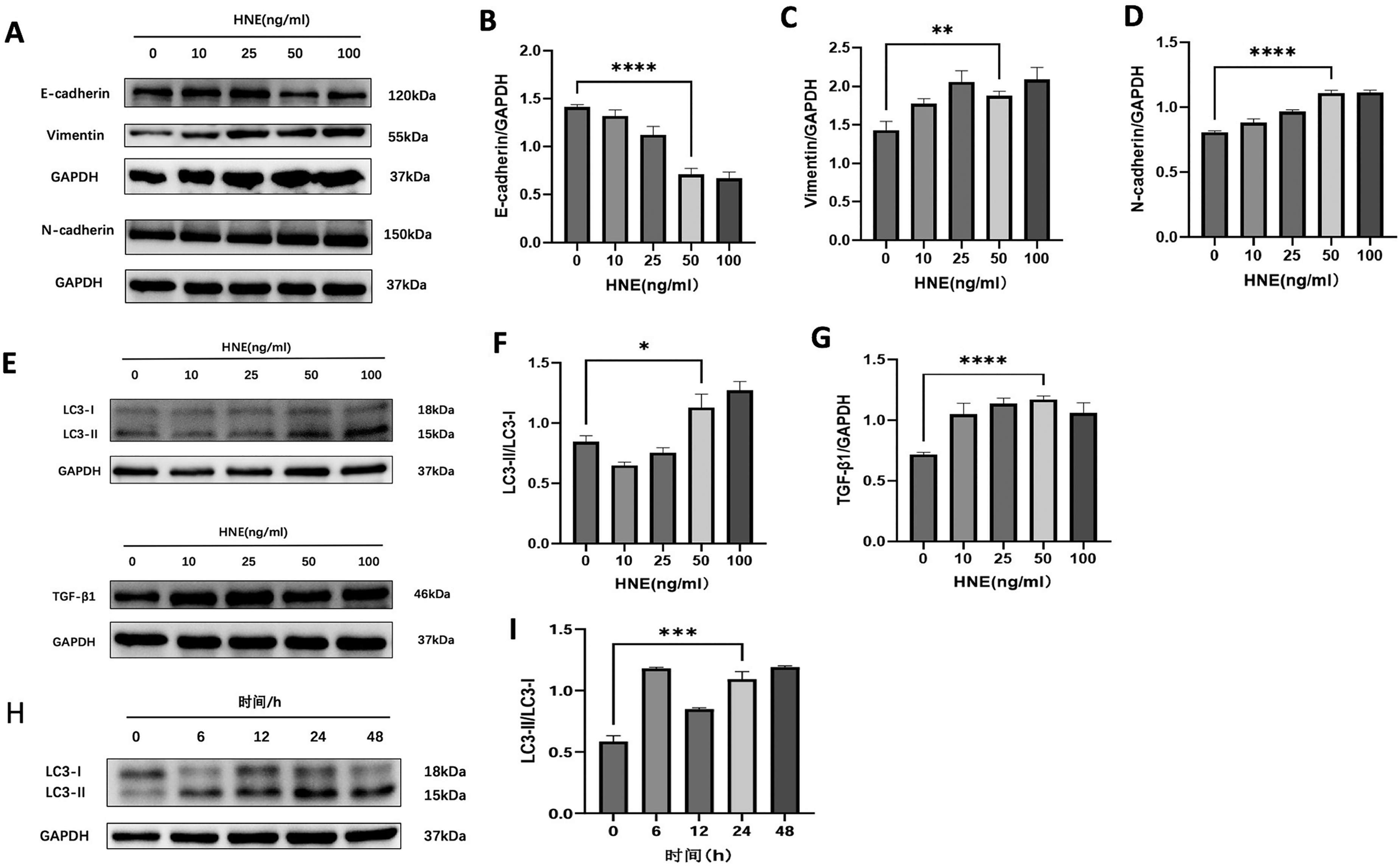
The expression levels of LC3, TGF-β1, and EMT-related proteins in nasal epithelial cells after stimulation with HNE at different concentrations and time points. (A)-(D) The expression of EMT-related proteins after stimulating with different concentrations of HNE (0, 10, 25, 50, and 100 ng/mL). Following HNE stimulation, E-cadherin (an epithelial marker) expression was downregulated, while the mesenchymal markers N-cadherin and Vimentin were upregulated. (E)-(G) The expression of TGF-β1 and autophagy-related proteins after stimulating with different concentrations of HNE (0, 10, 25, 50, 100 ng/mL). An upregulation of the autophagy-related protein LC3 and TGF-β1 was observed. H-I The expression of LC3 after stimulating with 50 ng/ml HNE over different time intervals (0, 6, 12, 24 and 48 h). Expression of LC3 increased significantly after 24 h of HNE stimulation. *P < .05; **P < .01; ****P < .0001. HNE, human neutrophil elastase; EMT, epithelial–mesenchymal transition; TGF-β1, transforming growth factor-β1.
To establish the optimal stimulation time, cells were exposed to 50 ng/mL HNE for 0, 6, 12, 24, and 48 h. Expression of LC3 increased significantly after 24 h of HNE stimulation (Figure 4H-1). A stimulation duration of 24 h was therefore selected for further experiments.
HNE Induces TGF-β1, Autophagy, and EMT Expression in Nasal Epithelial Cells
To investigate the effects of HNE on autophagy, EMT, and TGF-β1, nasal epithelial cells were stimulated with 50 ng/ml HNE. The results showed that the expression of the autophagy-associated protein LC3-II/LC3-I(ratio) and TGF-β1 protein increased (P < .05, Figure 5A-C). Concomitantly, the expression level of the epithelial marker E-cadherin decreased, whereas the expression levels of the mesenchymal markers N-cadherin and Vimentin increased (P < .05, Figure 5D-G).
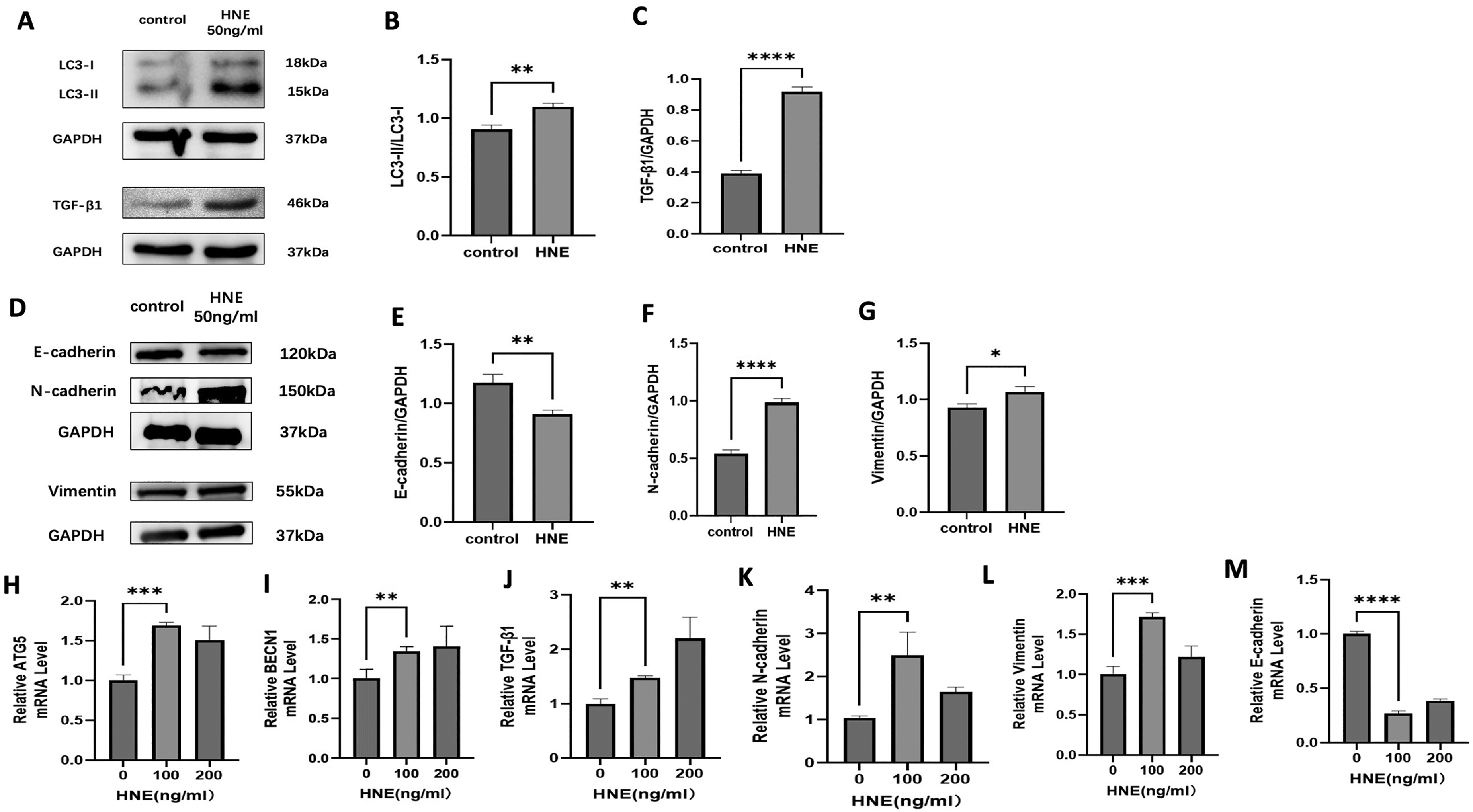
After HNE stimulation of nasal epithelial cells, the expression of TGF-β1, LC3, and EMT-related proteins and genes. (A) and (D) show the electrophoretic band patterns for the respective proteins. (B), (C), (E), (F), and (G) present quantitative analyses of band intensities. (H)-(M) show mRNA expression levels of TGF-β1, LC3, and EMT-related genes after stimulated with increasing concentrations of HNE (0, 100, 200 ng/ml). HNE stimulation significantly upregulated the expression of TGF-β1 and the LC3-II/LC3-I ratio (A)-(C). This was accompanied by a corresponding downregulation of the epithelial marker E-cadherin and upregulation of the mesenchymal markers N-cadherin and Vimentin (D)-(G). *P < .05; **P < .01; ****P < .0001. HNE, human neutrophil elastase; EMT, epithelial–mesenchymal transition; TGF-β1, transforming growth factor-β1.
Furthermore, stimulation of nasal epithelial cells with increasing concentrations of HNE (0, 100, 200 ng/ml) demonstrated that, compared to the control group (0 ng/ml), the HNE-treated cells exhibited increased mRNA expression of TGF-β1, N-cadherin, and Vimentin, accompanied by decreased E-cadherin mRNA expression (Figure 5J-M). Additionally, mRNA levels of the autophagy-related genes ATG5 and BECN1 were upregulated (Figure 5H,1).
Autophagy Inhibitors Inhibit EMT in Nasal Epithelial Cells
Following treatment of cells with the autophagy inhibitor 3-MA at varying concentrations, WB analysis was performed to assess the expression levels of LC3 and Vimentin (Figure 6A). Subsequently, cells treated with 500 μM 3-MA were compared to the control group to detect the expression of E-cadherin and N-cadherin (Figure 6B). As shown in Figure 6, treatment with the autophagy inhibitor 3-MA resulted in the inhibition of autophagy, as indicated by decreased expression of the autophagy-associated protein LC3. Concurrently, the expression of EMT-related protein E-cadherin increased, whereas the expression of N-cadherin and Vimentin decreased.
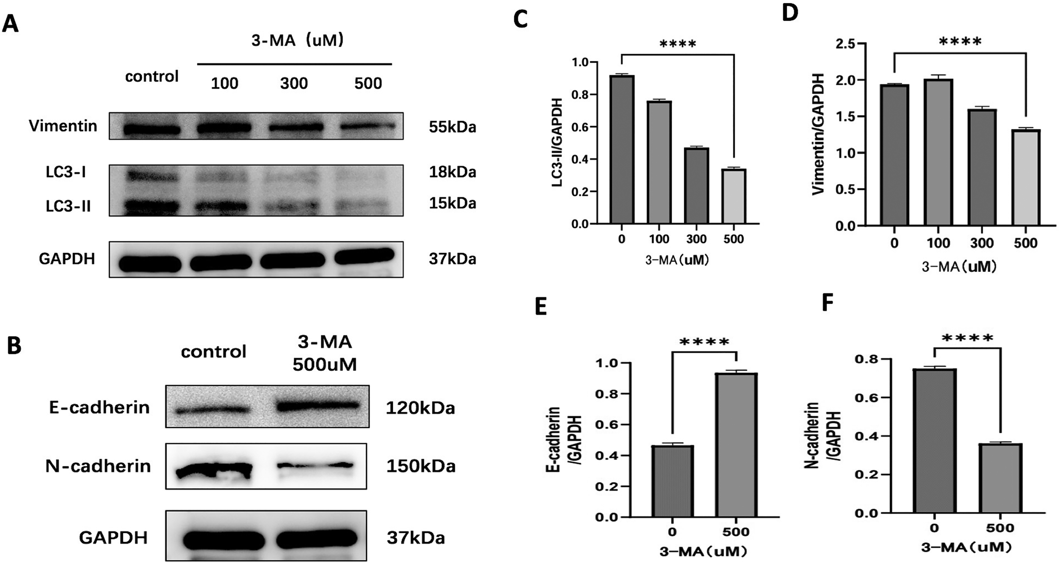
Western blot analysis of the expression of EMT-related proteins in nasal epithelial cells treated with autophagy inhibitors. (A) and (B) show the electrophoretic band patterns for the respective proteins. (C), (D), (E), and (F) present quantitative analyses of band intensities. Following treatment with increasing concentrations (100, 300, and 500 μM) of the autophagy inhibitor 3-MA, a decrease in the expression of LC3- and Vimentin proteins was observed (A, C, and D). Furthermore, at 500 μM, 3-MA reversed the EMT phenotype, as evidenced by upregulation of E-cadherin and downregulation of N-cadherin (B, E, and F). ****P < .0001. EMT, epithelial–mesenchymal transition; 3-MA, 3-methyladenine.
TGF-β1 Inhibitors Inhibit EMT in Nasal Epithelial Cells
Compared with the control group, after treatment of nasal epithelial cells with 50 μM TGF-β1 inhibitors HY-10431, the expression of E-cadherin was increased, while the expression levels of TGF-β1, N-cadherin, and vimentin proteins were decreased (P < .05) (Figure 7).
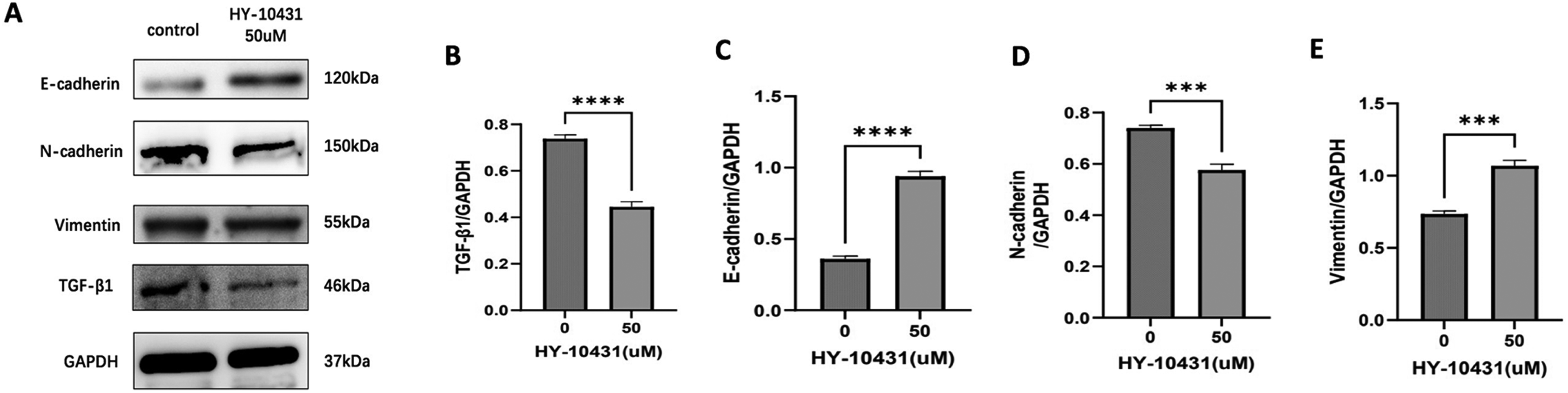
Western blot analysis of the expression of TGF-β1and EMT-related proteins in nasal epithelial cells treated with TGF-β1inhibitors. (A) shows the electrophoretic band patterns for the respective proteins. (B), (C), (D), and (E) present quantitative analyses of band intensities. Treatment of nasal epithelial cells with 50 μM HY-10431 (a TGF-β1 inhibitor) resulted in upregulated E-cadherin expression and downregulated expression of TGF-β1, N-cadherin, and vimentin proteins compared to the control group (A-E). ***P < .001; ****P < .0001. EMT: epithelial–mesenchymal transition; TGF-β1, transforming growth factor-β1.
HNE Induces EMT in Nasal Epithelial Cells Through Autophagy
Three groups of nasal epithelial cells were established: a blank control group, an HNE-treated group, and a group pretreated with autophagy inhibitor 3-MA for 6 h followed by HNE treatment. Western blot analysis revealed that, compared to the blank control group, HNE treatment significantly increased the expression of the autophagy-related protein LC3-II/LC3-I(ratio) (P < .05, Figure 8A, B). Concurrently, HNE stimulation decreased expression of the epithelial marker E-cadherin while elevating levels of the mesenchymal markers N-cadherin and Vimentin, as well as TGF-β1 (P < .05, Figure 8 A, C, D, E and F). However, pretreatment with 3-MA prior to HNE exposure significantly attenuated these effects. Specifically, compared to the HNE-only group, the 3-MA + HNE group exhibited reduced expression of LC3-II/ LC3-I(ratio), TGF-β1, N-cadherin, and Vimentin, alongside increased E-cadherin expression (P < .05). Consistent with the protein findings, in the 3-MA + HNE group, mRNA expression of N-cadherin and Vimentin was significantly decreased, and E-cadherin mRNA was increased compared to the HNE-only group (P < .05, Figure 8G-K).
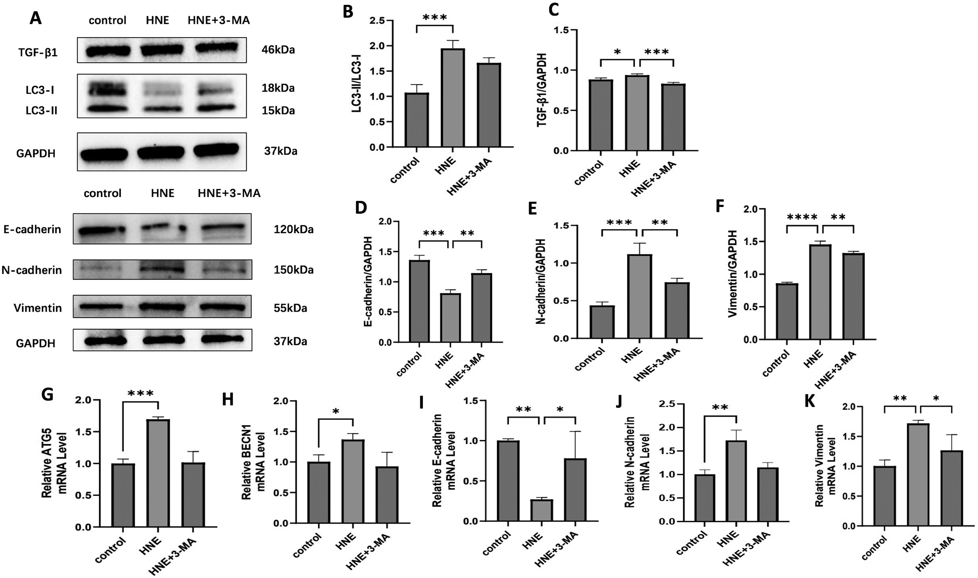
HNE induces EMT in nasal epithelial cells through autophagy. (A)-(F) Western blot analysis of the expression of TGF-β1, LC3, and EMT-related proteins in each group of cells. G-K qRT-PCR analysis of the mRNA expression of autophagy-related genes (ATG5, BECN1) and EMT-related markers (E-cadherin, N-cadherin, Vimentin) in different experimental groups. HNE treatment significantly increased the LC3-II/LC3-I ratio (A, B), downregulated E-cadherin, and upregulated N-cadherin, Vimentin, and TGF-β1 (A, C-F). Pretreatment with 3-MA attenuated these effects, reducing the LC3-II/LC3-I ratio and the expression of TGF-β1, N-cadherin, and Vimentin while restoring E-cadherin levels at both protein and mRNA levels (G-K). HNE, human neutrophil elastase; EMT, epithelial–mesenchymal transition; TGF-β1, transforming growth factor-β1.
Discussion
CRSwNP is a highly prevalent chronic inflammatory disease of the upper airway. In recent years, EMT has been identified in nasal polyp tissue17,18 and is recognized as a significant pathogenic mechanism in CRSwNP. Studies have established an intricate link between neutrophil (NEU)-driven inflammation and EMT in CRSwNP. 19 As we have reported, HNE, which is linked to CRSwNP development, could enhance mucin overexpression in nasal epithelial cells in vitro and in vivo.6,20 In this study, we demonstrated that stimulation with HNE induces the expression of TGF-β1 and activates autophagy in nasal epithelial cells, thus facilitating EMT in nasal epithelium cells. Therefore, elucidating the roles of TGF-β1 and autophagy in HNE-induced EMT is essential for understanding the pathogenesis of CRSwNP.
Autophagy is an intracellular degradation pathway. 21 However, dysregulated, sustained, or ineffective autophagy can damage respiratory epithelial cells, potentially exacerbating inflammation in the respiratory system. 22 LC3 is a key marker of autophagy, existing in two forms: LC3-I and LC3-II. Upon autophagy induction, LC3-I is converted to LC3-II, and elevated autophagic activity correlates with increased LC3-II expression. Therefore, the extent of autophagy is commonly evaluated by measuring the LC3-II to LC3-I ratio. 23 In our study, stimulation of nasal epithelial cells with HNE resulted in increased mRNA expression of the BECN1 and ATG5 genes, accompanied by elevated LC3 protein expression. However, research findings regarding the role of autophagy in CRS are inconsistent. Some studies report elevated LC3-II expression in nasal polyps,5,16 whereas others describe downregulated LC3-II expression in nasal polyps.24,25 In our histological analyses, we observed more autophagosomes and higher expression of the autophagy-related protein LC3 in tissues from patients with CRSwNP compared to controls.
TGF-β1 plays a crucial role in numerous biological processes. For instance, in fibrotic diseases such as pulmonary fibrosis, liver cirrhosis, and renal fibrosis, TGF-β1 overexpression drove excessive ECM deposition.26,27 Research on TGF-β1 in CRS has gained considerable attention in recent years, with multiple studies demonstrating its significant contribution to EMT in the pathogenesis of CRS.28–30 Consistent with this, our study detected elevated TGF-β1 protein expression in CRSwNP. Furthermore, treatment of nasal epithelial cells (RPMI-2650) with the TGF-β1 inhibitor HY-10431 resulted in increased expression of the epithelial marker E-cadherin and decreased expression of the mesenchymal markers N-cadherin and vimentin. These findings further support the critical role of TGF-β1 in promoting EMT in the nasal mucosa.
In this study, we observed elevated expression of HNE and TGF-β1 in CRSwNP, concomitant with enhanced autophagy and evidence of EMT. In vitro, stimulating nasal epithelial cells with HNE induced TGF-β1 upregulation and increased expression of the autophagy marker LC3. Furthermore, we observed reduced E-cadherin levels alongside elevated expression of N-cadherin and Vimentin, suggesting that HNE promotes an EMT-like transition. Pharmacologically inhibiting TGF-β1 (HY-10431) or autophagy (3-MA) partially attenuated EMT. These results indicate that HNE may promote EMT in CRSwNP via the TGF-β1/autophagy pathway. Additionally, this study had several limitations. The sample size was relatively small, and only a single epithelial cell line was used in the experiments. In future studies, we will increase the sample size and utilize multiple cell types to further investigate the role and mechanism of autophagy in EMT in CRSwNP.
Conclusion
In summary, our observations have identified that HNE could promote EMT via autophagy. Furthermore, TGF-β1 significantly mediates EMT in this context. Our findings suggest that autophagy or TGF-β1 may be a selective therapeutic target for EMT in patients with CRSwNP.
Footnotes
Acknowledgment
We express our sincere gratitude to all the teachers and students in the ENT laboratory for their guidance on the experiments.
Ethical Approval
This study was performed in line with the principles of the Declaration of Helsinki. This study was approved by the Medical Ethics Committee of First Affiliated Hospital of Nanchang University.
Funding
The authors disclosed receipt of the following financial support for the research, authorship, and/or publication of this article: This work was supported by the National Natural Science Foundation of China (82260217).
Declaration of Conflicting Interests
The authors declared no potential conflicts of interest with respect to the research, authorship, and/or publication of this article.