






Abstract
Background
Prior microRNA expression profiling investigations have identified hsa-miR-664b-5p as one of the markedly upregulated microRNAs in patients with allergic rhinitis (AR), suggesting its potential important role in the pathogenesis of this condition. This study focused on hsa-miR-664b-5p, elucidate its fundamental mechanistic basis along with its influence on cellular proliferation and apoptotic processes.
Methods
Using reverse transcription-quantitative polymerase chain reaction, the expression profiles of hsa-miR-664b-5p and EGR2 were examined in nasal mucosal samples obtained from individuals with AR. The diagnostic usefulness of hsa-miR-664b-5p was evaluated through receiver operating characteristic (ROC) analysis. In human nasal epithelial cell lines (RPMI 2650 and primary HNEPCs), apoptotic rates were assessed by flow cytometry, and proliferative capacity was measured using the cell counting kit-8 assay. The targeting interaction between hsa-miR-664b-5p and EGR2 was verified by dual-luciferase reporter assay to investigate its regulatory mechanism.
Results
Results confirmed that hsa-miR-664b-5p was significantly upregulated in AR nasal mucosal tissues, while EGR2 was downregulated, showing a strong inverse correlation. ROC analysis demonstrated that hsa-miR-664b-5p holds considerable diagnostic value for AR ( area under the curve = 0.875). Overexpression of hsa-miR-664b-5p was found to promote proliferation and inhibit apoptosis in human nasal epithelial cells. Through dual-luciferase reporter assays, EGR2 was identified as a direct target of hsa-miR-664b-5p. Rescue experiments further supported that hsa-miR-664b-5p influences epithelial cell growth and death by specifically repressing EGR2.
Conclusion
The hsa-miR-664b-5p/EGR2 axis exerts a crucial role in the pathogenesis of AR and could serve as a novel molecular target for the diagnosis and treatment of AR.
Introduction
Allergic rhinitis (AR) ranks among the most prevalent non-infectious allergic disorders globally, primarily driven by immunoglobulin E (IgE)-mediated immune responses. 1 It is a complex condition resulting from interactions between genetic factors, epigenetic regulation, and environmental exposures. In recent decades, the global incidence of AR has been on a continuous rise, now impacting more than 250 million individuals and rendering it a major public health issue. 2 The disease is manifested by symptoms including sneezing, nasal congestion, nasal itching, and rhinorrhea, with pathological features including eosinophil infiltration and increased nasal epithelial barrier permeability. 3 AR severely impacts patients’ social interactions, sleep, and work quality, and imposes a substantial socioeconomic burden. 4 Although current treatments—including antihistamines, intranasal corticosteroids, and immunotherapy—have advanced, approximately 20% of adult patients and 30% of pediatric patients exhibit inadequate responses to existing therapies, 5 patients continue to experience persistent symptoms, treatment-related side effects, or short-lived efficacy, which points to a clear unmet need in current clinical practice. Therefore, identifying novel targets for the diagnosis and treatment of AR and developing more effective and safer therapeutic strategies have become critical issues in both clinical and research settings.
MicroRNAs (miRNAs) are short, evolutionarily conserved non-coding RNA molecules, approximately 22 nucleotides long, that serve as crucial regulators of gene expression at the post-transcriptional level and have been implicated in the development of numerous diseases, that function as key post-transcriptional regulators involved in the pathogenesis of various diseases. 6 In allergic inflammatory diseases, miRNAs are recognized as important modulators. For instance, miR-155 and miR-223 are upregulated in the serum of AR patients and correlate with disease severity and inflammatory cytokine levels, indicating their potential as biomarkers. 7 Additionally, several studies suggest that molecules such as miR-615-3p and miR-205-5p play roles in respiratory inflammation. 8 However, most miRNA expression profiling studies remain preliminary, and their specific mechanisms in AR are not fully elucidated. Notably, hsa-miR-664b-5p is significantly highly expressed in AR, 9 yet its biological function and regulatory mechanisms in the disease pathogenesis await further exploration.
The early growth response (EGR) family includes key members such as EGR1, EGR2, EGR3, and EGR4, which function as immediate-early genes involved in regulating various physiological and pathological processes. 10 Among them, EGR2 is induced upon T-cell receptor activation and negatively regulates T-cell immune responses, thereby suppressing excessive inflammation. 11 Studies have found that EGR2 expression is downregulated in AR and may possess disease-suppressive functions. 12 Although multiple lines of evidence indicate that various miRNAs participate in AR pathogenesis through diverse mechanisms, 13 the specific function of hsa-miR-664b-5p and its regulatory relationship with EGR2 remain poorly understood.
Consequently, this study aims to investigate the expression pattern, biological functions, and molecular mechanisms of hsa-miR-664b-5p in AR, focusing on its role in regulating the proliferation and apoptosis of human nasal epithelial cell lines (HNEpC and RPMI 2650) through EGR2.
Materials and Methods
Study Subjects
The study was performed in line with the principles of the Declaration of Helsinki. Approval was granted by the Ethics Committee of Longyan First Affiliated Hospital of Fujian Medical University before the study began (No. LYREC2023-k017-01, February 17, 2023). The written informed consent has been obtained from the participants involved.
A total of 130 patients undergoing nasal septum or turbinate surgery were recruited from Longyan First Affiliated Hospital of Fujian Medical University between 2022 and 2023; nasal mucosal tissues were collected from these 130 patients. Among them, 70 patients who met the diagnostic criteria for AR were assigned to the AR group, and the remaining 60 patients without AR served as the control group. All participants (aged 18-60 years) underwent detailed evaluation, including clinical history and physical examination. Fasting peripheral venous blood was drawn from all subjects and subjected to the required tests. Participants who had received medication within 10 days prior to the test were excluded. Smokers, pregnant women, and lactating mothers were also excluded. The diagnosis of AR was established based on the patient's symptomatic history and a positive specific IgE (sIgE ≥ 0.35 kU/L) result from the skin prick test (SPT, Allergopharma, Hamburg, Germany). The result of the serum-specific IgE consists of D1, D2 and PHAD. D1 stands for Dermatophagoides pteronyssinus, D2 stands for Dermatophagoides farinae, and PHAD stands for phadiatop, which suggests a respiratory allergy to inhalant allergens.
The total nasal symptom score (TNSS) was used to evaluate nasal symptoms in study participants. This scoring system rates four symptoms—nasal congestion, sneezing, nasal itching, and rhinorrhea—each on a four-point scale (0-3): 0 indicating no symptoms, 1 mild tolerable symptoms, 2 bothersome but tolerable symptoms, and 3 severe symptoms that interfere with daily activities. The TNSS is calculated by summing the scores of all four symptoms, yielding a maximum total of 12 points. 14
Cell Culture
The immortalized human nasal carcinoma line RPMI 2650 (ATCC® CCL-30™) and primary human nasal epithelial cells (HNEpC, BNCC340481) were selected. Cells were maintained in RPMI 1640 medium (Gibco, Rockville, MD, USA), supplemented with 10% fetal bovine serum (FBS) and 1% penicillin-streptomycin, and incubated at 37 °C in a humidified 5% CO2 atmosphere.
Cell Transfection
Transfection procedures were carried out with Lipofectamine 3000 (Invitrogen, USA) to introduce hsa-miR-664b-5p mimic, inhibitor, miR-664b-5p mimic negative control (mimic-NC), miR-664b-5p inhibitor negative control (inhibitor-NC), non-targeting siRNA negative control (si-NC), or siRNA targeting EGR2 (si-EGR2) into HNEpC and RPMI 2650 cells, following the supplier's guidelines. After 6 h, the transfection mixture was replaced with fresh complete medium containing 10% FBS, and cells were incubated for an additional 48 h prior to downstream assays.
Quantitative Real-Time Polymerase Chain Reaction
Total RNA was isolated using TRIzol™ reagent (Takara, Dalian, China). Reverse transcription was performed to synthesize cDNA, and quantitative polymerase chain reaction (PCR) was executed on an Exicycler™ 96 Real-Time Quantitative PCR Instrument (Bioneer, Korea). GAPDH and U6 served as endogenous references for mRNA and miRNA quantification, respectively, with expression levels calculated via the 2−ΔΔCT method.
Western Blotting
Tissue samples or cells were lysed using RIPA lysis buffer containing 1% protease inhibitor (Pierce). Protein concentration was determined using a BCA assay kit (Pierce). Total proteins were separated by 10% SDS-PAGE and transferred to a PVDF membrane (Santa Cruz Biotechnology). After blocking with 5% non-fat milk, the membrane was incubated with primary antibodies at 4 °C overnight. Following three washes with TBST, the membrane was incubated with secondary antibody for 1 h, and protein bands were visualized using enhanced chemiluminescence solution. Primary antibodies: EGR2 antibody (#ab245228, dilution 1:1000, Abcam, UK); GAPDH antibody (#A19056, dilution 1:1000, ABclonal, China).
Cell Counting Kit-8
Log-phase HNEpC and RPMI 2650 cells were seeded into 96-well plates at a density of 5 × 10³ cells per well following transfection. Cell proliferation was monitored at 0, 24, 48, and 72 h by adding 10 µL of cell counting kit-8 (CCK-8) solution to each well, incubating for 2 h at 37 °C, and measuring optical density at 450 nm.
Detection of Cell Apoptosis by Flow Cytometry
Post-transfection, cells were washed with PBS, harvested by trypsinization and centrifugation, washed again, and resuspended in binding buffer. Apoptosis was evaluated with an Annexin V-FITC/PI detection kit (Beckman Coulter, Brea, CA) per the manufacturer's protocol, using flow cytometry to determine the apoptosis rate as the ratio of apoptotic cells to total cells.
Dual-Luciferase Reporter Gene Assay
For dual-luciferase reporter assays, HNEpC and RPMI 2650 cells were plated in 24-well plates 24 h before transfection. Wild-type and mutant 3′-UTR sequences of EGR2 were cloned into reporter plasmids by Genomeditech (Shanghai, China). Cells were co-transfected with these plasmids and either hsa-miR-664b-5p mimic, inhibitor, or control mimic, using Lipofectamine 3000. Luciferase activity was quantified 48 h post-transfection with a dual-luciferase detection kit.
Statistical Analysis
Statistical analyses were conducted using SPSS 24.0 and GraphPad Prism 9.0. Data are expressed as mean ± standard deviation. Differences between groups were analyzed with Student's t-test (for two groups) or one-way ANOVA (for multiple groups). The diagnostic performance of hsa-miR-664b-5p was determined by receiver operating characteristic (ROC) analysis. Correlation between hsa-miR-664b-5p and EGR2 expression was evaluated using Pearson's correlation coefficient, with P < .05 deemed statistically significant.
Results
Hsa-miR-664b-5p is Upregulated and Inversely Correlated with EGR2 in Allergic Rhinitis
This study enrolled 70 patients with AR and 60 healthy controls. No statistically significant differences were observed between the two groups in terms of age or gender distribution (P > .05). Serum levels of allergen-specific IgE (PHAD, D1, D2) and the TNSS were significantly elevated in the AR group compared to the control group (P < .0001). Detailed clinical characteristics are presented in Table 1. Relative to healthy controls, the expression level of hsa-miR-664b-5p was notably increased in the nasal mucosal tissues of AR patients, whereas the mRNA expression of EGR2 was significantly decreased (Figure 1A, 1B). Furthermore, Western blot analysis revealed a marked decrease in EGR2 protein levels in AR patient tissues relative to controls (Figure 1C). A significant inverse correlation was observed between hsa-miR-664b-5p and EGR2 expression in AR patients (r = -0.814; Figure 1D). ROC curve analysis indicated that hsa-miR-664b-5p has considerable diagnostic accuracy for distinguishing AR patients from healthy individuals, with an area under the curve (AUC) of 0.875, sensitivity of 93.3%, and specificity of 72.9% (P < .0001; 95%CI:0.813-0.936; Figure 1E).
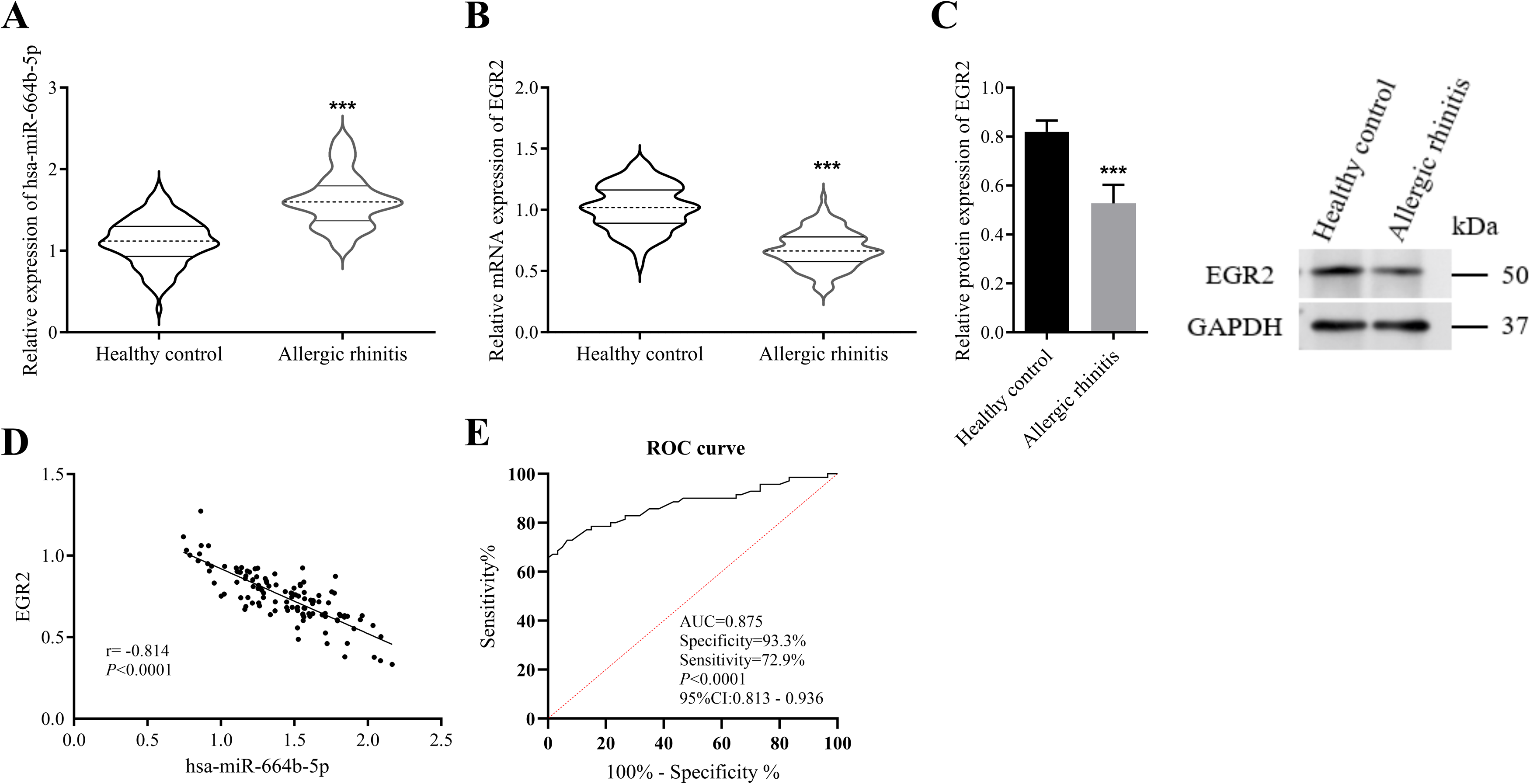
Hsa-miR-664b-5p expression and its regulatory role on EGR2 in allergic rhinitis. (A) Expression levels of hsa-miR-664b-5p in normal versus allergic rhinitis samples. (B) Expression of EGR2 mRNA in normal versus allergic rhinitis samples. (C) Expression of EGR2 protein in normal versus allergic rhinitis samples. (D) Correlation analysis between EGR2 and hsa-miR-664b-5p. (E) ROC curve analysis of the diagnostic efficacy of hsa-miR-664b-5p for allergic rhinitis (AUC = 0.875, 95%CI:0.813-0.936, P < .0001). ***P < .001. ROC, receiver operating characteristic; EGR, early growth response; AUC, area under the curve.
Clinical Characteristics of Healthy Controls and AR Patients.
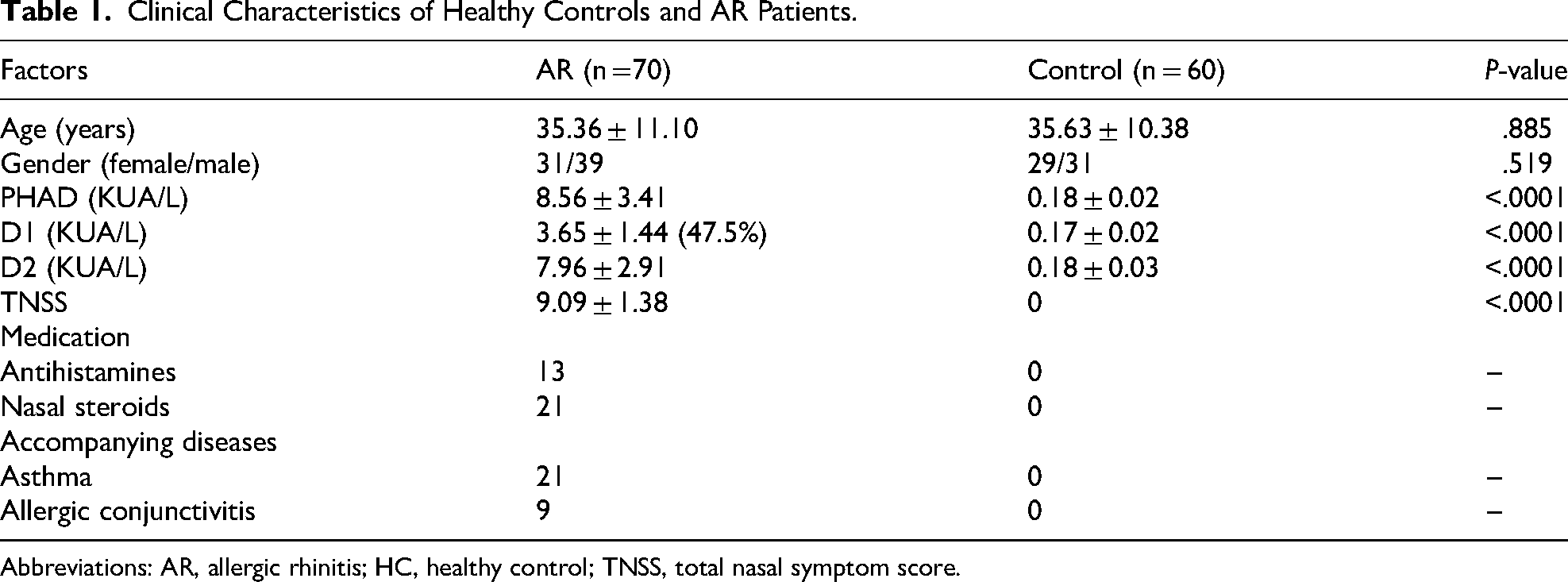
Abbreviations: AR, allergic rhinitis; HC, healthy control; TNSS, total nasal symptom score.
Hsa-miR-664b-5p Promotes Proliferation and Inhibits Apoptosis
As shown in Figure 2A, the expression of has-miR-664b-5p was significantly increased in the mimic group and decreased in the inhibitor group, compared with the control and NC groups. Functional assays revealed that has-miR-664b-5p overexpression promoted cell proliferation, whereas its knockdown suppressed proliferation in both cell lines, as measured by CCK-8 assay (Figure 2B, 2C). Furthermore, flow cytometric analysis demonstrated that has-miR-664b-5p overexpression inhibited apoptosis, while its knockdown enhanced apoptosis in HNEpC and RPMI 2650 cells (Figure 2D).
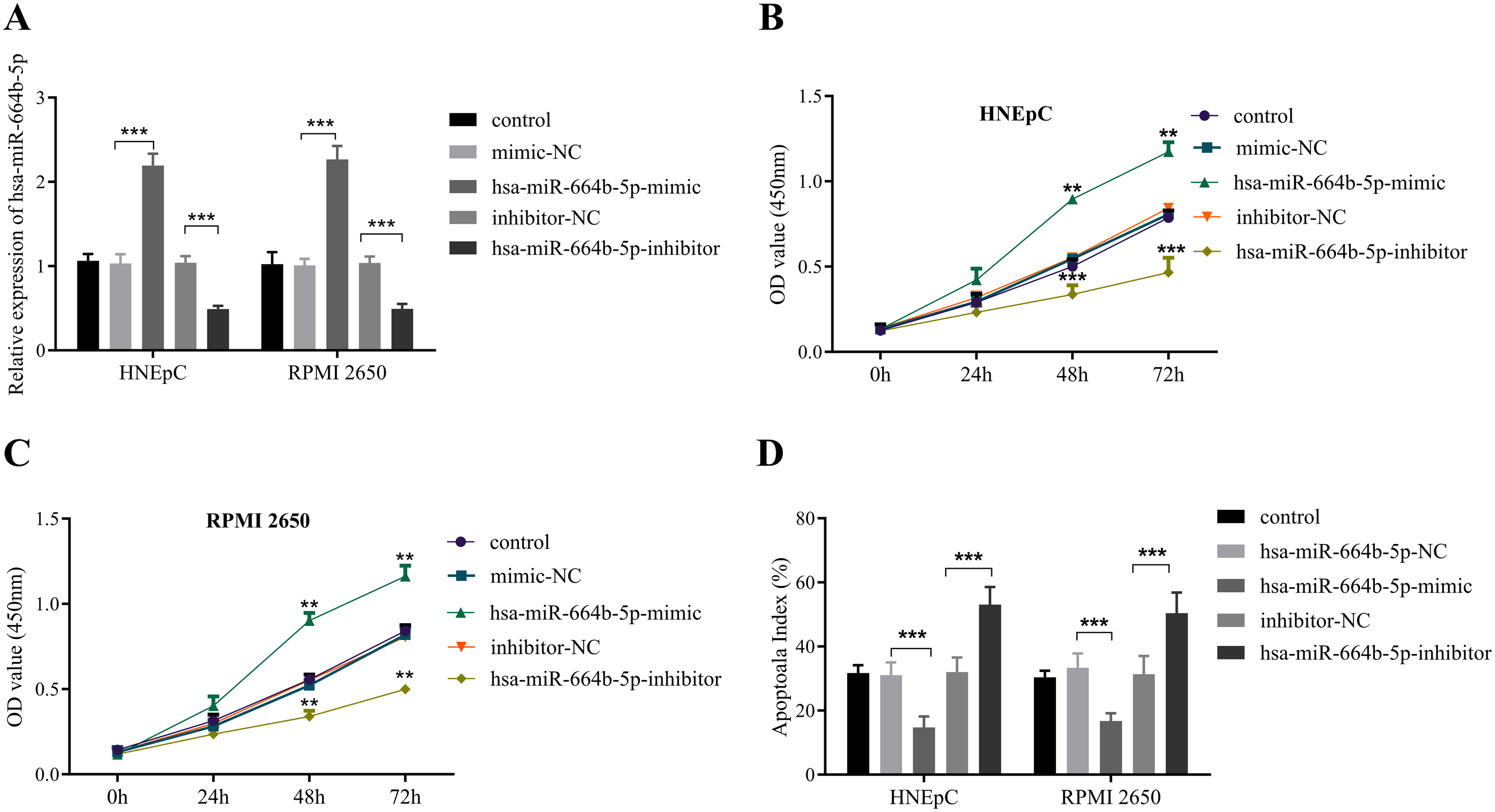
Effects of hsa-miR-664b-5p modulation in nasal epithelial cells. (A) Hsa-miR-664b-5p expression after transfection with mimic or inhibitor in HNEpC and RPMI 2650 cells. (B) CCK-8 assay showing that miR-664b-5p mimic promotes HNEpC cell proliferation. (C) CCK-8 assay showing that hsa-miR-664b-5p mimic promotes RPMI 2650 cell proliferation. (D) Apoptosis assay showing that hsa-miR-664b-5p mimic inhibits, while its inhibitor induces, cell apoptosis. **P < .01, ***P < .001. CCK-8, cell counting kit-8
EGR2 is a Direct Target of hsa-miR-664b-5p
Bioinformatics analysis indicated a putative binding site for hsa-miR-664b-5p in the 3′-UTR region of EGR2 (Figure 3A). Dual-luciferase assays confirmed that hsa-miR-664b-5p mimic significantly suppressed luciferase activity of the wild-type EGR2 3′-UTR reporter, but not the mutant version. Conversely, inhibition of hsa-miR-664b-5p enhanced the activity of the wild-type reporter (Figure 3B, 3C). These findings confirm that hsa-miR-664b-5p directly targets EGR2 through specific binding to its 3′-UTR.
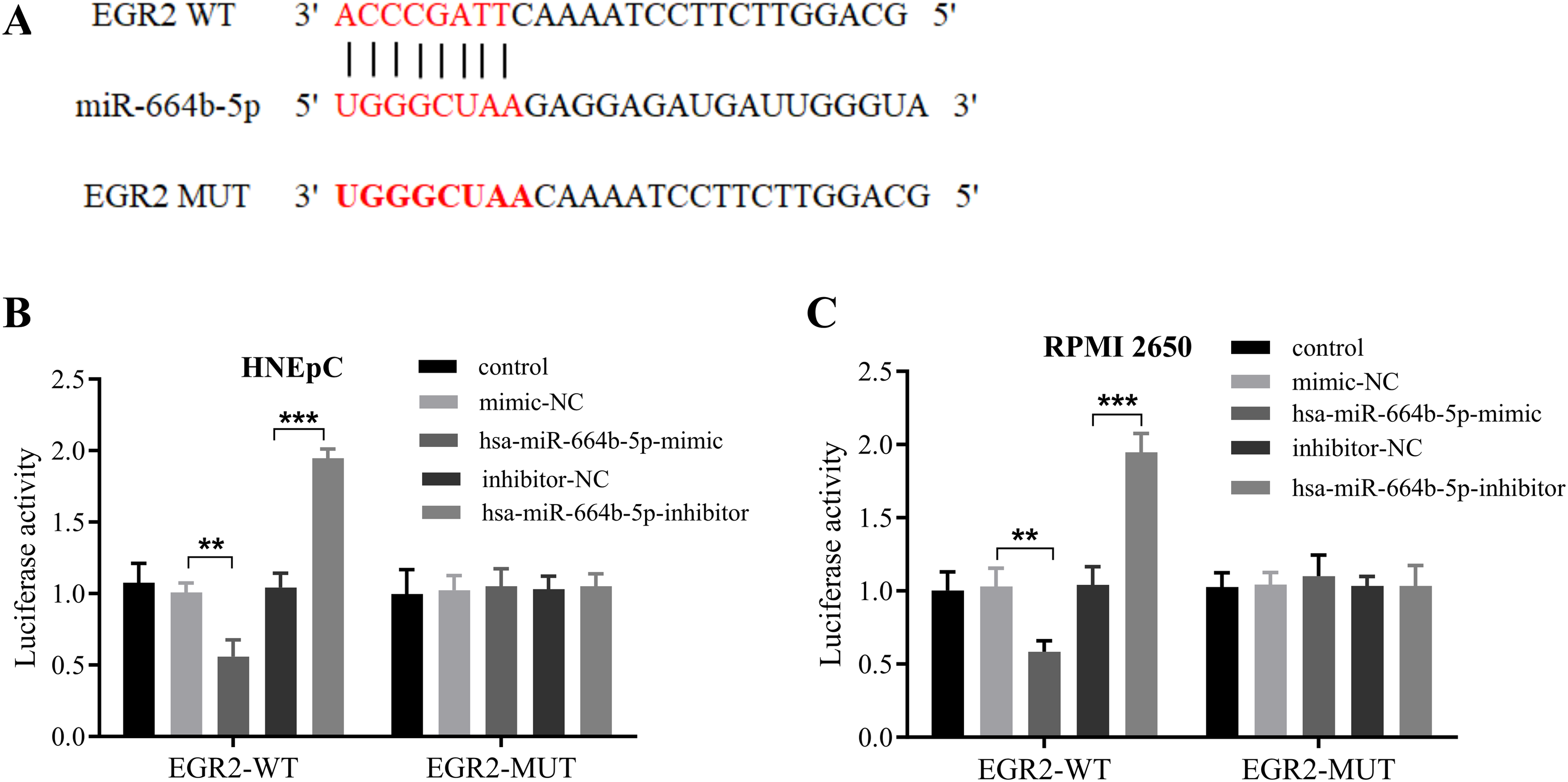
Hsa-miR-664b-5p directly targets and regulates EGR2. (A) Schematic representation of the predicted binding site for hsa-miR-664b-5p within the 3'-UTR of the EGR2 gene. (B) Relative luciferase activity of the EGR2 3'-UTR reporter (wild-type or mutant) in BRMI 2650 cells following modulation of hsa-miR-664b-5p expression. (C) Relative luciferase activity of the EGR2 3'-UTR reporter (wild-type or mutant) in primary HNEpC cells following modulation of hsa-miR-664b-5p expression. **P < .01, ***P < .001. EGR, early growth response.
EGR2 Knockdown Rescues hsa-miR-664b-5p Inhibitor Phenotypes
Functional rescue experiments further confirmed that hsa-miR-664b-5p regulates cell proliferation and apoptosis through EGR2. Results demonstrated that knockdown of hsa-miR-664b-5p significantly upregulated the mRNA expression of EGR2, which was reversed upon EGR2 silencing (Figure 4A, 4B). At the protein level, the knockdown of hsa-miR-664b-5p significantly upregulated EGR2 expression, an effect that was rescued following EGR2 silencing (Figure 4C, 4D). CCK-8 assays showed that hsa-miR-664b-5p knockdown attenuated cell proliferation, an effect that could be rescued by EGR2 knockdown (Figure 4E, 4F). Similarly, apoptosis assays indicated that the pro-apoptotic effect induced by hsa-miR-664b-5p knockdown was partially reversed by EGR2 knockdown (Figure 4G, 4H). In summary, hsa-miR-664b-5p directly targets EGR2 to control epithelial cell proliferation and apoptosis, playing a critical role in the pathogenesis of AR.
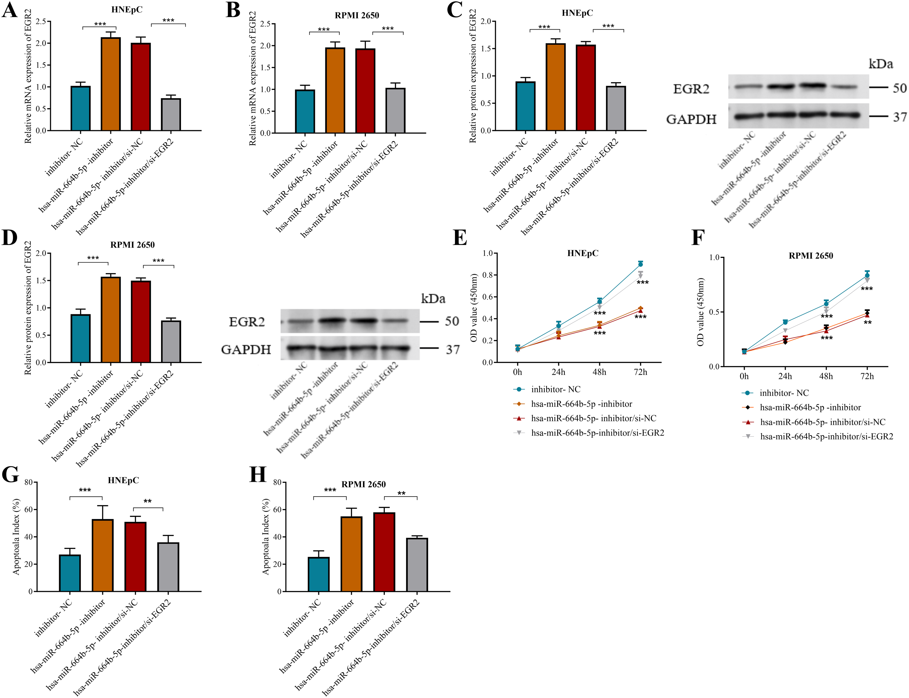
Rescue experiments. (A) EGR2 mRNA expression in HNEpC cells following the indicated treatments. (B) EGR2 mRNA expression in RPMI 2650 cells following the indicated treatments. (C) EGR2 protein expression in HNEpC cells following the indicated treatments. (D) EGR2 protein expression in RPMI 2650 cells following the indicated treatments. (E) Proliferation curve of HNEpC cells following the indicated treatments. (F) Proliferation curve of RPMI 2650 cells following the indicated treatments. (G) Apoptotic rates were measured by flow cytometry in HNEpC cells. (H) Apoptotic rates were measured by flow cytometry in RPMI 2650 cells. **P < .01, ***P < .001. EGR, early growth response.
Discussion
AR is a common inflammatory airway disease with a continuously increasing global prevalence-approximately 23% in Europe, 20%-30% in the United States, and 17.6% in China.15,16 Although current understanding of AR pathogenesis and treatment has advanced, the exact molecular mechanisms remain incompletely elucidated, and available therapies are not curative. 17 Therefore, exploring novel and effective treatments for AR is urgently needed. Emerging evidence indicates that miRNAs are instrumental in the differentiation of immune cells and the modulation of immune reactions. 18
The hsa-miR-664 family comprises important small non-coding RNAs, including hsa-miR-664a, hsa-miR-664b, and hsa-miR-664c. Owing to high sequence similarity among mature forms, they may exhibit functional redundancy. These miRNAs participate in regulating diverse biological processes such as cell proliferation and apoptosis.19,20 For example, miR-664a-5p has been shown to inhibit autophagy and induce apoptosis in renal tubular epithelial cells through the HIPK2/Calpain-1/GSα pathway 21 ; the lncRNA Punisher affects vascular smooth muscle cell apoptosis and neointima formation by regulating related miRNAs. 22 Notably, miR-664b-5p exhibits both tumor-suppressive and oncogenic activities in various cancers.23,24 For instance, it is downregulated in hepatocellular carcinoma and suppresses viability, migration, and invasion of HepG2 and SNU-475 cells while promoting apoptosis. 25 These findings imply that the hsa-miR-664 family plays a significant role in apoptosis regulation. It is noteworthy that the development of AR is associated with dysregulated apoptotic processes in nasal epithelial cells: decreased apoptosis can induce nasal mucosal hypertrophy, thereby leading to typical symptoms like nasal congestion and glandular hypersecretion 32.
This study found that hsa-miR-664b-5p was significantly upregulated in the nasal tissue of patients with AR. ROC analysis further confirmed the discriminative capacity of serum hsa-miR-664b-5p for AR, yielding an AUC of 0.875 (95% CI: 0.812-0.925), with a specificity of 93.3% and a sensitivity of 72.9%, supporting its favorable predictive value for AR. Compared with existing biomarkers, hsa-miR-664b-5p demonstrated distinct diagnostic characteristics. Previous studies reported that serum miR-135a achieved an AUC of 0.837 (95% CI: 0.772-0.889) for AR diagnosis, with a sensitivity (69.89%) comparable to that observed in the present study, though its specificity (82.89%) was lower than the 93.3% obtained here. 26 Another investigation evaluating miR-155 reported a markedly high AUC (0.99) and sensitivity (100%), yet its specificity was comparatively modest (71.1%). 27 The lower AUC of hsa-miR-664b-5p relative to that reported for miR-155 may be attributable in part to the relatively smaller sample size of the current study. Overall, serum hsa-miR-664b-5p shows promising potential as an auxiliary diagnostic biomarker for AR, with its high specificity representing a notable advantage.
The RPMI 2650 cell line is derived from a squamous cell carcinoma of the human nasal septum and maintains high stability and a near-diploid karyotype during long-term in vitro culture. 28 Studies have shown that these cells closely resemble normal human nasal epithelium in terms of karyotype, cytokeratin expression profiles, and the presence of mucin-like substances on the cell surface 29 The RPMI 2650 cell line has been developed as an in vitro model for research on AR. 30 The HNEpC cell line has been extensively used in studies investigating the inflammatory responses and barrier function of the nasal epithelium in AR. 31 Therefore, both RPMI 2650 and HNEpC cell lines were selected for the cellular experiments in this study. Functional experiments demonstrated that hsa-miR-664b-5p overexpression suppressed apoptosis and promoted proliferation in both RPMI 2650 and HNEpC nasal epithelial cells, whereas its knockdown enhanced apoptosis and inhibited proliferation, consistent with previous reports.32,33 Demonstrates the pro-proliferative and anti-apoptotic effects of hsa-miR-664b-5p.
miRNAs primarily exert their biological functions through the post-transcriptional regulation of gene expression, a mechanism mediated by complementary base-pairing with target mRNA. 34 EGR2 was predicted as a target of hsa-miR-664b-5p using TargetScan. Experimental data confirmed that EGR2 protein levels were significantly reduced in nasal tissues from AR patients compared to controls (P < .05) and inversely correlated with has-miR-664b-5p expression (r = -0.814). The targeting interaction was additionally confirmed via dual-luciferase reporter assays. Subsequent functional rescue experiments showed that inhibiting hsa-miR-664b-5p upregulates EGR2 expression, thereby suppressing proliferation and promoting apoptosis-effects that were reversed by EGR2 knockdown. These results clearly demonstrate the central role of the hsa-miR-664b-5p/EGR2 axis in AR-related epithelial dysfunction. Hsa-miR-664b-5p suppresses apoptosis and promotes proliferation in nasal epithelial cells via the EGR2 pathway.
This study has several limitations. As indicated by existing research, the development of allergic responses is closely associated with aberrant activation of immune signaling pathways. For instance, in house dust mite-induced AR, allergen exposure can upregulate CMPK2 expression, leading to mitochondrial DNA release, which in turn activates the NLRP3 inflammasome via the cGAS-STING pathway and promotes the production of inflammatory factors such as IL-1β. 35 The present study did not explore the specific role of this molecule in allergen-triggered immune signaling pathways. Further research should investigate changes in hsa-miR-664b-5p expression upon allergen exposure and its impact on downstream inflammasome activation and cytokine production in typical allergic response pathways. Another limitation is the RPMI-2650 cell line, although it serves as a useful tool for initial mechanistic studies, its tumor origin entails inherent biological differences from primary nasal epithelial cells of AR patients, which may affect the physiological relevance and extrapolation of the findings. Future studies incorporating animal models or clinical samples will help systematically evaluate the role of this molecule in linking epithelial abnormalities to immune dysregulation, thereby advancing a more comprehensive understanding of its function in the pathogenesis of AR.
In conclusion, this study reports the aberrant overexpression of hsa-miR-664b-5p in AR and confirms that it regulates nasal epithelial cell proliferation and apoptosis by targeting EGR2, elucidating its key role in AR pathogenesis.
Footnotes
Acknowledgements
Not applicable.

Ethical Considerations
The study was performed in line with the principles of the Declaration of Helsinki. Approval was granted by the Ethics Committee of Longyan First Affiliated Hospital of Fujian Medical University before the study began (No. LYREC2023-k017-01, 17 February 2023).
Consent to Participate
The written informed consent has been obtained from the participants involved.
Consent for Publication
Not applicable.
Authors’ Contributions
Conceptualization was done by JF, RD, YY, YH, GL, JW, and RL; data curation was contributed by JF, RD, YY, YH, GL, JW, and RL; formal analysis was done by YY and YH; funding acquisition was done by RL; investigation was done by YY and YH; methodology was done by JF, RD, YY, YH, GL, JW, and RL; project administration was done by RL; resources was done by GL and JW; software was contributed by GL and JW; supervision was done by RL; validation was done by GL and JW; visualization, roles/writing—original draft were done by YY and YH; writing—review and editing was done by JF, RD, and RL.
Funding
The authors received no financial support for the research, authorship, and/or publication of this article.
Declaration of Conflicting Interests
The authors declared no potential conflicts of interest with respect to the research, authorship, and/or publication of this article.
Data Availability
The datasets used and/or analyzed during the current study are available from the corresponding author on reasonable request.