
Abstract
Objective
The osteochondral allograft procedure uses grafts constructed larger than the recipient site to stabilize the graft, in what is known as the press-fit technique. This research aims to characterize the relationships between press-fit size, insertion forces, and cell viability in ovine and human osteochondral tissue.
Design
Human (4 donors) and ovine (5 animals) articular joints were used to harvest osteochondral grafts (4.55 mm diameter, N = 33 Human, N = 35 Ovine) and create recipient sites with grafts constructed to achieve varying degrees of press fit (0.025-0.240 mm). Donor grafts were inserted into recipient sites while insertion forces were measured followed by quantification of chondrocyte viability and histological staining to evaluate the extracellular matrix.
Results
Both human and ovine tissues exhibited similar mechanical and cellular responses to changes in press-fit. Insertion forces (Human: 3-169 MPa, Ovine: 36-314 MPa) and cell viability (Human: 16%-89% live, Ovine: 2%-76% live) were correlated to press-fit size for both human (force: r = 0.539, viability: r = −0.729) and ovine (force: r = 0.655, viability: r = −0.714) tissues. In both species, a press-fit above 0.14 mm resulted in reduced cell viability below a level acceptable for transplantation, increased insertion forces, and reduced linear correlation to press-fit size compared to samples with a press-fit below 0.14 mm.
Conclusions
Increasing press-fit size required increased insertion forces and resulted in reduced cell viability. Ovine and human osteochondral tissues responded similarly to impact insertion and varying press-fit size, providing evidence for the use of the ovine model in allograft-related research.
Keywords
Introduction
Fresh osteochondral allograft transplantation (OCAT) is a surgical repair technique for focal osteochondral lesions, often indicated for young and active patients, that is associated with positive clinical success rates. 1 OCAT replaces degraded cartilage and underlying bone with cadaveric osteochondral donor grafts.1,2 The goal is to recreate a stable articulating surface and ensure a sufficient number of viable cells in donor cartilage to maintain the extracellular matrix.2,3 A common threshold to predict the clinical success of donor cartilage is 70% viable cell density (VCD) or above at the time of transplantation, compared to donor cartilage cell viability on the day of procurement.4-6 The insertion method of osteochondral grafts requires a surgeon to tamp the graft into the recipient hole, 3 which has been reported to transmit high-impact forces to donor cartilage, leading to a significant reduction in cell viability.1,7
Impact forces are required because grafts are press-fit, which is created by harvesting an osteochondral graft slightly larger in diameter compared to the recipient site. The press-fit size is defined as the difference in diameter between the harvested graft and recipient site. 7 Press-fit ensures sufficient contact between graft and recipient tissues to minimize micro-motion and ensure initial stability until eventual graft integration. 7 Clinically, dowel grafts usually range from 15 to 35 mm in diameter with press-fit sizes between 0.3 and 0.5 mm.8,9 Using a bovine model, the magnitude of the insertion forces required to seat the graft were shown to be directly correlated to the press-fit size. 7 These authors investigated press-fit sizes of 0.00, 0.10, and 0.20 mm and found an increase in impact forces, crack development, and lower chondrocyte viability in donor tissue with increasing press-fit size. 7 Similarly, a combined bovine and porcine model observed that chondrocyte death varied logarithmically with impact energy and mean applied force during insertion of osteochondral grafts. 10
Previous osteochondral graft mechanics studies have used fresh cadaveric, 3 fresh-frozen cadaveric, 11 bovine,7,10 and porcine10,12,13 explant models. To the best of our knowledge, graft insertion impact mechanics have not been previously investigated in the ovine model, although sheep are widely considered appropriate for modeling surgical techniques related to OCAT due to similarities in anatomy and macroscopic appearance of the tibiofemoral joint.14-19 The structural, chemical, and physical differences between ovine and human tissues must be considered when using sheep as a preclinical surgical model. Biomechanically, ovine bone is more dense and stronger than human bone. In a comparison of femoral shafts, ovine bone was observed to have higher bone mineral density, 0.7 versus 0.4 g/cm3 in human, and higher fracture stress 13.22 MPa in ovine compared to 1.21 MPa in human. 20 Structural differences also exist with ovine subchondral bone observed to be more dense and less porous than human subchondral bone 21 with different bone microarchitecture. 22 Differences in material and structural properties are accompanied by differences in anatomy, primarily the thinner ovine cartilage, 0.4 to 0.5 mm on the femoral condyles,15,23 compared to 2.2 to 2.5 mm on the human femoral condyles.15,23 Despite these tissue differences, ovine tibiofemoral joints are anatomically similar to humans, 18 and the ovine model is cost-effective and accessible compared to other large animal models used in osteochondral research. 19
This research aimed to determine the relationship between press-fit size, insertion forces and chondrocyte viability and to compare these relationships between human and ovine tissues. It was hypothesized that the similarities between ovine and human osteochondral tissues would result in similar responses to changes in press-fit size.
Materials and Methods
All procedures were conducted with approvals from the Institutional Review Board of Sinai Health System (REB 19-0080-E) and Institutional Animal Care Committee of the University of Guelph (AUP #3974).
Sample Sources
Femoral condyles and tibial plateaus from human donors that exceeded storage duration limits for transplantation at Mount Sinai Allograft Technologies tissue bank (14- to 30-day storage, 4 donors, 20-34 years old, 99.7 ± 23.6 kg), and fresh tibial plateaus, femoral heads and talus joints from female Rideau-Arcott sheep (Fresh, 5 sheep, 6 years old, 67.7 ± 16.4 kg) were used.
Sample Preparation
Human femoral condyles and ovine tibial plateaus were used to harvest cylindrical osteochondral grafts using a 4.5 mm inner diameter manual core harvester (Smith & Nephew, Watford, United Kingdom). After harvest, the average diameter of all grafts (N = 68) was 4.55 ± 0.04 mm and average length was 10.53 ± 0.52 mm. To create a robust range of press-fit sizes (press-fit size = donor graft diameter − recipient site diameter), human (N = 28) and ovine (N = 28) graft diameters were modified manually using sandpaper (60-grit) with final diameter reported as the average of 6 to 8 measurements acquired at locations along the graft length. Samples assigned to the control group (Human N = 5, Ovine N = 7) were extracted using the same methods but did not undergo diameter modification. Recipient sites were created in human tibial plateaus and ovine talus and femoral head joint surfaces using a CNC machine (Model 5410, Sherline, Vista, California, USA) to mitigate drill wobble. Precise holes, 4.37 mm in diameter, were drilled perpendicular to the articular surface to depths matching graft lengths. Tissue for recipient sites was species and donor matched.
Insertion Force Application and Quantification
The graft was manually placed into the recipient site, with saline-soaked gauze placed on top of the graft prior to impact to recreate clinical conditions.
24
Impacts, delivered with a metal dowel and mallet, were measured with a dynamic load cell (Model 218C—10,000 lb Charge Output Dynamic Force Sensor with Model 421A11—Industrial Charge Amplifier. PCB Electronics, Depew, New York, USA) (
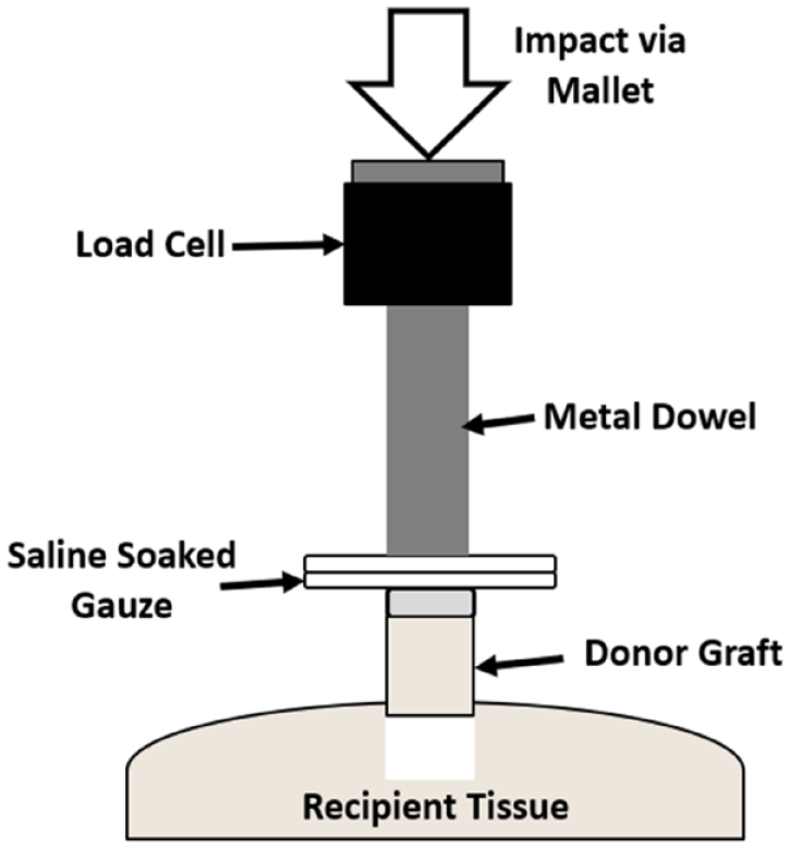
Diagram depicting mallet strike application and measurement of impact forces.
Mallet strikes were applied in succession, until recipient and donor cartilage surfaces were visually aligned. The number of strikes required to insert the graft was recorded (# strikes). Stress applied to the cartilage, calculated as force normalized by surface area of the dowel tip (MPa), and impulse, calculated as force integrated with respect to time (N·ms), were recorded for each strike. Stress and impulse refer to summation of stress and impulse for the total number of strikes required for insertion. Average stress and impulse were calculated using summative values divided by number of strikes. After insertion, samples were stored at 4°C in an airtight container lined with saline-soaked gauze for 5 days. This extended storage period was chosen due to Borazjani et al. 3 demonstrating that impact-related chondrocyte death becomes more prominent after 48 to 72 hours.
Cell Viability Analysis
After storage, a wet saw (Isomet, Buehler, Lake Bluff, IL, USA) was used to separate the osteochondral graft from recipient tissue while preserving cartilage and 1 to 2 mm of subchondral bone. Each graft was bisected and stained concurrently with Calcein AM (4 mM, Millipore-Sigma, Burlington, MA, USA) and Ethidium Homodimer-1 (2 mM, Millipore-Sigma, Burlington, MA, USA). Tissue was imaged using an inverted confocal microscope (Nikon Ti-E, Nikon Inc., Minato, Tokyo, Japan) with images captured 25 to 75 µm above the bisected surface. Analysis was performed on the central 50% width of each graft, to avoid damage on the graft periphery due to harvesting, 25 using full thickness images, defined as images with both surface and tidemark visible. Cells were counted using an in-house validated MATLAB script (MATLAB 2019a, MathWorks, Natick, MA, USA). Cells positive for both stains were counted as dead and subtracted from the live count. VCD (cells/mm2), dead cell density (DCD, Cells/mm2), and % live cells (% Live) were calculated. Cell viability in the surface region (SR), defined as the top 10% of the cartilage thickness, was quantified because this region is the most affected by impact stress. 11 In addition, % live cells compared to control (% Live [CTC]) was calculated by dividing percent live cells of the sample by percent live cells of the donor-matched control. Cartilage thickness (mm) was measured at five equidistant points along the sample using ImageJ (U. S. National Institutes of Health, Bethesda, MD, USA).
Histological Analysis
After fluorescent staining and imaging, all samples underwent histological processing. Osteochondral cores were fixed in 10% neutral buffered formalin, decalcified in ethylenediaminetetraacetic acid at room temperature for 1 week, then sectioned into 5-µm thick sections. Sections were stained with Safranin-O and Fast green (Millipore-Sigma, Burlington, MA, USA) to visualize proteoglycan content. Images of sections were captured using an inverted brightfield microscope (Osteoimager, Bioquant, Nashville, TN, USA). Histological sections were graded by two blinded observers using a modified OARSI histopathology scoring system for Safranin-O appearance (Saf-O Grade) from 0 to 6, representing uniform staining (Grade 0) to loss of staining in all cartilage (Grade 6). Stained sections were also graded for articular surface appearance (Surface Grade) from 0 to 6, representing normal surface appearance with no cracks (Grade 0) to cracks or fissures penetrating the full thickness (Grade 6). 26 Modifications to the scoring systems were made to analyze only histological sections. Only a subset of ovine samples (ovine controls N = 2, ovine press-fit samples N = 15) were available for histological grading with the others lost during processing due to their thin cartilage. All human samples (human controls N = 5, human insertion samples N = 28) were analyzed for histology.
Data Analysis and Statistics
An interspecies comparison was performed by one-way analysis of variance (ANOVA) with the independent variable of species and dependent variables of press-fit size, # strikes, stress, average stress, impulse, average impulse, VCD, dead cell density, total cell density, % live cells, % live cells (SR), % live cells (CTC), cartilage thickness, safranin-o grade, and surface grade. Bivariate Pearson’s correlation analysis was performed to evaluate linear relationships between dependant variables listed above and the independent variable of press-fit size. To determine an appropriate cut-off for press-fit size, a value of 0.14 mm was selected that corresponded to the 70% viability threshold for OCAT4-6 and a one-way ANOVA used to compare the dependant variables grouped above (Human N = 16, Ovine N = 10) and below (Human N = 10, Ovine N = 17) the cut-off value. Correlation analysis was performed with species groups split using the 0.14 mm cut-off to investigate if relationships were maintained above and below the cut-off. The Shapiro–Wilk and Levene’s tests were used to confirm assumptions of normality and equal variance, and Tukey’s post-hoc test was used for multiple group comparisons. Statistics were performed using SPSS (SPSS v.28, IBM, Armonk, NY, USA), and 95% confidence intervals (CI) are reported. Data for group mean comparisons is presented as: (Mean Difference [Lower CI, Upper CI], p-value). For correlations, data are presented as: (Pearson’s r [Lower CI, Upper CI] p value). Graft samples were assumed to be independent due to topographic variability of cellularity and composition, 27 no influence from one graft to another with respect to treatment, and use of respective controls for comparisons.
Results
Insertion Force
Press-fit size spanned a similar range for both ovine and human samples, with median press-fit size of 0.133 mm and 0.150 mm, respectively ( Table 1 ). Insertion force variables, including # strikes, impulse, and average impulse, were not significantly different between species, suggesting a similar change in momentum is required to achieve insertion of the grafts ( Table 1 ). However, average stress per impact was significantly different between species, with human samples experiencing approximately 3.5 times higher stress per impact compared to ovine samples, Mean Difference = 11.1 MPa (95% CI = 8.3, 14.0 MPa), p < 0.0001. Similarly, total applied stress was approximately 5.7-fold higher in human compared to ovine, 90.7 (58.0, 123.5) MPa, p < 0.0001, samples. In human tissue, indirect strikes during insertion were qualitatively higher. An indirect strike is defined as an impact with a significant force not transferred to the cartilage surface, which occurred when the dowel moved from the cartilage surface during impact.
Press-Fit Size and Graft Insertion Mechanics Results Over the Range of Press-Fit Sizes Tested for Ovine and Human Samples.
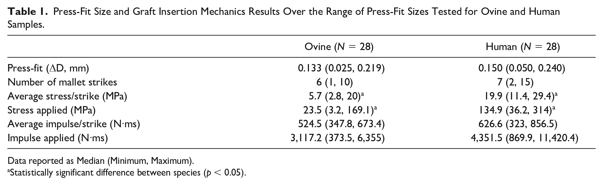
Data reported as Median (Minimum, Maximum).
Statistically significant difference between species (p < 0.05).
Chondrocyte Viability and Histological Analysis
Interspecies differences were observed between control groups for: VCD, 616.1 (481.5, 750.7) cells/mm2, p < 0.0001, TCD, 556.6 (416.6, 696.5) cells/mm2, p = 0.0001, and cartilage thickness, 1.85 (1.40, 2.31) mm, p < 0.0001, (
Table 2
). Interspecies differences were also observed in press-fit sample groups for variables of VCD, 509.4 (397.9, 621.0) cells/mm2, p < 0.0001, TCD, 655.5 (536.1, 774.8) cells/mm2, p < 0.0001, DCD, 146.0 (26.4, 265.7) cells/mm2, and cartilage thickness, 1.91 (1.71, 2.11) mm, p < 0.0001. DCD was not significantly different between species control groups, 59.5 (−87.8, 206.9) cells/mm2, p = 0.3891. Percent live cells and % live cells (SR) were significantly higher in ovine compared to human tissue in control, 31.3 (12.9, 49.7) %, p = 0.0035, SR: 27.9 (3.1, 52.7) %, p = 0.0331, and sample groups, 20.1 (8.7, 31.5) %, p < 0.0001, SR: 16.4 (5.7, 27.1) %, p = 0.0035, likely due to storage duration differences. Cell viability of press-fit samples, when normalized to viability of control samples (% Live Cells [CTC]), showed both species had similar proportions of live chondrocytes after insertion and storage, 1.31 (−16.1, 18.7) %, p = 0.8809. Cell death was observed qualitatively to occur primarily in the superficial region for press-fit below 0.2 mm and through the full thickness for press-fit above 0.2 mm (
Chondrocyte Viability and Histological Grading Results for Human and Ovine Press-Fit Samples and Non-impacted Controls Samples.
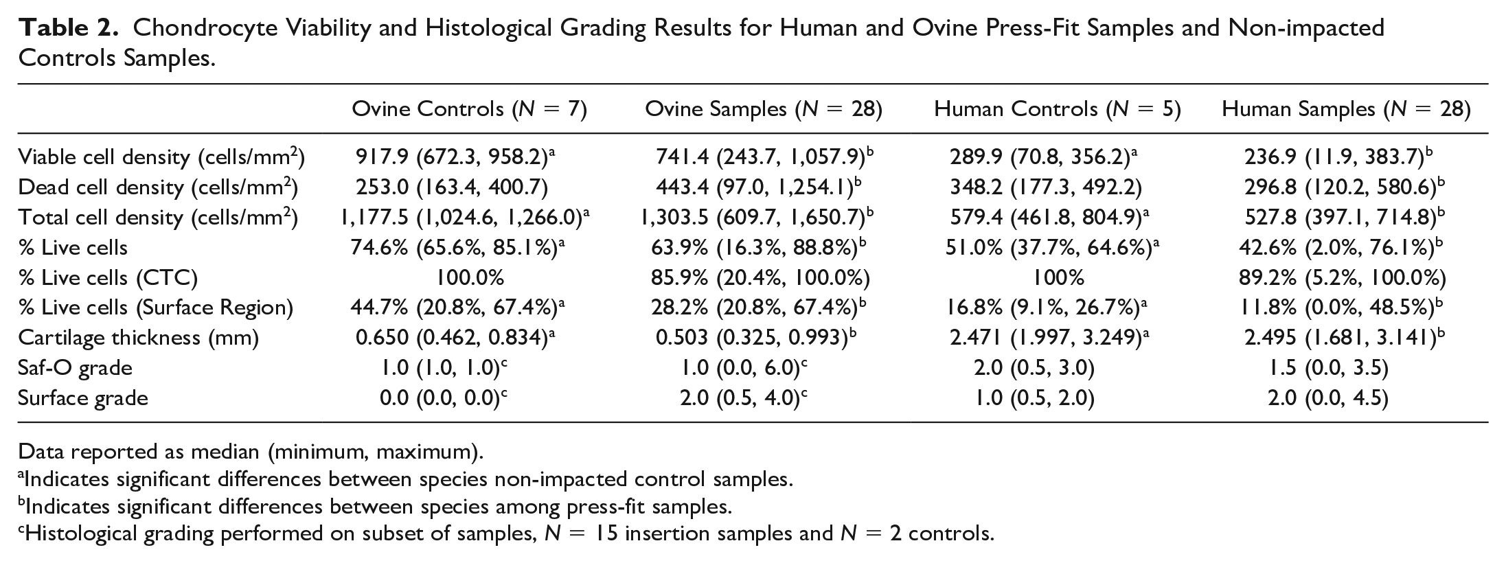
Data reported as median (minimum, maximum).
Indicates significant differences between species non-impacted control samples.
Indicates significant differences between species among press-fit samples.
Histological grading performed on subset of samples, N = 15 insertion samples and N = 2 controls.
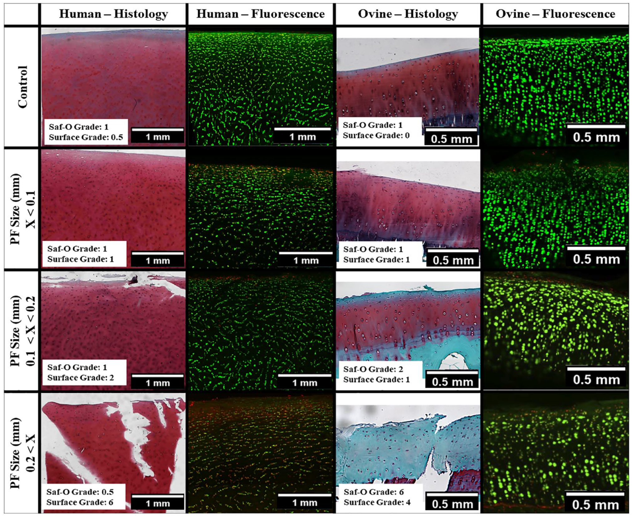
Representative images of human and ovine histological sections stained with safranin-O and fast green stains and confocal microscopy images stained with calcein AM (green, live cells) and ethidium homodimer (red/yellow, dead cells). Images shown for control samples, press-fit sizes below 0.1 mm, press-fit sizes between 0.1 and 0.2 mm, and press-fit sizes above 0.2 mm.
Histological grading showed both species exhibited cracks and surface irregularities with increased press-fit size ( Table 2 ), although having fewer ovine samples available for histological grading may have limited the ability to detect significance among surface grades. Safranin-o staining was qualitatively observed to have reduced stain presence at higher press-fit values for ovine tissue. No trends were observed in safranin-o staining in human tissue, likely due to increased variability in scores, ranging from 0.5 to 3.0, resulting from the lengthy storage conditions prior to testing ( Table 2 ).
Correlation Analysis
For both species, VCD, DCD, % live cells, % live cells (CTC), stress and impulse were linearly correlated to press-fit size (
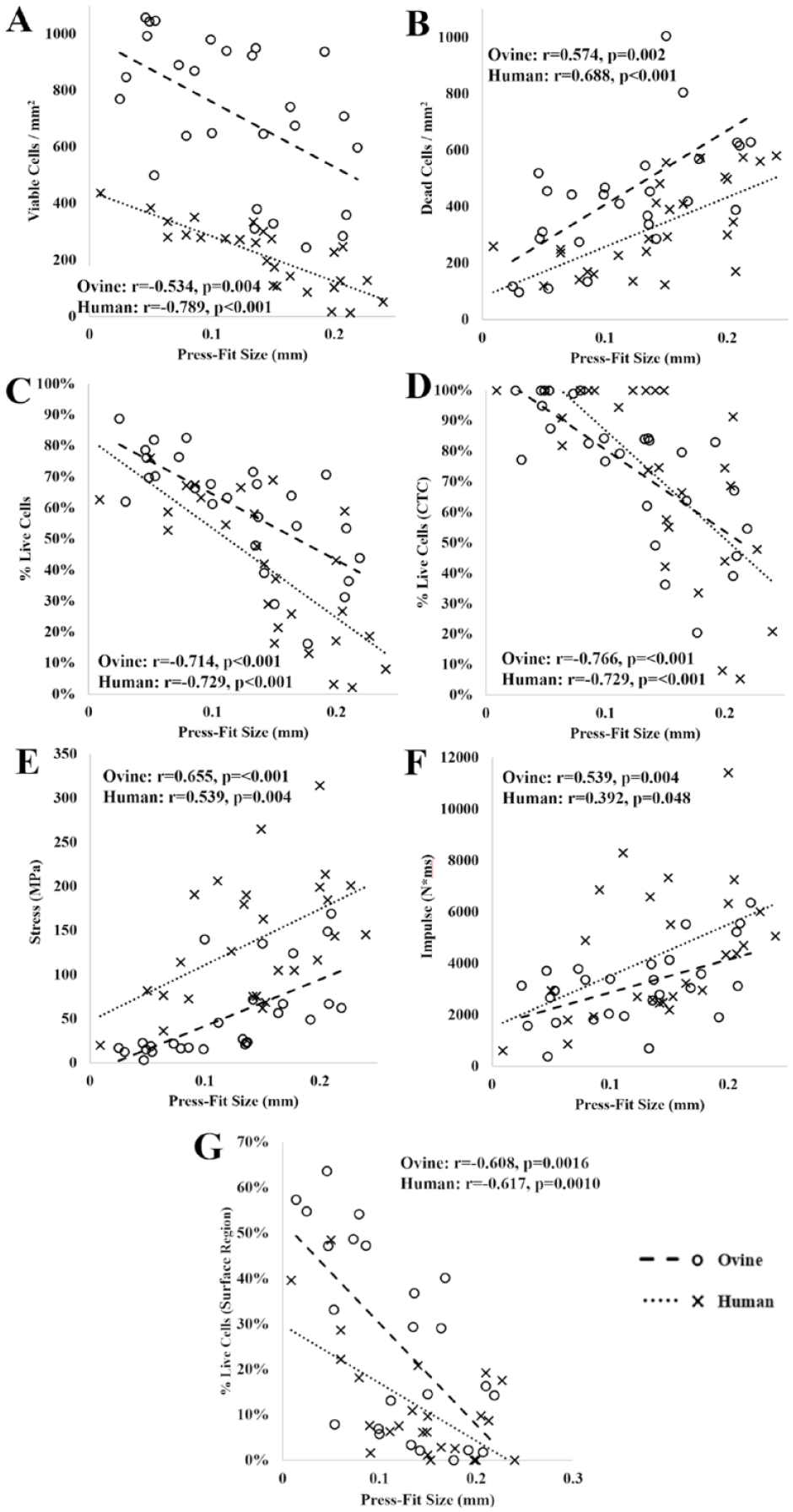
Human and ovine sample correlations between press-fit size (mm) and (
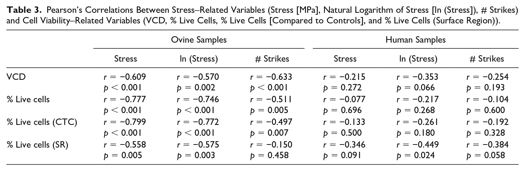
Significant correlations were observed between the natural log of stress and total stress versus chondrocyte variables for ovine samples. Human samples were observed to have a significant correlation between the natural log of stress and % live (SR), but not % live (CTC), which may be due to increased variability in stress observed in human samples ( Table 3 ).
Pearson’s Correlations Between Stress–Related Variables (Stress [MPa], Natural Logarithm of Stress [ln (Stress]), # Strikes) and Cell Viability–Related Variables (VCD, % Live Cells, % Live Cells [Compared to Controls], and % Live Cells (Surface Region)).
Press-Fit Size Cut-Off
These data were further analyzed to identify a press-fit size above which VCD would be insufficient for transplantation based on the 70% threshold for successful transplantation.28,29 The % live cells (CTC) variable was used to investigate a press-fit size cut-off due to its ability to account for differences in total cell density and initial viability. This analysis determined a press-fit size cut-off of 0.150 mm for human samples and 0.135 mm for ovine samples and these values were averaged to 0.14 mm to permit statistical comparisons between data above and below the cut-off (
There were significant differences in most variables of chondrocyte viability, insertion mechanics and histological grading ( Table 4 ) above the press-fit size of 0.14 mm compared to fresh controls. In ovine tissue, VCD, 327.9 (108.9, 547.0) cells/mm2, p = 0.0063, % live cells, 32.3 (16.5, 48.0) %, p < 0.0001, and % live cells (SR), 30.5 (5.6, 55.4) %, p = 0.0228, were significantly higher for fresh control samples compared to samples above the cut-off. In human tissue VCD, 159.0 (73.7, 244.3) cells/mm2, p < 0.0001, % live cells, 24.1 (5.0, 43.2) %, p = 0.0161, and % live cells (SR), 10.8 (2.9, 18.7) %, p = 0.0104, were also significantly higher in the control group compared to samples above the cut-off. In both human and ovine tissues, no variable was significantly different between samples below the cut-off and fresh controls.
Results of Cell Viability, Graft Insertion Mechanics, and Histological Grading Grouped Above and Below 0.14 mm Press-Fit Size Cut-Off (PF).
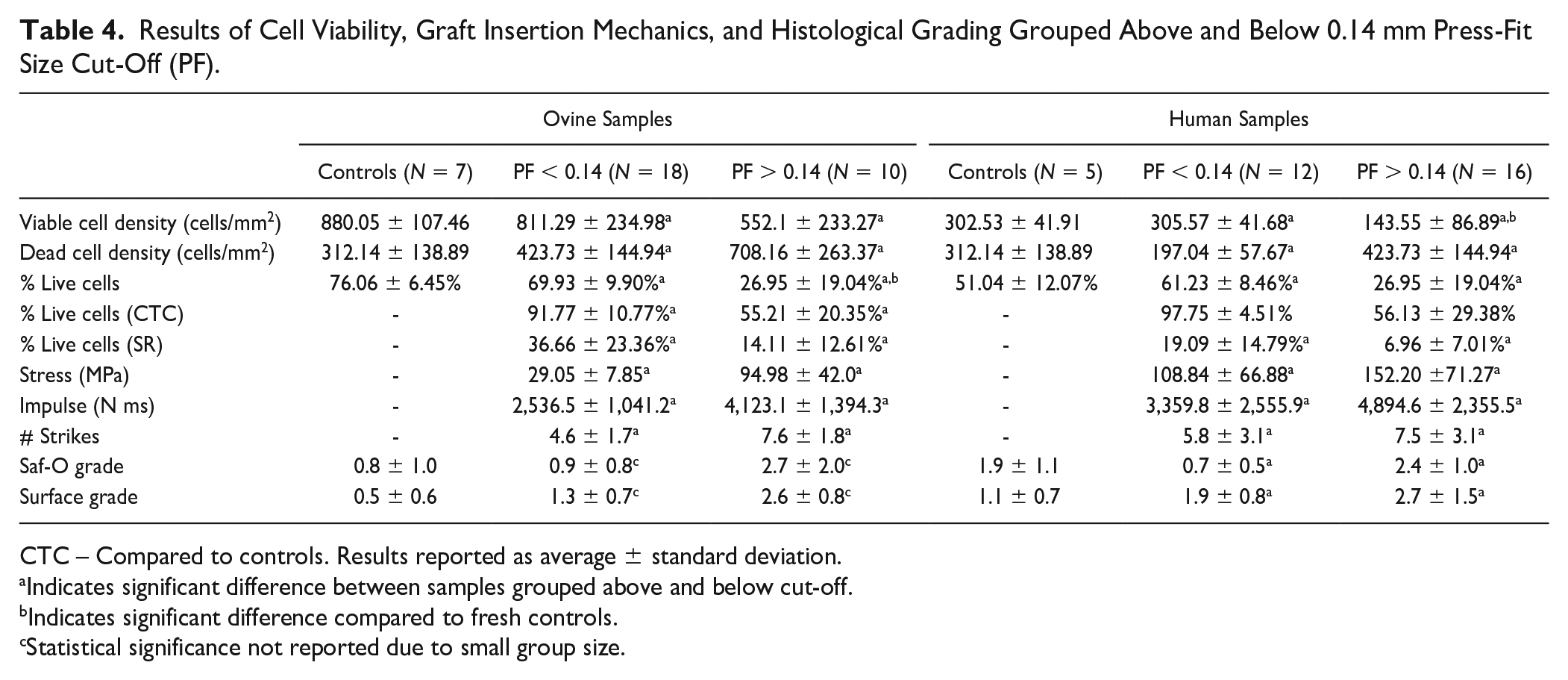
CTC – Compared to controls. Results reported as average ± standard deviation.
Indicates significant difference between samples grouped above and below cut-off.
Indicates significant difference compared to fresh controls.
Statistical significance not reported due to small group size.
Pearson’s correlation analysis conducted separately on samples grouped above and below the press-fit size cut-off (
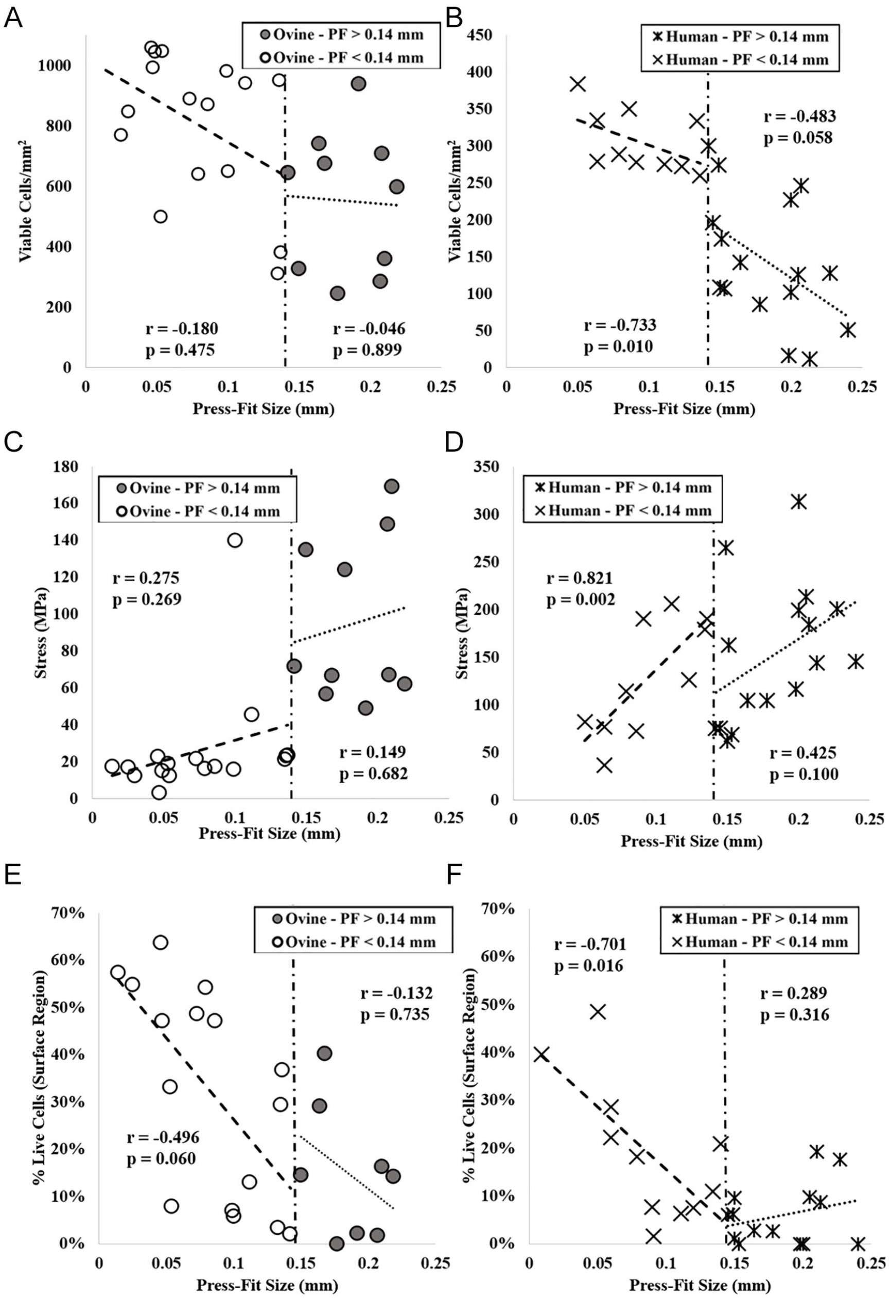
Comparison of variables below and above 0.14 mm press-fit size cut-off for; (
Discussion
Press-Fit Size Correlates With Viability and Force
In human and ovine tissues, press-fit size was positively correlated to graft insertion mechanics and negatively correlated to cell viability (
The current research expands upon Borazjani et al.
3
that tested press-fit at intervals of 0.0, 0.1, and 0.2 mm, by investigating smaller intervals of press-fit within a similar range (Human: 0.050-0.240 mm, Ovine: 0.025-0.219 mm). Smaller increments allowed for estimation of a cut-off value of 0.14 mm, above which chondrocyte viability decreases below acceptable levels, as determined by the 70% chondrocyte viability threshold for OCAT.4-6 Both species showed a reduction in correlation to press-fit size and cell viability as well as increased insertion stress when grouped above this cut-off, compared to samples below the cut-off (
Translation of Experimental Results to Surgical Technique
Impact forces applied to cartilage during graft insertion are typically at the high-end of physiological forces experienced in human and ovine joints. Experiments in fresh cadaveric tissues have shown that the magnitude of forces needed for graft insertion can cause chondrocyte death, primarily in the superficial region. 3 The average stress applied per mallet strike in this study was of a similar magnitude ( Table 1 ), with stresses in the physiological range for sheep (5-8 MPa) 32 and above the physiological range experienced in the human knee (5-12 MPa);33,34 however, far fewer mallet strikes were required ( Table 1 ) compared to estimates of the clinical procedure. 11 Differences in surgical technique are hypothesized to introduce variability to the magnitude of forces applied; therefore, it is difficult to state the magnitude of typical insertion forces and whether these cause significant chondrocyte death. This research presents evidence of the relationship between press-fit, insertion force and cell viability, but magnitudes of force and cell death are more variable in the clinical setting.
A press-fit cut-off of 0.14 mm determined using the 70% viability threshold,4-6 demonstrated differences in correlations with both chondrocyte viability and stress above and below 0.14 mm (
Interspecies Comparison
Interspecies osteochondral tissue differences are well documented, with thicker human cartilage and differences in bone architecture.21,22 Thicker human cartilage was expected to affect transmission of impact loads and magnitude of cell death, which can be observed in the micrographs (
Limitations
Study limitations include discrepancies between the force measured and the force delivered to human grafts. We propose that thicker human cartilage ( Table 2 ) and interspecies differences in bone properties 20 contributed to mallet deflections, resulting in weaker correlations and increased measured stress in human samples compared to ovine. In addition, this study was unable to assess the effects of impact insertion on the graft periphery because chondrocyte viability assessments were performed only on the central regions of the graft width to exclude cell death resulting from graft harvest.25,35 Finally, this study was unable to assess the effects of donor storage time on cartilage response to impact stress because, although human samples were obtained from donor tissues stored for different durations, there was an absence of grafts covering the full range of press-fit sizes from each donor. Differences among donors were minimized by normalizing to donor-specific controls.
Findings from this study are applicable to a certain extent in the clinical setting. The two common techniques for OCAT are the shell technique, where grafts are manually cut to match the recipient site, and the dowel technique, which uses grafts prepared using commercial cutting guides. 36 The results of this study are more applicable to the dowel technique, as manufacturers tune instruments for precise sizes. In this study, grafts 4.5 mm in diameter were used, which is similar in size to grafts used in autograft transplantation, 37 but smaller than allografts which range from 15 to 35 mm in diameter. 8 Estimations of interface stress in a press-fit system 38 supports the conclusion that increasing graft diameter requires greater press-fit size to achieve the same interface stress. These estimations assume solid structure and do not consider interspecies structural differences of subchondral bone. 21
Conclusion
Increasing press-fit size of both human and ovine osteochondral grafts increased the likelihood of cartilage being subjected to high-impact forces that reduce cell viability below levels acceptable for transplantation. Chondrocyte viability fell below 70% compared to controls when using press-fit size greater than 0.13 to 0.15 mm for both human and ovine grafts. A cut-off press-fit size value between 0.13 and 0.15 mm is suggested for osteochondral grafts of similar diameter; however, further investigation is needed to translate the findings to the clinical scenario with larger diameter osteochondral grafts. Despite differences in tissue structure and characteristics, both ovine and human tissues responded similarly to press-fit size, providing evidence for the use of the ovine model for allograft-related research.
Footnotes
Acknowledgments and Funding
Special thanks to David Murray & Brendan Wood for their contributions to this research. The author(s) disclosed receipt of the following financial support for the research, authorship, and/or publication of this article: RPS was supported by a Canadian Graduate Scholarship (Master’s) from the Natural Sciences and Engineering Research Council of Canada. The study sponsors had no role in the study design, design, analysis and interpretation of data; in the writing of the manuscript; and in the decision to submit the manuscript.
Declaration of Conflicting Interests
The author(s) declared no potential conflicts of interest with respect to the research, authorship, and/or publication of this article.
Ethical Approval
All procedures were conducted with approvals from the Institutional Review Board of Sinai Health System (REB 19-0080-E) and Institutional Animal Care Committee of the University of Guelph (AUP #3974).