
Abstract
Objective
Knee osteoarthritis (KOA) is a complex degenerative joint disease and a major cause of joint dysfunction. This study aimed to explore the function of hsa_circ_0007482 on inflammation, proliferation, differentiation, and apoptosis in KOA.
Design
Real-time quantitative polymerase chain reaction (PCR) was performed to detect the expression of circ_0007482, inflammatory factors, and differentiation-related molecules in KOA chondrocytes and interleukin (IL)-1β–stimulated chondrocytes. The correlation between the circ_0007482 expression and inflammatory factors was analyzed by the Pearson method. KOA cell model was established using IL-1β for 24 hours. The proliferation activity of chondrocytes was evaluated by CCK-8 assay, and cell apoptosis rate was assessed by flow cytometry. The downstream miRNA of circ_0007482 was validated using dual-luciferase reporter assay.
Results
The circ_0007482 expression was elevated in both KOA cartilage tissues and IL-1β–treated chondrocytes and positively correlated with inflammatory factors expression. In comparison to the control group, IL-1β treatment diminished chondrocyte proliferation abilities and increased cell apoptosis and inflammatory factors IL-6, IL-8, and tumor necrosis factor (TNF)-α mRNA expression. Inhibition of circ_0007482 partially improved IL-1β–induced inflammatory reaction. Circ_0007482 could negatively regulate the expression of miR-558.
Conclusions
Interfering of circ_0007482 might partially promote cell proliferation and differentiation, while inhibit cell apoptosis to improve joint injury by regulating miR-558 in IL-1β–treated chondrocyte cell model.
Introduction
Osteoarthritis (OA), also known as osteoarthropathy, is a common clinical degenerative disease of bone and joint swelling, pain, deformity, and complex pathogenesis.1,2 OA can develop in any joint, but most commonly affects the knees, hips, hands, and feet. As a common chronic disease in the elderly population, knee osteoarthritis (KOA) is mainly characterized by degeneration of articular cartilage and subchondral bone hyperplasia. 3 The prevalence and incidence of KOA are increasing year by year due to many influence factors.4,5 The typical pathological changes of KOA include destruction of cartilage, formation of osteophyte, and hyperplasia of synovial. 6 Cartilage is a major component of the joint and has a buffering effect to relieve stresses acting on the joint. Chondrocytes in cartilage can produce collagen and glycosaminoglycans to form a concentrated and highly coordinated extracellular matrix (ECM) that plays a key role in the progression of OA. 7 In addition, inflammation is a key feature associated with progression of OA. 8 At present, the treatment of KOA mainly focuses on improving the clinical symptoms of patients (relieving pain and controlling inflammation), and there is still a lack of effective treatment therapies. Therefore, understanding the pathogenesis of KOA is crucial for the treatment of KOA.
Circular RNA (circRNA) is a type of endogenous non–coding RNA that has a closed-loop structure, which can regulate the progression of a variety of diseases and participate in cellular activities, such as growth, differentiation, and apoptosis.9,10 Numerous circRNAs are aberrantly expressed in OA and play their role through miRNA sponge, regulation of pathways, and direct targeting of protein molecules.11-13 Hsa_circ_0007482, also known as circCOL5A1, is located at chr9:137582757-137593179 and generated by back-splicing of exons 13 to 19 of the Collagen type V alpha 1 chain (COL5A1) gene. COL5A1, one of the ECM-related genes, is more prevalent in OA subchondral bone tissues compared with normal tissues and may be a biomarker for OA. 14 Circ_0007482 was upregulated in aseptic loosening synovial tissues after total hip arthroplasty. 15 Whereas the expression and influence of circ_0007482 in KOA has not been reported.
circRNAs were indicated as functional sponges for miRNAs in diseases.16,17 Circ_0007482 could interact with miR-27a-3p, miR-6512-3p, miR-4483, and miR-3918 in keloid 18 and other miRNAs in Ni et al. 15 miR-558 has regulatory role in the processes of many diseases, such as chronic obstructive pulmonary disease and OA.19,20 miR-558 expression was found to be lower in OA tissues than in normal articular cartilage. 21 However, whether circ_0007482 has correlation with miR-558 remains unclear.
Therefore, in this study, 15 patients with meniscus injuries and 25 patients with KOA were selected to detect the expression of circ_0007482 in KOA tissue specimens. The functional implications and potential mechanism of circ_0007482 in modulating the pathophysiology of KOA were elucidated.
Materials and Methods
Tissue Specimens
All procedures were performed in line with the principles of the Declaration of Helsinki. This study was giving permission by the ethics committee of Zhejiang Provincial People’s Hospital, approval number: 2021(025), and informed consent was obtained from the participants. A total of 15 patients with meniscus injuries (normal control) and 25 patients with KOA admitted to our hospital from March 2021 to June 2023 were selected in this study. All patients met the inclusion criteria of this study: (1) patients with KOA were diagnosed according to the clinical diagnostic criteria of American College of Rheumatology; (2) patients with grade 3 meniscus injuries had no other underlying diseases except meniscus injury, (3) patients with other joint disease, such as rheumatoid arthritis, ankylosing spondylitis, joint involvement, infection, and tumor, were excluded. Articular cartilage tissue samples from the knee joint were collected after total knee arthroplasty surgery or arthroscopic resection surgery and were stored in liquid nitrogen.
Cell Line Culture and Cell Model Conduction
Human chondrocyte CHON-001 cell line was gained from ATCC and cultured in DMEM (Gibco, Thermo Fisher Scientific, Waltham, MA) with 10% fetal bovine serum (FBS) (Gibco). The culture condition was 37°C and 5% CO2. The chondrocyte CHON-001 cells were treated with a medium containing 10 ng/ml interleukin (IL)-1β for different time intervals, which was recorded as the IL-1β group (KOA in vitro model). At the same time, the normal cultured chondrocytes were recorded as the control group.
Cell Transfection
CHON-001 chondrocytes in the logarithmic growth phase were collected and seeded in 6-well plates (1 × 10⁴ cells/well). Once cell growth confluence reached 60% to 70%, cell transfection was performed using Lipofectamine 3000 Transfection Reagent (Invitrogen, Waltham, MA). Circ_0007482 siRNA (si-circRNA-1, si-circRNA-2, si-circRNA-3), siRNA negative control (si-NC), miR-558 mimic, and mimic NC were synthesized by Sangon Biotech (Shanghai, China). After 24 hour culture of CHON-001 with 10 ng/ml IL-1β, either si-circRNA or si-NC was added. Cell transfection efficacy was measured after 48 hours.
Total RNA Extraction and Quantitative Real-Time Polymerase Chain Reaction
RNA extraction experiments were performed in the absence of RNase. Tissue samples were washed 3 times with sterile phosphate-buffered saline (PBS) containing 1% double antibody. The normal cartilage tissues and KOA cartilage tissues (50 mg) were placed directly into a mortar, ground to a power under liquid nitrogen, ground by adding Trizol reagent, and the grinding solution was put into a 2 ml centrifuge tube for total RNA extraction. Reverse transcription was performed using Evo M-MLV RT Kit with gDNA Clean for quantitative polymerase chain reaction (qPCR) kit. SYBR Green Premix Pro Taq HS qPCR Kit was used in Quantitative Real-Time Polymerase Chain Reaction (RT-qPCR). The relative expression of target genes was calculated by 2−ΔΔCt method.
RNase R Experiment
RNA was extracted from cells by the Trizol method. Total RNA (2 ng) was taken, one was used as control, the other was added 6 U RNase R (3 U/pg), placed in a 37°C, 5% CO2 volume fraction incubator digestion for 15 minutes through reverse transcription to form cDNA. RT-qPCR was performed to detect the relative expression of circ_0007482 and COL5A1.
CCK-8 Assay
The transfected cells (2000 cells/well) were seeded in 96-well plates for cell proliferation detection using cell counting kit-8 (CCK-8; Dojindo, Japan). Over a period of 3 days, 10 μl of CCK-8 reagent was added to each well every 24 hours. After 1 hour of further cultivation, the absorbance at 450 nm was measured to assess the proliferation abilities.
Apoptosis Assay
Cells in the logarithmic growth phase were selected, washed 3 times with precooled PBS, resuspended in 500 μl PBS, added with 5 μl Annexin V-fluorescein isothiocyanate and 5 μl propidium iodide, and incubated at room temperature for 10 minutes. Then, the apoptosis rate was detected by the FACS Calibur flow cytometry.
Dual-Luciferase Reporter Assay
Wild type (WT) or mutant (MUT) circ_0007482 was cloned into the pmirGLO plasmid receptor, and miR-558 mimics or mimic NC was transferred into CHON-001 at the same time. After 48 hours of co-culture, the dual-luciferase reporter assay system was used to measure the dual-luciferase activity.
Statistical Analysis
GraphPad Prism 9.0 software was used for statistical analysis; the experimental data were expressed as mean ± SD. Two independent sample Student t tests were used for comparison between 2 groups. One-way analysis of variance (ANOVA) was used for comparison among multiple groups. If the data set was normally distributed (measured by Kolmogorov-Smirnov test), the Pearson correlation analysis was used for correlation analysis. *P < 0.05, **P < 0.01, and ***P < 0.001 were considered statistically significant.
Results
Circ_0007482 Expression in KOA Cartilage Tissue
To comprehend the expression characteristics of circ_0007482 in articular cartilage tissue of KOA patients, the expression of circ_0007482 in KOA and normal articular cartilage tissues was detected. As shown in
Figure 1A
, circ_0007482 expression in cartilage tissues of KOA patients was increased in contrast to that in normal cartilage tissues. Furthermore, the COL5A1 mRNA was also measured and showed an elevated expression in KOA cartilage tissues in contrast with normal cartilage tissues (
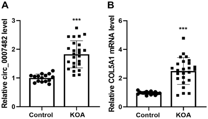
Circ_0007482 and Collagen Type V alpha 1 chain (COL5A1) mRNA expression in articular cartilage tissues was measured by quantitative real-time PCR (RT-qPCR). (
Correlation Between Upregulated Circ_0007482 and Inflammatory Factors
To explore the expression level of inflammatory genes in articular cartilage tissues of patients with KOA, this study measured the IL-6, IL-8, and tumor necrosis factor (TNF)-α mRNA levels in cartilage tissues of KOA patients and in normal articular cartilage tissues of patients in the control group by RT-qPCR. The results in Figure 2A-C displayed that the mRNA levels of IL-6, IL-8, and TNF-α were higher in KOA tissues than in normal cartilage tissues.
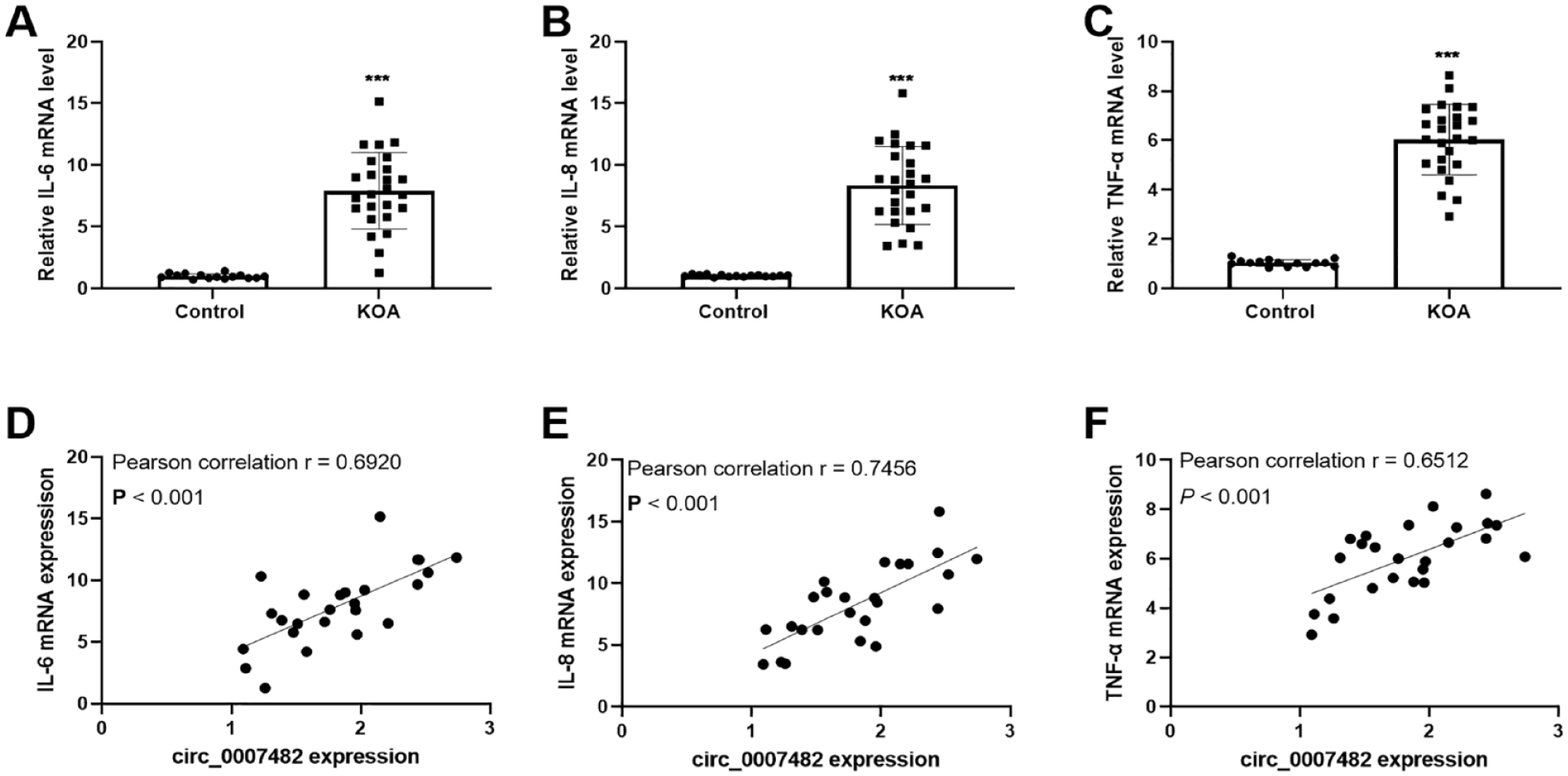
There is a correlation between upregulated circ_0007482 and inflammatory factors. (
The Pearson correlation analysis was used to assess the relationship between circ_0007482 and KOA-related inflammatory factors. As depicted in Figure 2D-F , circ_0007482 was positively correlated with the mRNA levels of IL-6, IL-8, and TNF-α.
Silencing of Circ_0007482 Enhanced Chondrocyte Viability and Repressed Apoptosis After Interleukin-1β Injury
RNase R treatment experiments were used to confirm the cyclization of circ_0007482. The relative expression of circ_0007482 did not significantly change in the control and RNase R-treated chondrocytes CHON-001, whereas the COL5A1 mRNA levels were downregulated in the RNase R-treated group (
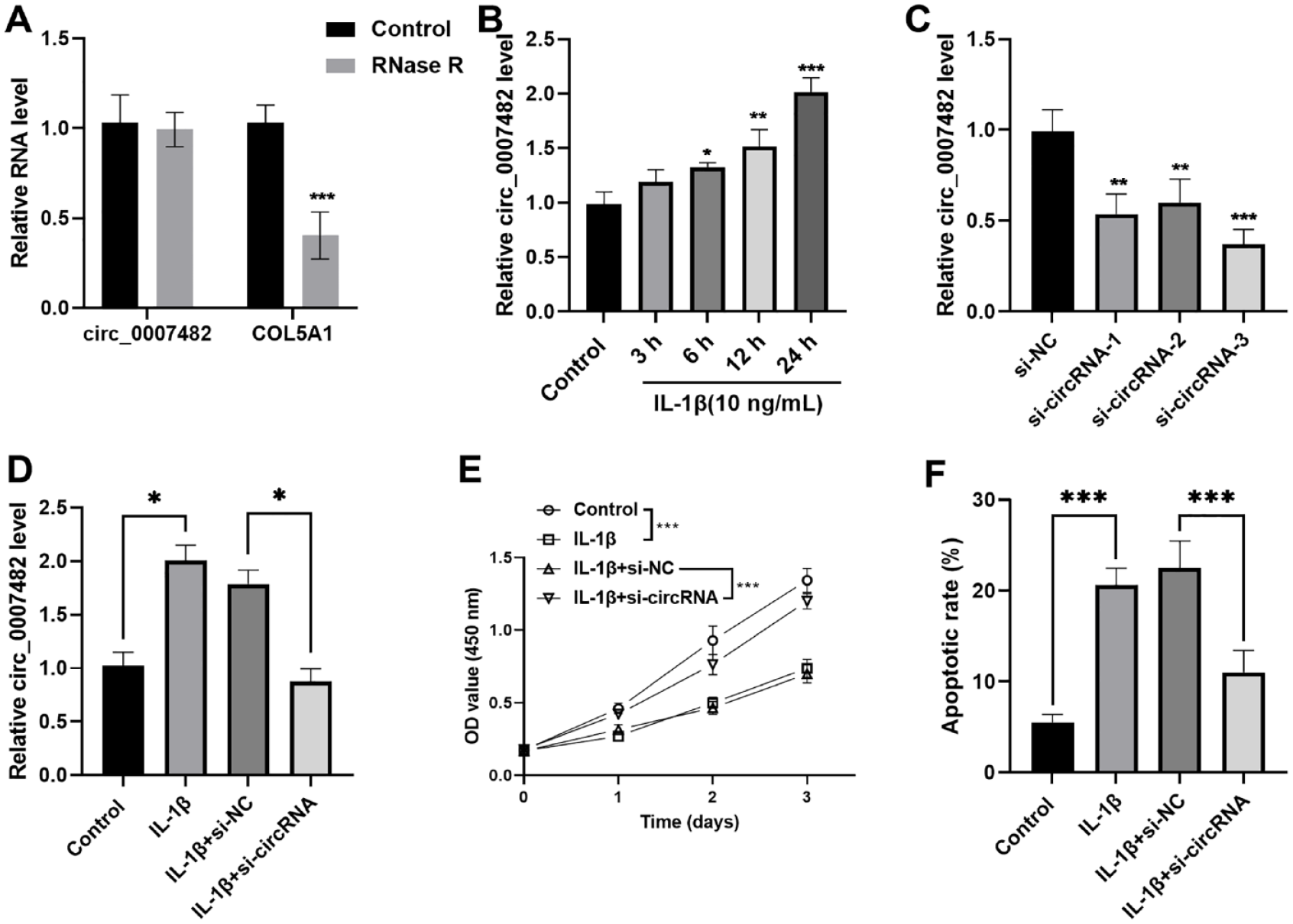
Effect of circ_0007482 on IL-1β–treated CHON-001 cell proliferation and apoptosis. (
Effect of Circ_0007482 Downregulation on the Inflammatory Factors and Differentiation of Chondrocytes
The inflammatory factors including IL-6, IL-8, and TNF-α mRNA expression were measured in KOA cell model. IL-1β–treated chondrocytes exhibited higher IL-6, IL-8, and TNF-α mRNA levels compared with the control group. In contrast with the IL-1β + si-NC group, silencing circ_0007482 decreased the inflammatory factors expression (
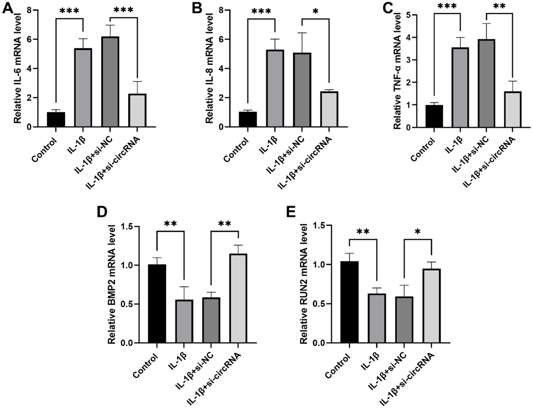
Low expression of circ_0007482 repressed inflammatory factors and promoted differentiation of CHON-001 chondrocytes. (
Circ_0007482 Targeted and Bound to miR-558 in Chondrocytes CHON-001 Cells
The potential circ_0007482-targeted miRNAs were predicted through the CircInteractome website (http://circinteractome.nia.nih.gov). Based on the binding score over 90, 7 miRNAs were selected and measured in CHON-001 cells transfected with circ_0007482 siRNA. Among these miRNAs, miR-558 displayed the most significant expression changes (
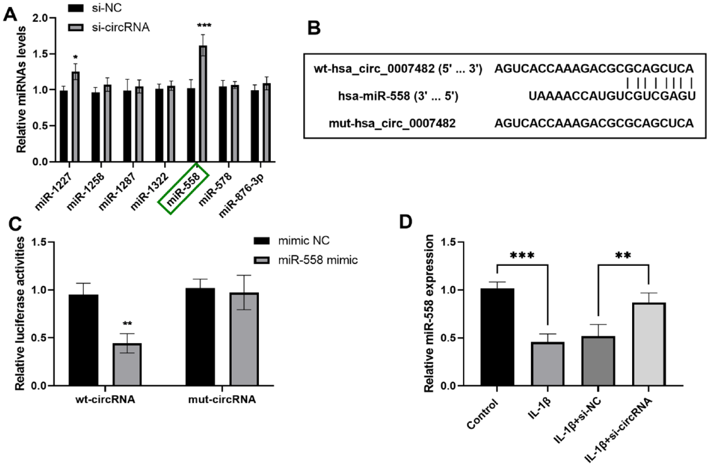
miR-558 was a potential target of circ_0007482. (
Discussion
This study indicated that circ_0007482 expression was elevated in both KOA tissues and the OA cell model. Silencing circ_0007482 could facilitate chondrocyte cell proliferation and differentiation and repress cell apoptosis and chondrocyte cell inflammation. In addition, miR-558 may be the target miRNA of circ_0007482.
With the in-depth study of genetics and the development of advanced biotechnology, such as RNA sequencing and bioinformatics methods, numerous non-coding RNAs have been identified.22,23 Research increasingly confirmed that circRNA has crucial biological functions, including miRNA sponging, functional protein trapping, and translation functions, and that the dysfunction of circRNA is involved in the occurrence and progression of various diseases.24,25 Increasing studies identified circRNAs in OA progression and the regulation of disease state, such as circRNA HIPK3 and circ_0128846.26,27 The aberrant expression of circ_0007482 was involved in the pathogenesis of pterygium and keloid scarring.18,28 In this study, overexpressed circ_0007482 was detected in the cartilage tissues of KOA cases, which presented that circ_0007482 might act as a promoting factor in the progression of KOA.
IL-6, IL-8, and TNF-α are inflammatory factors associated with OA and are the main inflammatory mediators in the pathogenesis of KOA. 29 The levels of inflammatory factors can reflect the degree of OA lesions to a certain extent. There is a synergistic effect between the inflammatory factors, which can lead to the destruction of articular cartilage and accelerate the process of OA by mediating the inflammatory response together. 30 The mRNA expression of IL-6, IL-8, and TNF-α in KOA articular cartilage was higher than that in normal articular cartilage, which verified the correlation between inflammatory factors and the progression of KOA. Furthermore, circ_0007482 was positively correlated with the trend of inflammatory factors, which hinted that circ_0007482 might be significantly correlated with the KOA progression.
Abnormal proliferation and differentiation of chondrocytes are the main characteristics affecting joints in KOA diseases. 31 CircRNA is differentially expressed in OA. For instance, circ_0002715 was upregulated in OA tissues, and interfering with circ_0002715 could inhibit inflammation and apoptosis in OA chondrocytes induced by IL-1β by sponging miR-127-5p. 32 This study explored the cell function using CHON-001 cells and IL-1β act on CHON-001 cells to mimic the KOA cell model in vitro. BMP2 and Runx2, which are crucial for chondrocyte differentiation, have increased expression when circ_0007482 is downregulated. These data suggest that the downregulation of circ_0007482 could promote cell differentiation. Interference with circ_0007482 not only facilitated IL-1β-stimulated CHON-001 cell proliferation and differentiation, but also repressed inflammation and apoptosis. Similarly, some circRNAs, such as ciRS-7, circSERPINE2, and circ_0045714, are able to inhibit the pathogenesis of OA. 33 These data confirmed the crucial role of circRNAs in the pathogenesis of OA.
The online CircInteractome website predicted the potential targets of circ_0007482. miR-558 was verified to be a target miRNA of circ_0007482. miR-558 could be targeted by circ_0043610 or circ_0083756 and take part in the progression of several diseases, such as preeclampsia or intervertebral disc degeneration.34,35 Importantly, miR-558 expression was found to be lower in OA tissues than in normal articular cartilage. 21 Another study indicated that circTBX5 could bind to miR-558 to mitigate IL-1β–stimulated chondrocyte apoptosis, ECM degradation, and inflammation. 36 Consistently, herein, miR-558 was downregulated in IL-1β–induced chondrocyte CHON-001 cells. In addition, circ_0007482 could negatively regulate miR-558 expression. These data revealed that circ_0007482 might participate in regulating chondrocyte proliferation, differentiation, and apoptosis of KOA by modulating miR-558 expression. However, the precise regulatory role and specific mechanism of circ_0007482 in the onset and progression of KOA require further verification in future in vivo studies. Besides, whether inhibition of circ_0007482 has effect on the other circRNAs remains unknown, which also need to be explored in future.
In summary, circ_0007482 can partially ameliorate the IL-1β–stimulated chondrocyte injuries by mediating miR-558. This study explored the pathogenesis of KOA from the perspective of circRNA dysregulation, providing the basis for circ_0007482 as a valuable marker for KOA.
Footnotes
Acknowledgments and Funding
The authors have no acknowledgments. The author(s) disclosed receipt of the following financial support for the research, authorship, and/or publication of this article: This study was funded by General Program of Zhejiang Provincial Health Commission (Nos 2022492562 and 2023583754).
Declaration of Conflicting Interests
The author(s) declared no potential conflicts of interest with respect to the research, authorship, and/or publication of this article.
Ethical Approval
All procedures were performed in line with the principles of the Declaration of Helsinki. This study was giving permission by the Ethics Committee of Zhejiang Provincial People’s Hospital and informed consent was obtained from the participants.